High resolution photon emission computed tomographic imaging tool

Inactive Publication Date: 2006-09-12
NEUROLOGICA CORP
View PDF14 Cites 12 Cited by
  • Summary
  • Abstract
  • Description
  • Claims
  • Application Information

AI Technical Summary

Benefits of technology

[0007]The present invention utilizes a collimator with a sharp focus within the object, and steeply tapered holes that provide extremely high sensitivity-resolution characteristics. This requires that the detector be translated and moved radially-but, with sufficient numbers of detectors to provide 360 degree scanfield coverage, no rotational movement is necessary. By its nature, namely the constan

Problems solved by technology

Furthermore, it is immune to various gamma camera effects caused by errors in fi

Method used


Image

  • High resolution photon emission computed tomographic imaging tool
  • High resolution photon emission computed tomographic imaging tool
  • High resolution photon emission computed tomographic imaging tool

Examples


Embodiment Construction

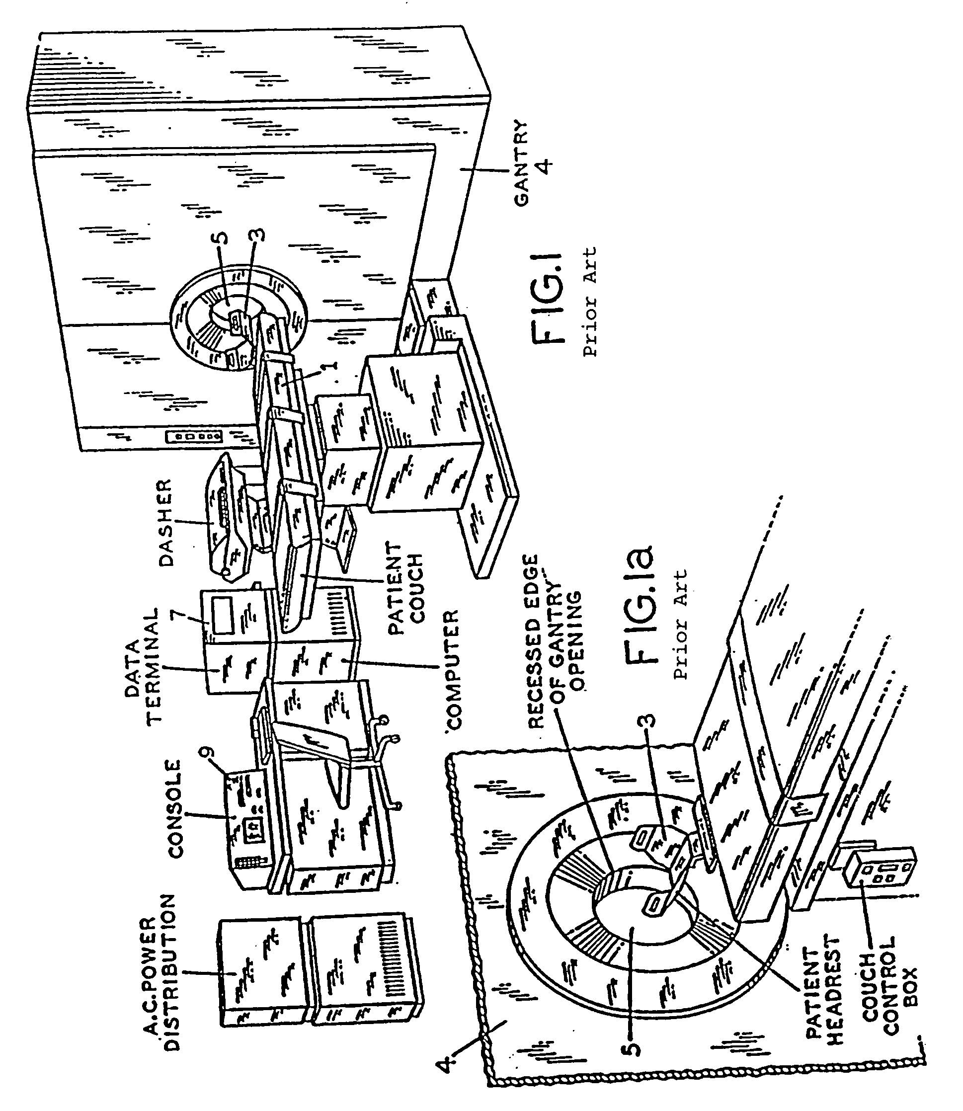
[0029]With reference to FIG. 1, a patient's couch is indicated at 1 which is provided with controls, not shown, for raising and lowering the couch 1, and for moving the headrest 3, of couch 1, in and out of the opening 5 of the gantry indicated at 4. Within gantry 4, as hereinafter more fully described, there is arranged, in a unique and novel manner, a plurality of scanning detectors, having highly focused collimators, from which electrical signals are obtained which are readily processed, e.g. by a general purpose computer, and enable a display at console 9 of a transverse section of the brain of a radionuclide administered patient, which display exhibits high sensitivity quantification and spatial resolution. The patients couch 1 is moveable in and out of the opening 5 of the gantry 4 to provide for the scanning of a plurality of transverse sections.

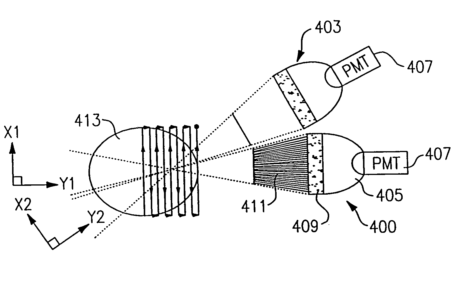
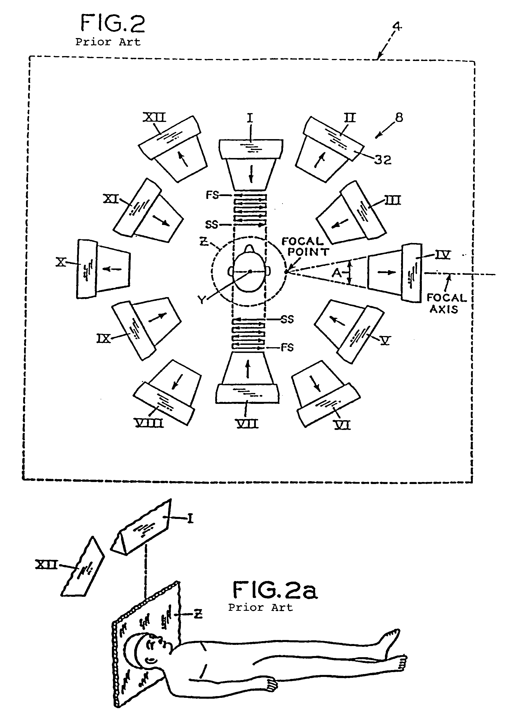
[0030]With reference to FIG. 2, this figure shows at 8 an essentially schematic representation of the arrangement of scanning detect...


PUM


Abstract

An brain scanning apparatus having a scanner device and a host computer. The scanner radiation detectors detect radiation emitted from a desired portion, or slice, of a brain source. The scanner device contains microprocessors and code which control the movement of the radiation detectors and performs the data acquisition over a number of slices and transmits this data to the host computer. The host computer initiates a scan by sending desired setup parameters to the scanner device and instructing the scanner to begin collecting data. During the scan, acquired data is sent to the host computer and spooled to a hard disk. The computer can be instructed to perform a slice by slice reconstruction of the source brain while the scan is taking place in order to produce a 2-dimensional reconstruction of the mapped brain. A complete 3-dimensional reconstruction of all the compiled slices is performed and visually displayed after acquisition of all the required slices.

Description

FIELD OF THE INVENTION[0001]The present invention provides a very high resolution single-photon emission computed tomographic (SPECT) imaging tool for brain function research and brain disease diagnosis. It is intended to complement other functional imaging modalities such as positron emission tomography (PET), functional magnetic resonance imaging (fMRI), electroencephalography (EEG) and event-related potential (ERP), magnetoencephalography (MEG), and new, near-infrared optical imaging.BACKGROUND OF THE INVENTION[0002]In the last quarter century the use of brain imaging for the treatment and understanding of diseases and genetic flaws has grown dramatically following the introduction of Tomographic X-Ray (CT) in 1972 followed in 1982 by magnetic resonance in the gene (MRI). The reason for this growth and importance in brain imaging is that neurologists, psychotherapist, and neuro-scientists utilize and attached substantial importance to high resolution, three dimensional anatomical...

Claims


Application Information

Patent Timeline
IPC IPC(8): G01T1/166G01T1/164
CPCG01T1/1644A61B6/037G01T1/2985
Inventor STODDART, HUGH A.STODDART, HUGH F.
Owner NEUROLOGICA CORP
Why Eureka
Try Eureka