Methods for monitoring and optimizing central venous pressure and intravascular volume
a technology of intravascular volume and measurement method, applied in the field of medicine, can solve the problems of inability to accurately assess the intravascular volume status of patients' physical examination and medical history, failure of the heart's pumping mechanism, and failure of the pumping force of the heart, so as to remove the disadvantages of invasive cvp measurement and maintain the accuracy of such invasive measurements
- Summary
- Abstract
- Description
- Claims
- Application Information
AI Technical Summary
Benefits of technology
Problems solved by technology
Method used
Image
Examples
example 1
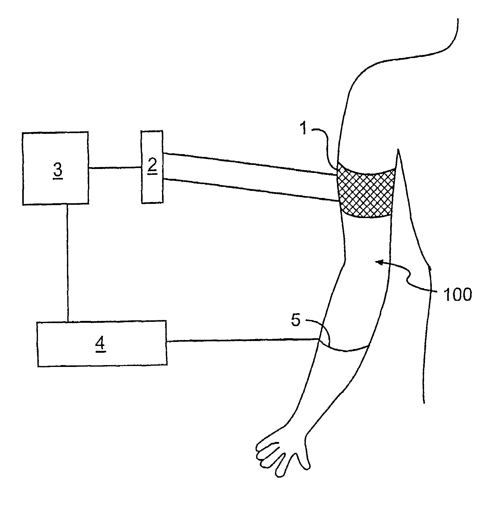
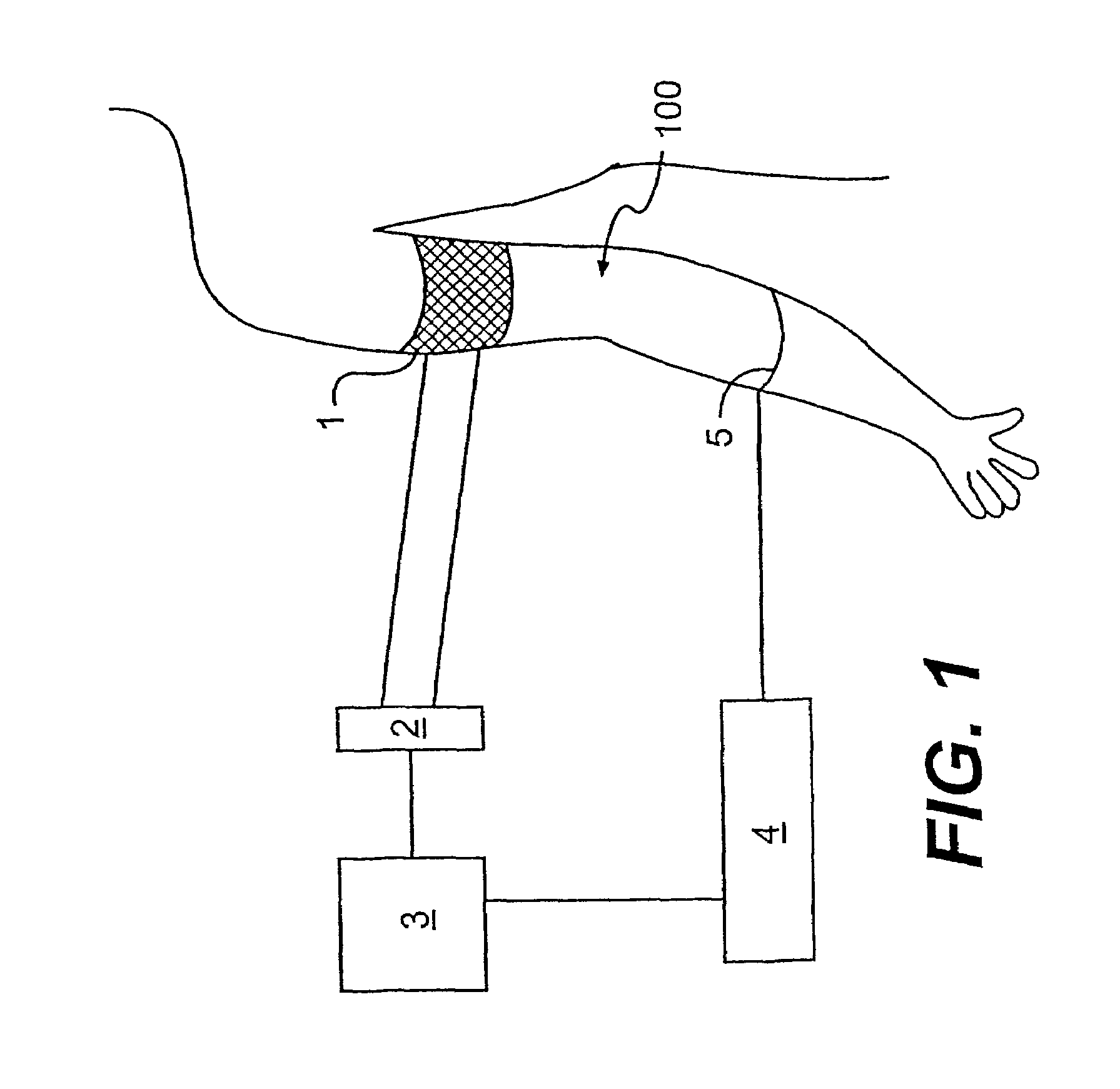
[0086]It was observed that previously it had been shown that pressure within the axillary vein of the upper extremity is nearly equal to that of CVP at the level of the superior vena cava and right atrium. It further was observed that previously it had been demonstrated that intraluminal activity vein pressure can be accurately estimated from pressures obtained with pressure cuffs applied to the upper arm, which surround the axillary vein. It was theorized as follows. When applying sufficient pressure over the axillary vein, it occludes preventing venous return. With arterial flow still constant, the forearm swells at a rate proportional to the arterial inflow. The resultant change in forearm volume can be accurately monitored with several types of plethysmography. Rather than calculate the value of forearm swelling, it was proposed that the forearm volume will increase beyond a critical threshold (approximately 0.5% volume) when the axillary vein is occluded. As a result, it should...
example 2
[0096]14 human patients were undergoing invasive monitoring of CVP as part of their routine care. From each patient, simultaneously to taking of invasively measured CVP, non-invasive CVP was taken from the upper extremity using a system according to FIG. 1 herein. Data from these 14 subjects (four readings from each subject) are depicted in FIG. 4. In this series noninvasive CVP was taken from the upper extremity and is compared to the invasively measured CVP. Notably, the correlation is over 0.9 (specifically, 0.97). In no instance would the values have differed to the magnitude that different patient management would have been indicated for the noninvasively measured CVP versus the invasively measured CVP.
example 3
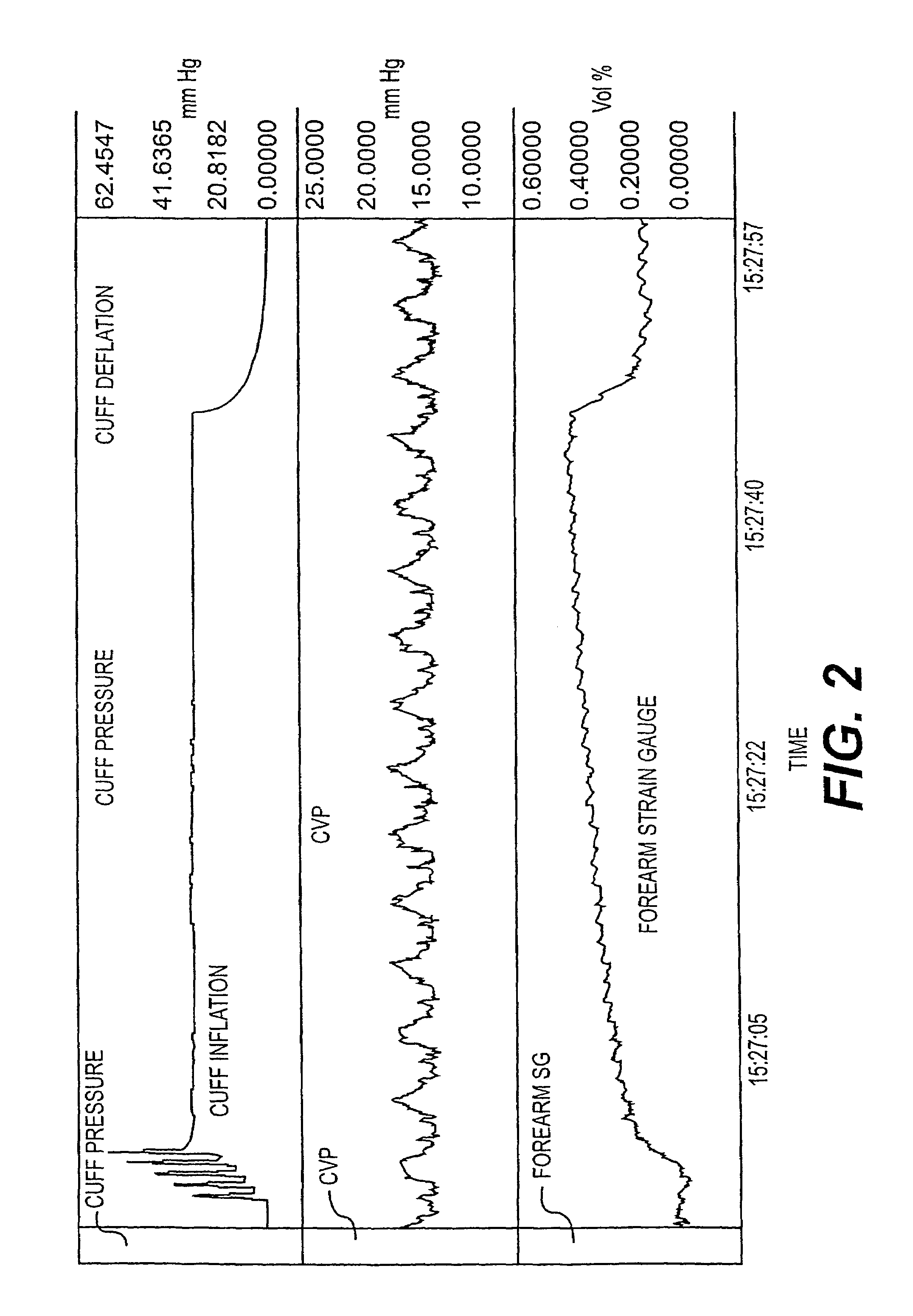
[0097]FIG. 3 is a detailed analysis of the recording from FIG. 2 during deflation of the vascular cuff. The steepest portion of the slope (derivative) was located. The cuff pressure at this time was noted. In this case it was exactly the same as the mean CVP noted during the time indicated between the two solid bars in the CVP tracing.
PUM
 Abstract
Description
Claims
Application Information
 - R&D
- Intellectual Property
- Life Sciences
- Materials
- Tech Scout
- Unparalleled Data Quality
- Higher Quality Content
- 60% Fewer Hallucinations
Browse by: Latest US Patents, China's latest patents, Technical Efficacy Thesaurus, Application Domain, Technology Topic, Popular Technical Reports.
© 2025 PatSnap. All rights reserved.Legal|Privacy policy|Modern Slavery Act Transparency Statement|Sitemap|About US| Contact US: help@patsnap.com


