Method for detecting proteins under mutual interaction
a mutual interaction and protein technology, applied in the field of methods of detecting proteins, can solve the problems of inability to prepare antibodies corresponding to the entirety of genes, lack of feasibility in antibody specificity, and inability to detect proteins in the presence of a single protein
- Summary
- Abstract
- Description
- Claims
- Application Information
AI Technical Summary
Problems solved by technology
Method used
Image

Examples
reference example 1
Protein Synthesis from Genes (35S-Met Labeling)
[0096]Conducted with a cell-free protein synthesis kit (Promega Co. kit). The experimental conditions were as follows:
[0097]
Reticulocyte lysate12.5μLTNT buffer1μLTNT T7 polymerase0.5μLAmino acid mixture (Minus Met) 1 mM0.5μL35S-Met1.0μLRNasin (40 units / μL)0.5μLDNA template* + H2O9.0μL(total 25μL)*) Each DNA template was added as follows: for luciferase (1.0 ρg), for SV40 large T (1.0 μg), for p53 (0.5 μg), for fos (0.5 μg), for jun (1.0 μg).
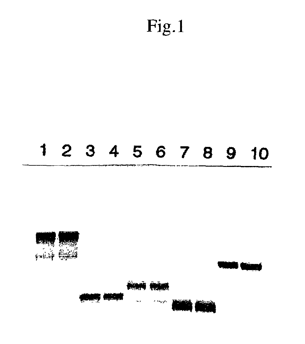
[0098]After reacting for 1.5 hr at 30° C., the reaction was stopped by cooling with ice. 4 μL of TE4 and 5 μL of 2× sample buffer (total 10 μL) were added to 1 μL of the reaction solution and the mixture was heated for 3 min at 100° C. and subjected to SDS polyacrylamide electrophoresis (30 mA, about 1.5 hr of migration). After fixing, Coomassie dyeing, and drying, the RI band was detected with BAS2000.
[0099]The results are shown in FIG. 1. In the figure, the DNA templates employed were as follows: l...
reference example 2
[0100]Protein Synthesis from Genes (Biotin Labeling and Recovery of Biotinylated Protein)
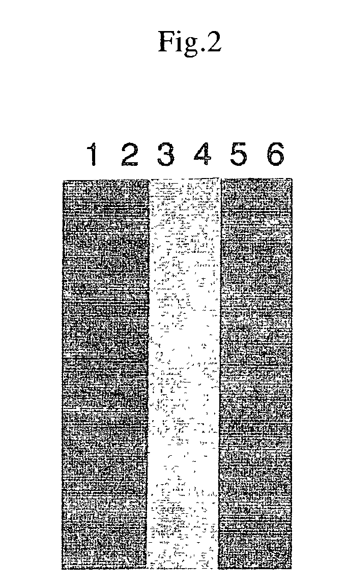
[0101]Similar to the above-described 35S-Met labeling; 1 μL of 1 mM amino acid mixture was added in place of the 1 mM amino acid mixture (Minus Met) and 1 μL of biotinylated lysine tRNA (Transcent™ tRNA) was added in place of 35S-MET. When SDS polyacrylamide electrophoresis was conducted, a PVDF membrane was blotted and biotinylated protein was detected with a biotin detection kit (Behringer Co.)
[0102]The results are shown in FIG. 2. The plasmids employed in the figure were as follows: lanes 1 and 2: for SV40 large T; lanes 3 and 4: for p53; and lanes 5 and 6: for luciferase. Lanes 2, 4, and 6 are the results of the supernatant recovered with the addition of streptoavidin beads following the reactions. The results show that a specific protein was synthesized for each DNA template and that the biotin-labeled proteins were reacted with streptoavidin beads and recovered. Although not described here...
example 1
Detection of Protein—Protein Interaction (Model System)
[0103]Reaction solutions of the RI-labeled protein of Reference Example 1 and the biotinylated protein of Reference Example 2 were mixed together in various combinations once the reactions had ended. That is, 2.5 μL of each reaction solution was mixed and placed on ice for 60 min. A 15 μL quantity of streptoavidin (Dynabeads) diluted with TBST-1 percent skim milk was added, and the mixture was stirred for 30 min at 4° C. Magnetic force was employed to recover the beads, thorough washing was conducted with TBST, and then the RI of the beads was measured. The results are given in Table 1. These results show that for SV40 large T and p53, and fos and jun, the interaction of which is well known, the isolation of one protein permitted the detection of the other. The interaction between SV40 large T was also detected.
[0104]
TABLE 1BiotinylatedRI-Labeled Protein (c.p.m.)ProteinFosJunLucLarge TP53Fos177675172156169Jun707204131176187Luc(a...
PUM
| Property | Measurement | Unit |
|---|---|---|
| temperature | aaaaa | aaaaa |
| temperature | aaaaa | aaaaa |
| fluorescent | aaaaa | aaaaa |
Abstract
Description
Claims
Application Information
 - R&D
- Intellectual Property
- Life Sciences
- Materials
- Tech Scout
- Unparalleled Data Quality
- Higher Quality Content
- 60% Fewer Hallucinations
Browse by: Latest US Patents, China's latest patents, Technical Efficacy Thesaurus, Application Domain, Technology Topic, Popular Technical Reports.
© 2025 PatSnap. All rights reserved.Legal|Privacy policy|Modern Slavery Act Transparency Statement|Sitemap|About US| Contact US: help@patsnap.com