
Self-renewing pluripotent hepatic stem cells

a hepatic stem cell and self-renewing technology, applied in the field of stem cells, can solve the problems of non-transformed hepatic stem cells and their progeny, and achieve the effect of reducing the number of stem cells

Inactive Publication Date: 2006-12-19
REPROCELL
View PDF0 Cites 11 Cited by
  • Summary
  • Abstract
  • Description
  • Claims
  • Application Information

AI Technical Summary

Benefits of technology

"The present invention provides a new type of stem cell that can be isolated and cloned from the liver. These cells have the ability to differentiate into hepatocytes and cholangiocytes, which are the two main types of cells in the liver. These cells can be obtained by fractionating cells from the liver and using special assays to identify them. The cells can be cultured and transplantated into mammals, providing a potential therapy for diseases of the digestive system. The invention also includes methods for screening and testing the effects of substances on these stem cells. Overall, the invention provides new tools for research and potential treatments for liver-related diseases."

Problems solved by technology

These methods result in the production of non-transformed hepatic stem cells and their progeny.

Method used


Image

  • Self-renewing pluripotent hepatic stem cells
  • Self-renewing pluripotent hepatic stem cells
  • Self-renewing pluripotent hepatic stem cells

Examples


example 1

Flow Cytometric Fractionation of Fetal Mouse Liver Cells

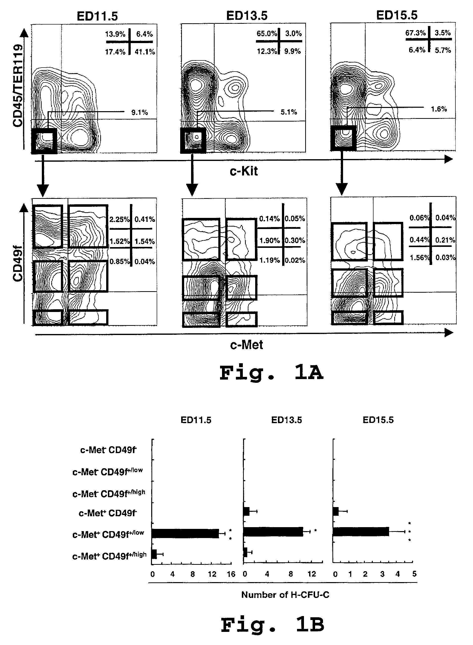
[0102]In this example, we fractionated H-CFU-H according to the method that we have previously reported (Suzuki et al., 2000), and thereafter we attempted to sort for cells expressing c-Met, the hepatocyte growth factor (HGF) receptor for further fractionation.

(Fractionation of “H-CFU-C” by Flow Cytometry) Cell preparation

[0103]Single cell suspensions of liver cells were prepared from Balb / cA ED13.5 fetal mice (CLEA, Tokyo, Japan). For sequential analysis, we used embryonic day (ED) 11.5, 13.5, 15.5, and neonatal (1 day after birth) mice. For sorting, we used mechanical pipetting in staining medium (PBS containing 3% FBS). Cell viability after each treatment was exceeded 90% as assessed by trypan blue dye exclusion.

Cell Staining and FACS Analysis

[0104]Dissociated liver cells were incubated at 4° C. for 30 minutes with biotinylated anti-CD45 and TER119 mAb (PharMingen, San Jose, Calif.), and anti-c-Met mAb (Upstate Biotechnology...

example 2

Characterization of c-Met+ CD49f+ / low c-Kit− CD45− TER119− cells

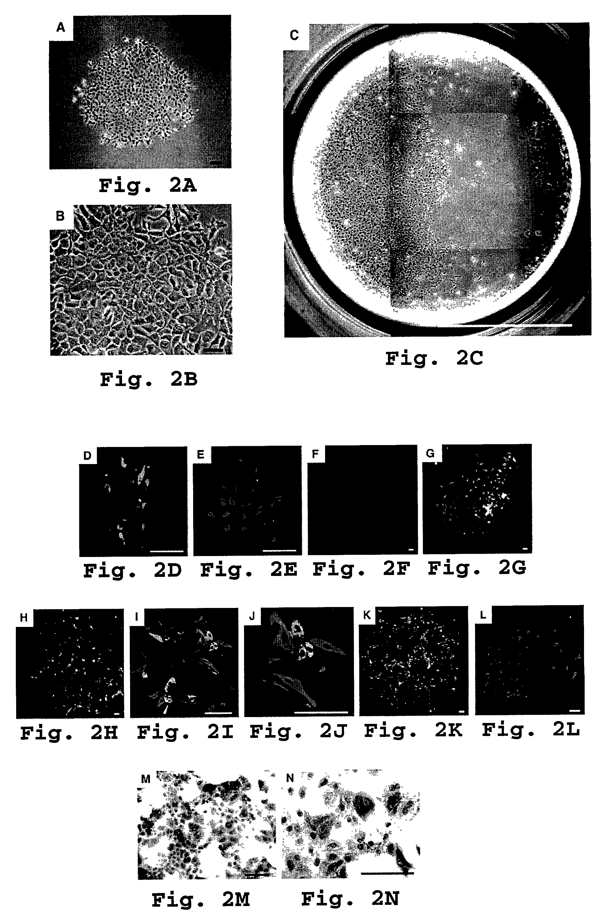
[0113]In the present example, we characterized of c-Met+ CD49f+ / low c-Kit− CD45− TER119− cells.

[0114]High enrichment in H-CFU-C achieved in Example 1 permitted efficient culture of clone-sorted c-Met+ CD49f+ / low c-Kit− CD45− TER119− cells for analyses of self-renewal and differentiation potential.

[0115]For low density culture analysis, sorted cells were plated on laminin-coated 6-well plates (Becton Dickinson) at a density of 30 cells / cm2 and cultured in our fresh standard medium (1:1 mixture of DMEM and F-12 (Sigma, Chemical Co., St. Louis, Mo.) with 10% fetal bovine serum (JRH BIOSCIENCES, Lenexa, Kans., γ-insulin (1 μg / ml) (Wako, Tokyo, Japan), dexamethasone (1×10⁻⁷ M) (Sigma), nicotinamide (10 mM) (Sigma), L-glutamine (2 mM) (Gibco BRL, Gaithersburg, Md.), β-mercaptoethanol (50 μM) (Sigma), HEPES (5 mM) (Wako), and penicillin / streptomycin (Gibco BRL)). For single cell culture analysis, we used standard medium 50% su...

example 3

Clonal Expansion and Self-Renewal Capability of H-CFU-C in Culture

[0121]We next used sub cloning experiments to test the self-renewal potential of H-CFU-C. Single c-Met+ CD49f+ / low c-Kit− CD45− TER119− cells were clone-sorted and individually cultured. This yielded several large H-CFU-C colonies (see FIG. 2 C), which we then replated onto new culture dishes. Over 2 to 3 weeks, about half of subcultured clones gradually expanded, finally to become confluent. These clonally expanding subcultured cells then again underwent clone-sorting and single cell culture.

[0122]We then conducted detection of hepatocyte or cholangiocyte marker gene expression by RT-PCR as described previously in Suzuki at al., 2000. Briefly, a cloning ring (Iwaki Glass, Tokyo, Japan) was placed on the colony and total RNA was prepared by gentle pipetting cells with ISOGEN (Nippon Gene, Tokyo, Japan). Prior to reverse transcription, 0.8 μl of oligo-d(T)1218 primers was added to the total RNA solution. The reaction m...


PUM

PropertyMeasurementUnit
pHaaaaaaaaaa
pHaaaaaaaaaa
weightaaaaaaaaaa

Abstract

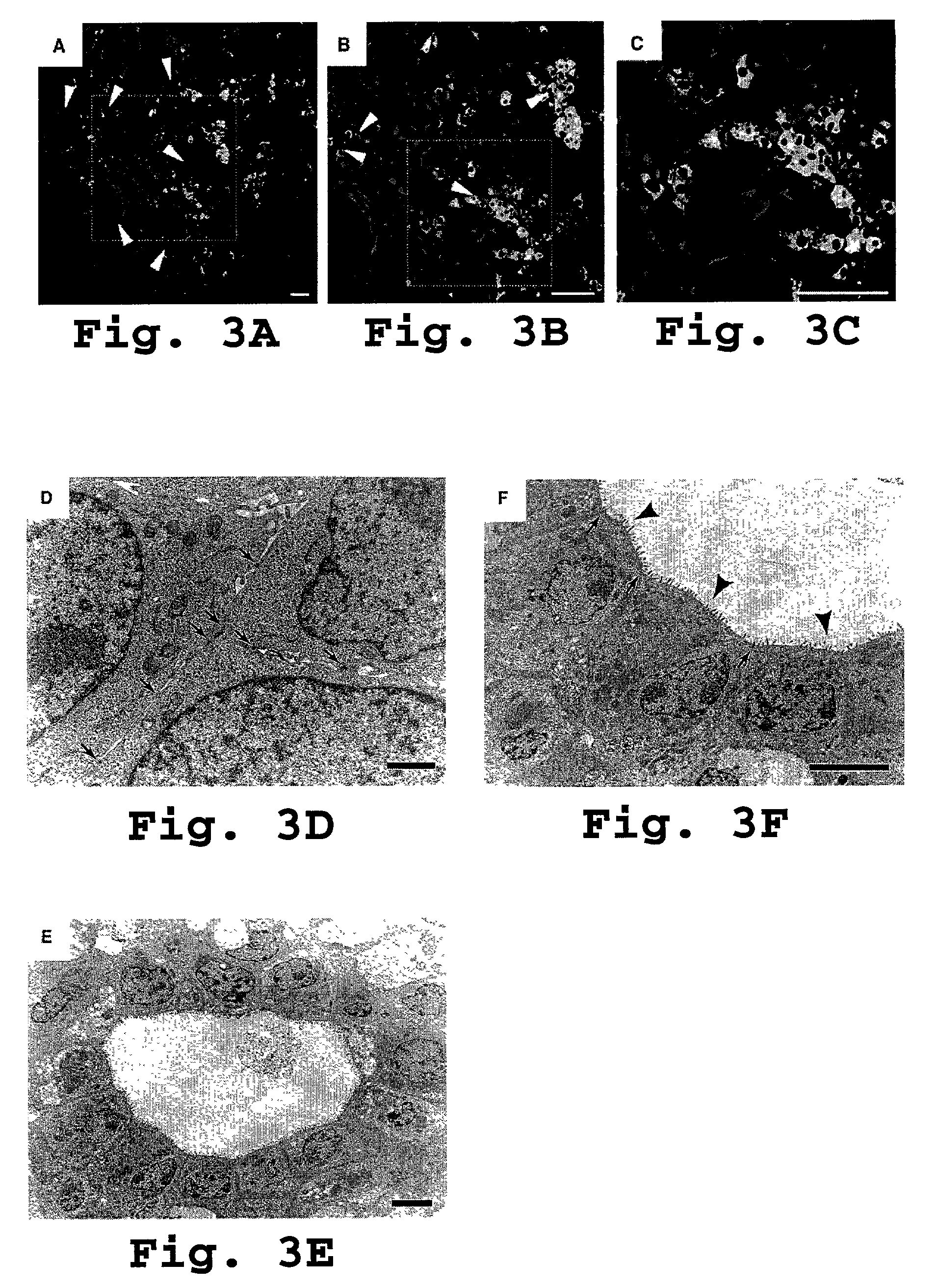
The present invention provides clonal pluripotent hepatic stem cells using flow cytometry and in vitro single-cell-based assays. These cells possess multilineage differentiation potential and self-renewing capability. These cells may be clonally propagated in culture, to continuously produce hepatocytes and cholangiocytes as descendants while maintaining primitive stem cells. When expanded cells are transplanted into recipient animals, they morphologically and functionally differentiated into hepatocytes and cholangiocytes, with reconstitution of hepatocyte and bile duct structures. Furthermore, these cells differentiated into pancreatic ductal and acinar cells or intestinal epithelial cells when transplanted into pancreas or duodenal wall. Thus, the self-renewing multipotent stem cells persist in the developing mouse liver and can be induced to become cells of other organs of endodermal origin under appropriate microenvironment, providing new insight into therapies for diseases of the digestive system.

Description

BACKGROUND OF THE INVENTION[0001]1. Field of the Invention[0002]The present invention relates to stem cells. More specifically, the present invention relates to self-renewing pluripotent hepatic stem cells.[0003]2. Description of the Related Art[0004]The enormous regenerative capacity of the liver after partial hepatectomy or chemical injury is well known. In rodents, liver weight returns to normal within a few weeks even after loss of up to two-thirds of total liver mass (Fausto, N., and E. M. Webber. 1994. Liver regeneration: The liver. I. M. Arias, J. L. Boyer, N. Fausto, W. B. Jakoby, D. A. Schachter, and D. A. Shafritz, editors: Raven Press, N.Y., ed. 3, 1059–1084.). Remarkable regenerative potential is also retained in hematopoiesis. Hematopoietic stem cells(HSCs) certainly exist in bone marrow where they self-renew and differentiate along all hematopoietic lineages. Sophisticated isolation methods have recently identified a highly probable HSC candidate; a single such cell ca...

Claims


Application Information

Patent Timeline
Patent Type & Authority Patents(United States)
IPC IPC(8): C12N5/06C12N5/10A01N63/00C12N5/08A61K35/12C12N5/071C12N5/074
CPCC12N5/0672C12N5/0676A61K35/12C12N2506/02C12N2506/14Y02A50/30
Inventor NAKAUCHI, HIROMITSUSUZUKI, ATSUSHITANIGUCHI, HIDEKIFUKAO, KATASHI
Owner REPROCELL
Learn More