Apparatus and method for drainage

a technology of apparatus and drainage, applied in the direction of wound drainage, suction drainage container, catheter, etc., can solve the problems of preventing infection and promoting healing, tubular surgical drains are prone to blockage and other forms of obstruction, and the passage of discharge may be blocked or obstructed, so as to minimize the restriction of flow

Inactive Publication Date: 2007-02-27
MCCRAW JOHN B
View PDF4 Cites 167 Cited by
  • Summary
  • Abstract
  • Description
  • Claims
  • Application Information

AI Technical Summary

Benefits of technology

[0018]An apparatus for drainage of a wound or surgical site of a human or animal fits anatomically and passes from inside the wound or surgical site through the wound or surgical opening extending percutaneously externally for accumulation and removal of fluids. The apparatus for drainage preferably has a plurality of fibers each flexible and having an internal end, an external end and between them a middle. It is desired that each fiber is elongate with a thickness and a length so the fiber thickness is substantially less than its length for flexibility. The plurality of fibers forms preferably a loose bundle having gathered together external ends leaving internal ends unrestrained for spreading divergently inside the wound or surgical site. As will be explained herein the middle being a loose bundle allows fiber shifting and guidance of fluid flow out of the wound or surgical site while minimizing restriction of flow.
[0019]A collector is preferably positioned in fluid communication with the gathered together external ends of the loose bundle of the plurality of fibers. The collector preferably accumulates bodily fluid guided along and amongst the fibers from the internal ends along the middle and to the gathered together external ends during drainage of fluid from inside the wound or surgical site. The plurality of fibers at their unrestrained internal ends desirably spreads divergently about the inside wound or surgical site for guiding fluid. A source of vacuum in an active system connects to draw fluid from the collector that is positioned in fluid communication with the gathered together external ends. The plurality of fibers in the preferred embodiment are each a filament, for example, Nylon suture but they may be selected from the group of structures consisting essentially of monofilaments, strings, strands, hollow fibers or any combination thereof. The plurality of fibers may be made from a biocompatible material selected from the group consisting essentially of polymers, cellulose, natural substances or any combination thereof. The biocompatible material can be bio-absorbable but must maintain its structure over the at least the period during which the drainage is desired. The plurality of fibers may include an anticoagulant for resisting the clotting of fluid passing there about, along and among. Anticoagulants such as heparin, saline solution or even water adhered to absorbed in or about the loose bundle of the plurality of fibers may be provided to minimize the fluid coagulation or clotting on or to the fibers increasing the time over which fluid may easily drain. The plurality of fibers may include an anti-microbial for deterring the passing of microbes there about, along and among. The source of vacuum if not regulated may have a regulator connected between it and the collector for adjusting the amount of vacuum applied to the gathered together external ends.

Problems solved by technology

There are a number of disadvantages of tubular surgical drains including the difficulties of avoiding infection and promoting healing inherent in a device inserted in a wound cavity.
Tubular surgical drains tend to be prone to blockage and other forms of obstruction, e.g., when the tube opening contacts body tissue the passage of discharge may be plugged or obstructed.
As with tube-type drains, fabric drains suffer from a number of disadvantages, such as susceptibility to sloughing or fragmentation, which may promote irritation and associated infection, and their drainage capacity maybe limited to the fluid retention capacity of the fabric, i.e., the drainage is not continuous.
Using hydrophilic fibers, e.g., cellulose derived and yarns may aggravate flow problems, due to their water binding properties.
A consequence of the drainage capacity limitation is that fabric-type surgical drains must ordinarily be replaced on relatively short intervals.
Conventional fabric-type drain materials are often so loosely woven that they fragment which increases the risk of infection.
Moreover, natural fibers like cotton, and other cellulose, are themselves prone to sloughing or fragmentation that also increase the risk of infection.
In addition, depending on the fibers selected, known fabric-type surgical drains may decompose over time, again necessitating more frequent replacement, and irritating of the wound cavity.
Another limitation of known fabric-type surgical drains is that, once positioned, they may be difficult to relocate or retrieve.
While this type of drain has the advantage of ease of manipulation and may have better drainage characteristics than the other general types of surgical drains, it may also suffer from their disadvantages.
Gauze is prone to fragmenting leaving many loose threads when it is cut and additional parts of the fabric may fall away under minimal pressure in the form of lint.
The use of the sheath or glove to contain lint is not completely effective or convenient for use in an operating room setting.
Furthermore, drainage with this type of surgical drain may be limited to the fluid retention capacity of the fabric and evaporation.
As with other known surgical drains, these drains have lint and their drainage capacity may be limited to the fluid retention capacity of the fabric.
Another problem encountered with the using of tubular drains is that drainage is localized about the internal end of the tube particularly when vacuum is applied externally.
Localization of drainage is not effective to remove the detritus and accelerate healing of the wound or surgical site.
Moreover, the vacuum applied to the drain tube can draw the adjacent internal tissue into the internal end resulting in restriction of flow and perhaps collapse of the tubular passageway stopping drainage and requiring withdrawal and removal and replacement of the drain tube.
Additional trauma to the wound or surgical opening and interference with the healing process are detrimental and undesired consequences.
Yet another difficulty with tubular drains is their relatively rigid circular cross section.
If suturing is performed to secure the tube, it pulls and is uncomfortable during drainage and removable.

Method used


Image

  • Apparatus and method for drainage
  • Apparatus and method for drainage
  • Apparatus and method for drainage

Examples


Embodiment Construction

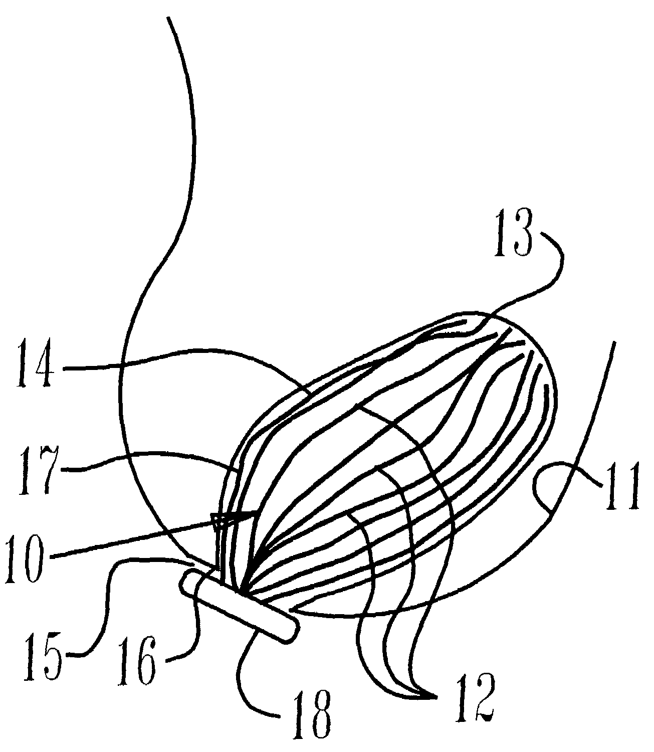
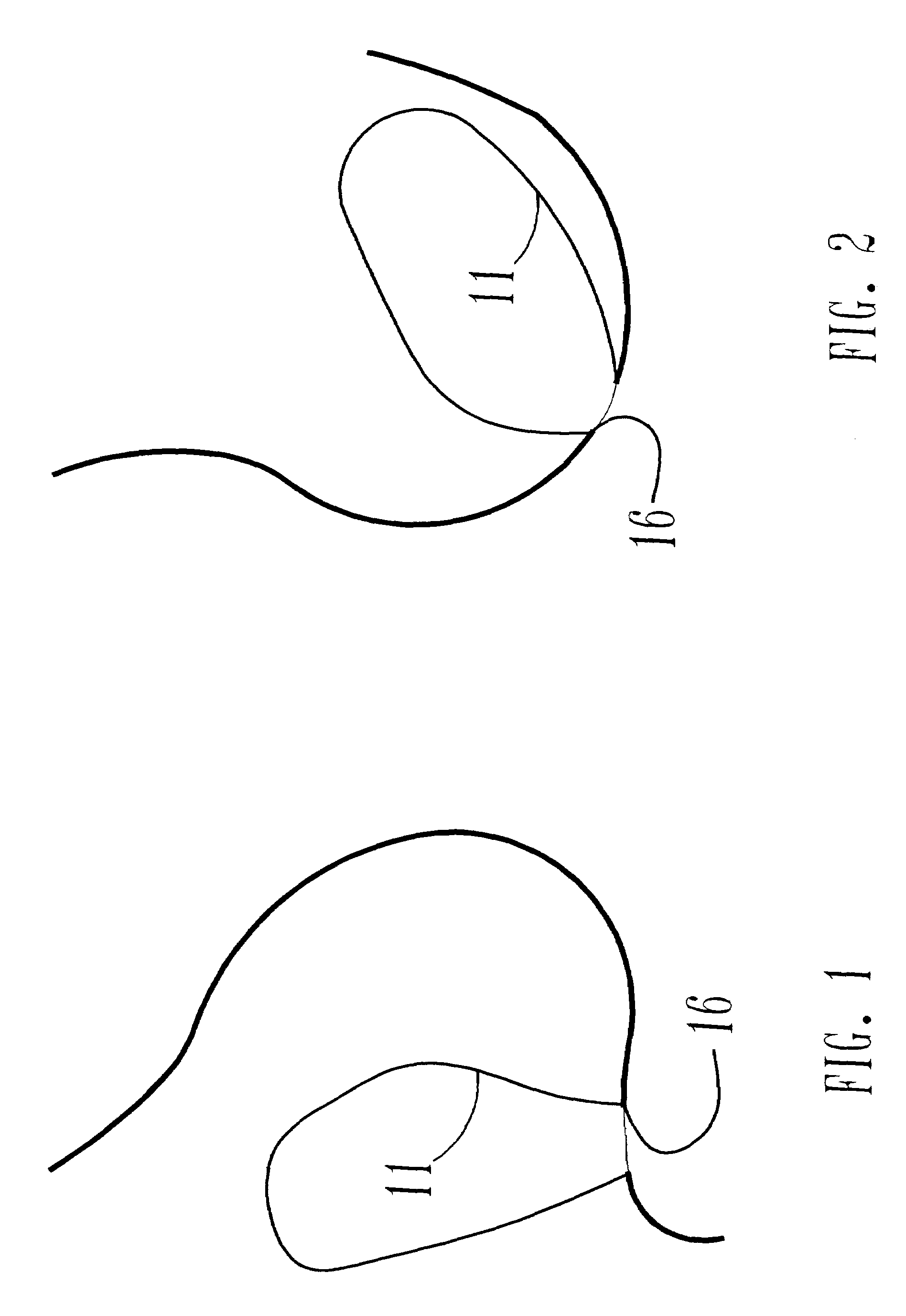
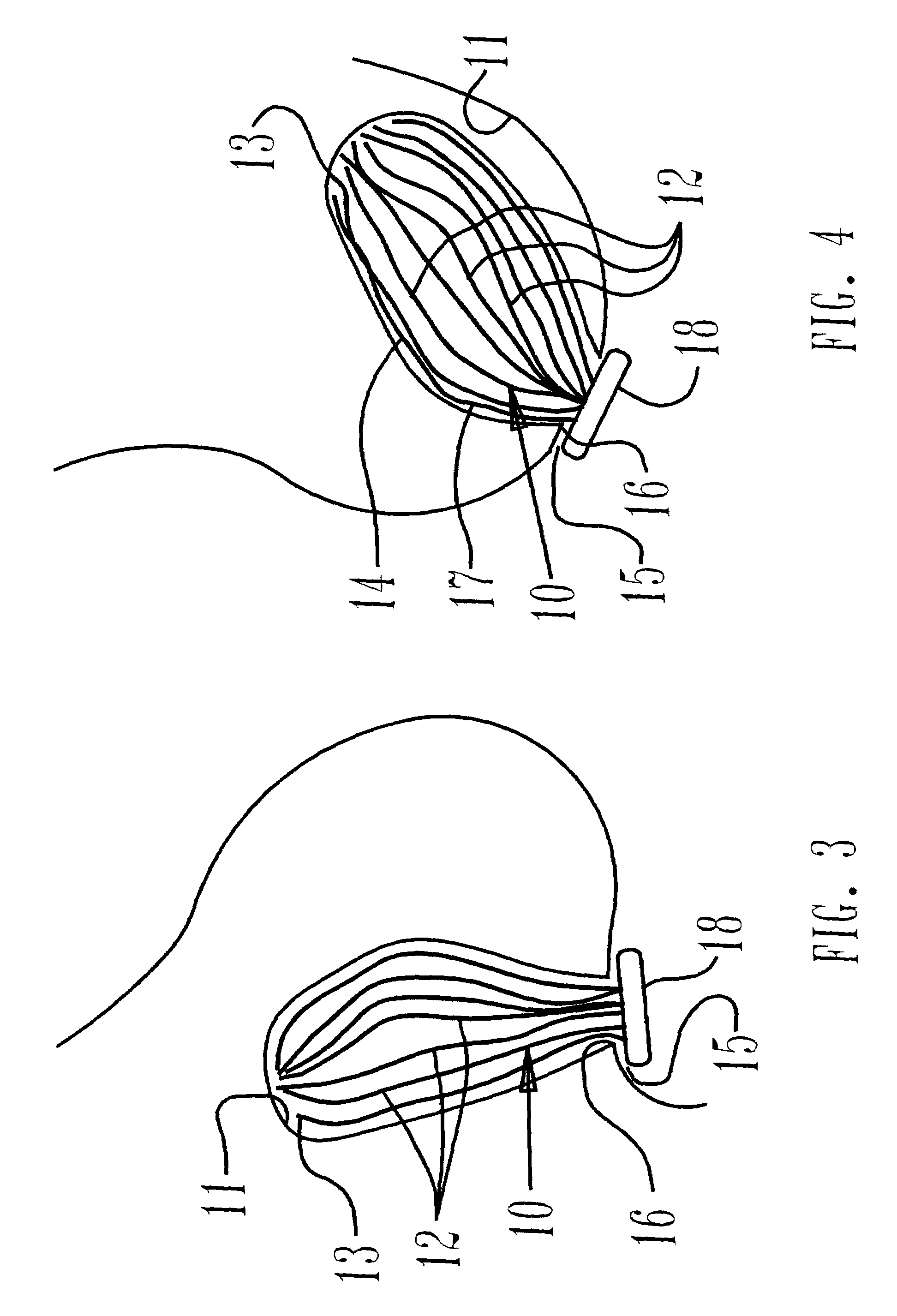
[0039]An apparatus for drainage 10 of a wound or surgical site 11 accommodates a wound or surgical site 11 anatomically, passes from inside the wound or surgical site 11 percutaneously and extends externally for accumulation of fluids, see FIGS. 1 to 4. The apparatus for drainage 10 has a plurality of fibers 12 and each is elongate and has an internal end 13, middle 14 and an external end 15. It is desired that each elongate fiber 12 be of a thickness about 0.5 to 2 mm and a length perhaps 4 to 8 cm so its thickness is substantially less than its length for flexibility. Preferably each fiber 12 is medical grade suture material e.g., Nylon and is long enough to extend from inside the wound or surgical site 11 through a wound or surgical opening 16 to the external accumulation of the surgical fluids, as depicted in FIGS. 3 and 4. The plurality of fibers 12 form a loose bundle 17 having gathered together external ends 15 and with internal ends 13 unrestrained for spreading divergently ...


PUM


Abstract

An apparatus for drainage of a wound or surgical site of a human or animal fits anatomically, passes from inside the wound or surgical site percutaneously or covering an open wound surface and extends externally for accumulation of fluids. A plurality of fibers each with an internal end, middle and an external end is elongate with a thickness and a length wherein its thickness is substantially less than its length for flexibility. The fibers form a loose bundle with gathered external ends and with internal ends unrestrained for spreading divergently inside the wound or surgical site. A collector is positioned in fluid communication with the gathered external ends for accumulating bodily fluid guided along and amongst the fibers from the internal ends along the middle and to the external ends during drainage. Vacuum connects to draw fluid from the collector. A method has steps of providing fibers with the internal ends unrestrained, the external ends gathered in a collector to accumulate drained fluid. The method guides bodily fluid along and amongst the fibers from the internal ends along the middle and to the gathered together external ends during drainage. The fibers spread divergently for accumulating bodily fluid inside the wound or surgical site. A method of manufacture of the apparatus has the steps of accumulating a plurality of fibers with internal ends, a middle and external ends in a loose bundle but gathering together in a collector at their external ends while leaving the internal ends unrestrained.

Description

CROSS-REFERENCE TO RELATED APPLICATIONS[0001]None.STATEMENT REGARDING FEDERALLY SPONSORED RESEARCH[0002]Not applicable.SEQUENCE LISTING[0003]Not applicable.BACKGROUND OF THE INVENTION[0004]The purpose of a drain is to evacuate fluid or gas that is present or likely to accumulate in a localized cavity. Typically, drains are used in an abscess cavity, to treat pneumothorax, or evacuation of blood, serum or lymph from a wound. Drains also remove pus or transudate or alternately to irrigate cavities. The process of drainage eliminates the dead space in a cavity. Herein and throughout this disclosure the cavity will be referred to as the wound or surgical site no matter what the reason for the cavity to be drained. Drainage systems are closed if they capture the fluids with tubing coupled to a closed container or canister and are open if fluid is accumulated in gauze or corrugated rubber sheet. Canisters and collapsible vessels use vacuum or the restoration force of the collapsed vessel ...

Claims


Application Information

Patent Timeline
Patent Type & Authority Patents(United States)
IPC IPC(8): A61M1/00A61F13/00A61M5/00A61M5/32A61B17/06A61M27/00
CPCA61M27/00A61M1/0088A61B17/06166A61M1/0003A61M1/0023A61M1/84A61M1/92A61M1/71A61M1/74A61M1/96A61M1/60A61M1/916
Inventor MCCRAW, JOHN B.
Owner MCCRAW JOHN B
Why Eureka
Try Eureka