Device for magnetic immobilization of cells

a cell and magnetic technology, applied in the field of cell magnetic immobilization devices, can solve the problems of not being particularly suited to the immobilization or micromanipulation of individual cells, mechanical holding methods tend to obscure or damage the target cells, and the sorting of cells into pools does not allow the investigator to experiment on or manipulate individual cells, etc., to achieve strong yet more uniform external magnetic field, increase the effect of magnetic force and small for

Inactive Publication Date: 2007-10-23
SURFACE LOGIX INC
View PDF87 Cites 62 Cited by
  • Summary
  • Abstract
  • Description
  • Claims
  • Application Information

AI Technical Summary

Benefits of technology

[0025]FIG. 7(A) shows a magnetic particle in a weak external magnetic field with very little magnetic field gradient. The magnetic particle will experience very little force. (B) shows the same magnetic particle in the same magnetic field as (A) in a position where there is a magnetic field gradient which increases with proximity to the magnetic pole. The particle experiences increasing magnetic force. (C) shows a stronger yet more uniform external magnetic field with little or no gradient in the center. (D) illustrates how “highly-magnetically-permeable material” having a high relative magnetic permeability (μr) generates a localized magnetic field gradient in the uniform external magnetic field.

Problems solved by technology

However, sorting cells into pools does not allow the investigator to experiment on or manipulate individual cells.
Such mechanical holding methods tend to obscure or damage the target cells.
Although current magnetic-based cell separation methods afford certain advantages in performing medical or biological analyses based on biospecific affinity reactions involving colloidal magnetic particles, the systems developed to date are not particularly suited for immobilization or micromanipulation of individual cells.
It has been found that the steel wool matrix of such prior art high-gradient magnetic separation (HGMS) devices often causes non-specific entrapment of biological entities other than target entities.
The entrapped non-magnetic components cannot be removed completely without extensive washing and resuspension of the particles bearing the target substance.
Moreover, sizes of the columns in many of the prior art HGMS devices require substantial volumes of test media, which poses an impediment to their use in performing various laboratory-scale separations.
In addition, the steel wool matrix may damage sensitive cell types.
Furthermore, immobilizing target cells on steel wool does not allow the investigator to experiment on or manipulate individual target cells.
As above, immobilizing target cells on bent metal pins does not allow the investigator to immobilize, array and / or isolate individual target cells.
Magnetic-based isolators are also used to purify nucleic acid or proteins from mixed samples, however, none can immobilize, array and / or isolate cells for study.

Method used


Image

  • Device for magnetic immobilization of cells
  • Device for magnetic immobilization of cells
  • Device for magnetic immobilization of cells

Examples


example 1

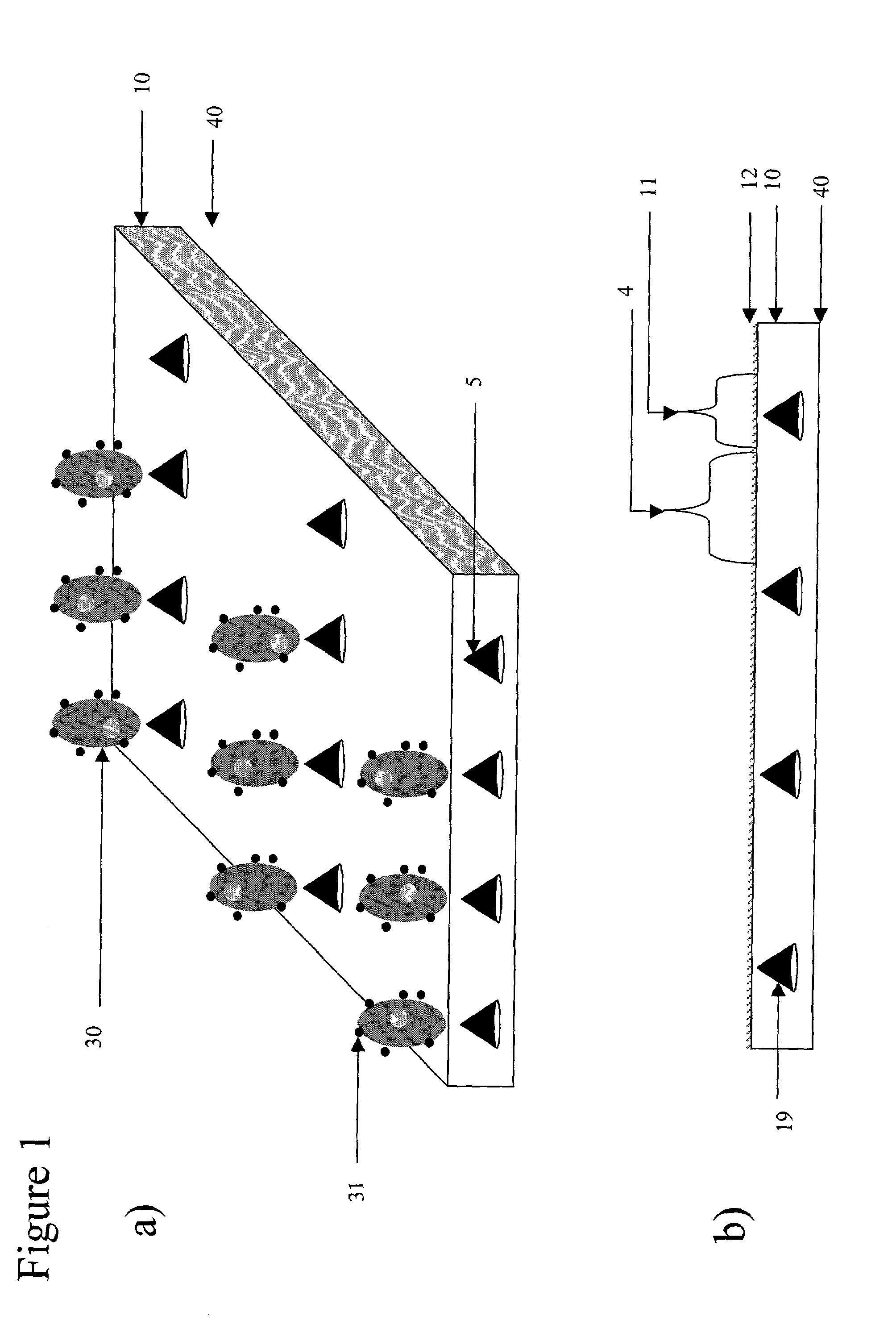
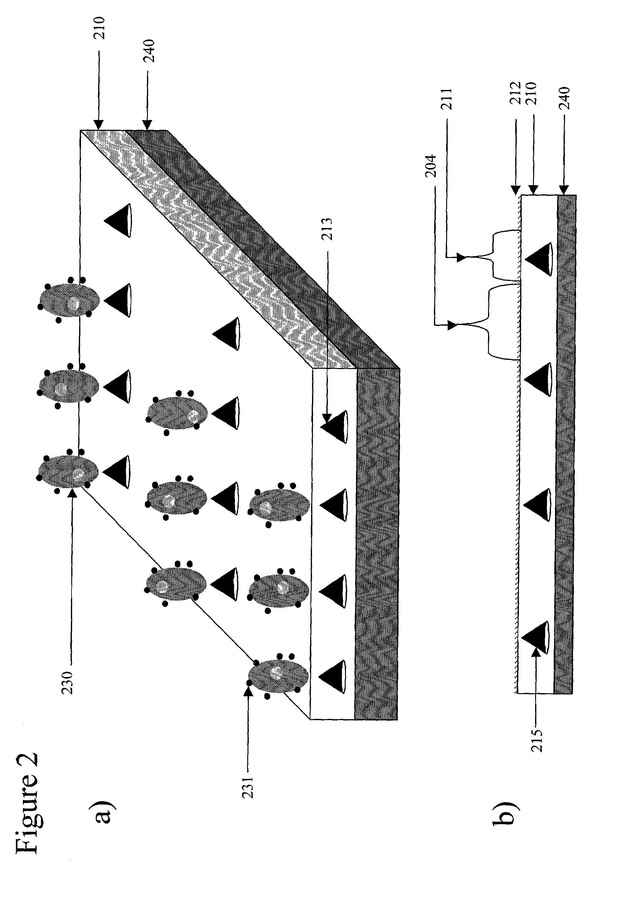
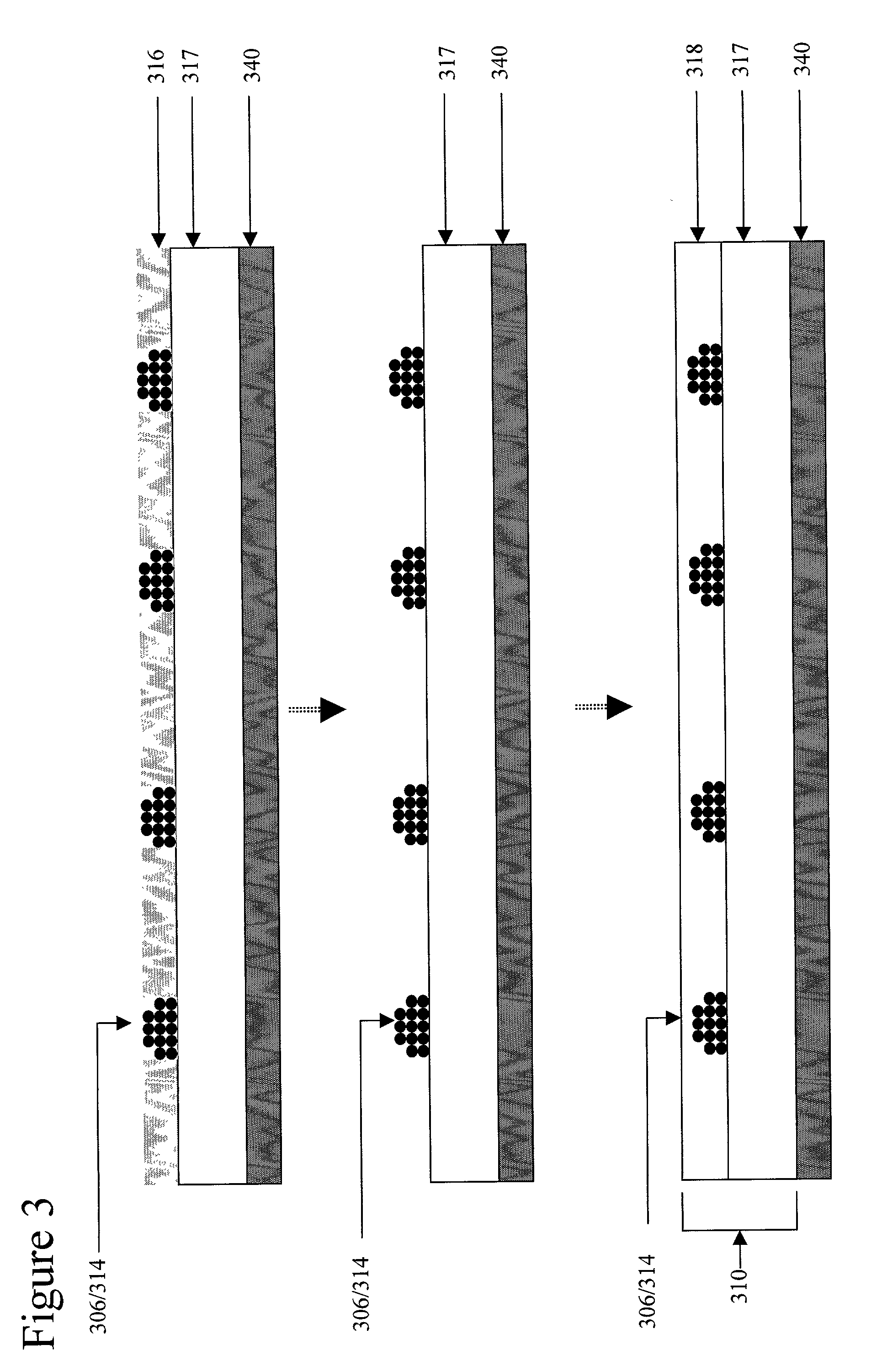
[0101]Several permanent neodymium iron boron magnets each with a strength of 12,000 Gauss, are embedded in a PDMS substrate. A magnetic bead-containing phosphate buffered saline (PBS) solution is introduced to a cell panning device mated to the substrate. 2 ml of magnetic beads in suspension (Spherotech™ 9 μm diameter beads at a density of 1×10⁶ beads / μl suspension) are uniformly distributed by cell panning. The coupled substrate and cell delivery device are rotated for a period of 30 minutes at 0.5 RPM. The substrate with immobilized beads is washed with cell-free PBS. The local magnetic field gradient derived from each of the permanent magnets captures several thousand beads on the PDMS substrate above the embedded magnets, forming the desired array on the substrate.

example 2

[0102]Several permanent neodymium iron boron magnets each with a strength of 12,000 Gauss, are embedded in the PDMS substrate. 1 ml Spherotech™ (1840 Industrial Dr., Suite 270 Libertyville, Ill. 60048-9467) 9 μm diameter magnetic beads at a density of 1×10⁶ beads / μl suspension coated with the bioaffinity ligand streptavidin are pre-incubated with a biotinylated anti-mouse anti-syndecan antibody at a final antibody concentration of 50 μg / ml. The antibody conjugated beads are then washed to PBS remove unbound antibody. 1 ml hybridoma cells (2×10⁵ / μl) in RPMI media are then incubated with 1 ml antibody conjugated Spherotech™ 9 μm diameter magnetic beads at a density of 1×10⁶ beads / μl suspension 2 ml of the magnetic bead and cell containing RPMI media (Sigma; www.sigma-aldrich.com) is introduced to the cell panning device mated to the substrate. The coupled substrate and cell delivery device are rotated for a period of 3 hours at 3 RPM. The substrate with immobilized cells is washed wit...

example 3

[0103]Several localized magnetic field gradients are derived from several small permanent magnetic pins embedded in PDMS with their tips coplanar with the substrate. The permanently magnetic pins generate a several localized magnetic field gradients, each with an attractive force of about 5×10⁻¹³ Newtons on cells associated with magnetic material passing over the substrate. 1 ml Spherotech™ 9 μm diameter magnetic beads at a density of 1×10⁶ beads / μl suspension coated with the bioaffinity ligand streptavidin are pre-incubated with a biotinylated anti-mouse anti-syndecan antibody at a final antibody concentration of 50 μg / ml. The antibody conjugated beads are then washed to PBS remove unbound antibody. 1 ml hybridoma cells (2×10⁵ / μl) in RPMI media are then incubated with 1 ml antibody conjugated Spherotech™ 9 μm diameter magnetic beads at a density of 1×10⁶ beads / μl suspension 50 μl of the magnetic bead and cell containing RPMI media (Sigma) is introduced into the in-port of the cell ...


PUM

PropertyMeasurementUnit
sizeaaaaaaaaaa
cell diameteraaaaaaaaaa

Abstract

The present invention relates to an apparatus and methods that immobilize one or more cells associated with magnetic material on a substrate on which are located one or more magnetic receptacle(s). Alternatively, in another aspect the present invention, the device arrays cells associated with magnetic material on a substrate having a pattern of magnetic receptacles disposed thereon. The size of the magnetic receptacle(s) determines the number of target cells that it is capable of immobilizing. The size of the magnetic receptacle is defined by the strength of a localized magnetic field gradient. The localized magnetic field gradient may be derived from 1) permanent magnets embedded in the substrate or alternatively, the localized magnetic field gradient may be derived from an 2) external magnet whose strength is focused by objects of highly-permeable-magnetic material which create localized magnetic field gradients. The invention apparatus comprises a removable cell delivery device and a substrate, which has one or more magnetic receptacles disposed thereon.

Description

CROSS-REFERENCE TO RELATED APPLICATIONS[0001]This application claims the benefit of U.S. Provisional Application No. 60 / 334,593, filed Dec. 3, 2001, and U.S. Provisional Application No. 60 / 307,843, filed Jul. 27, 2001.FIELD OF THE INVENTION[0002]The present invention relates to methods and devices that are useful in immobilizing, arraying and / or isolating cells individually. In particular, the present invention relates to methods and devices that are useful for immobilizing, arraying and / or isolating cells using a magnetic source.BACKGROUND OF THE INVENTION[0003]Many biological techniques employed in biotechnology, microbiology, clinical diagnostics and treatment, in vitro fertilization, hematology and pathology, require such processes as identification, separation, culturing, immobilization and / or manipulation of a target entity. Typical target entities include cells or microbes within a fluid medium such as culture fluids, environmental samples, blood or other bodily fluids. It is...

Claims


Application Information

Patent Timeline
Patent Type & Authority Patents(United States)
IPC IPC(8): C12M1/12C12M1/34C12N11/02C12N11/08B01L3/00C12M1/26C12N5/00C12N5/06C40B60/14G01N33/543G01N35/00
CPCB01L3/5085C12M33/00G01N33/54326B01J2219/00317B01J2219/00655B01J2219/00659B01J2219/00743G01N35/0098B01L2400/043C40B60/14B01L3/5088
Inventor CASAGRANDE, ROCCOWANG, EVELYNKIRK, GREGORYNUSSBAUM, MICHAELKIM, ENOCHRAPHEL, AARON
Owner SURFACE LOGIX INC
Why Eureka
Try Eureka