Microscope system and methods for intracellular studies

a microscopy and intracellular technology, applied in the field of in vitro polynucleotide delivery applications, can solve the problems of limited visualization technology of dynamic behavior of complexes inside cells, inability to study the dynamic behavior of living cells, and high system cost, and achieve high resolution

Inactive Publication Date: 2008-05-06
NITTO DENKO CORP
View PDF3 Cites 13 Cited by
  • Summary
  • Abstract
  • Description
  • Claims
  • Application Information

AI Technical Summary

Problems solved by technology

Technology that provides visualization of dynamic behaviors (paths 1-6, FIG. 1) of the complexes inside cells has been limited.
However, this method does not allow the study of the dynamic behavior of living cells.
Other workers have used confocal microscopy to study intracellular trafficking, but the system is expensive and observation could not be longer than 30 minutes.
Fluorescence microscopy has been used to observe a whole cell, but resolution inside the cell was poor.

Method used


Image

  • Microscope system and methods for intracellular studies
  • Microscope system and methods for intracellular studies
  • Microscope system and methods for intracellular studies

Examples


examples

[0063]Gene carriers were synthesized according to the procedure in U.S. application Ser. No. 10 / 375,075, filed Feb. 25, 2003, which is incorporated herein by reference and described below, or purchased from Polysciences, Inc. FITC-Fluorescence-5-X and Rhodamine Red were purchased from Molecular Probes. The conjugation of fluorescent dye to gene carriers was performed by the literature's procedure (Godbey, et al. PNAS 96, 5177-5181 (1999)).

[0064]A Zeiss axiovert S100 microscope and a Quantix Q-57 backthinned, cooled CCD camera were used. The Rhodamine was imaged with a Chromatechnologies 41002 filter set. The FITC-Fluorescence-5-X and the GFP were imaged with Achroma technologies 41028 filter set optimized for Yellow Fluorescent Proteins (YFP) but suitable for said excitation and emission. A 40× oil immersion, NA 1.4, objective and either a 1.6× or 2.0× optivar additional magnification were used.

[0065]Cell lines and cultures used in the following examples were prepared as follows: Ka...

examples 1-4

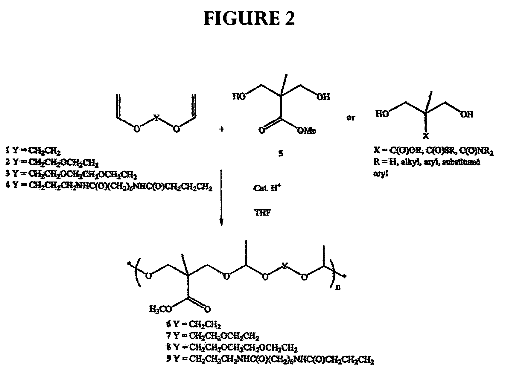
[0067]The following description for the synthesis of polyacetal is illustrative: Di(ethylene glycol) divinyl ether (1.39 g, 8.76 mmol) and bis-(2-hydroxymethyl)methyl propionate (1.30 g, 8.76 mmol) were mixed in tetrahydrofuran (THF) (10 mL) with molecular sieves (1.0 g) at room temperature and stirred for 20 min. A catalytic amount of toluensulfonic acid monohydrate (TSA, 0.015 g, 0.08 mmol) was added and stirring was continued for four days. The reaction mixture was quenched with sodium bicarbonate (1 mL, 5% in water) or triethylamine (1 mL). Water (10 mL) was added and the organic phase was extracted with ethylacetate (3×10 mL). The extracts were combined, dried with sodium sulfate, filtered, and concentrated by rotary evaporation. The residue was dried under high vacuum to give polyacetal (2.65 g, 8.65 mmol, 98%) as an oil.

examples 5-11

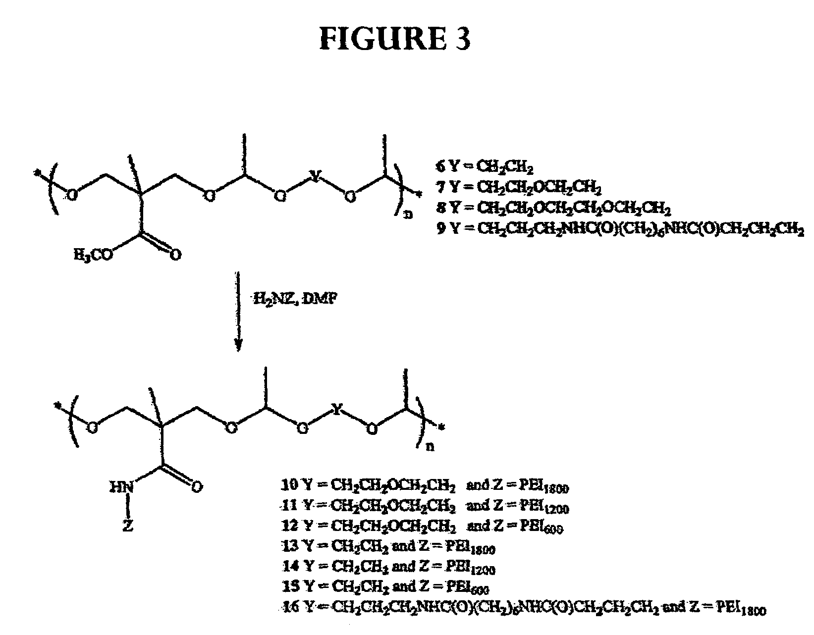
[0068]The following description for the synthesis of polyacetal is illustrative: To poly(ethylenimine) (PEI1800) (30 g, 16.7 mmol) was added a solution of polyacetal (0.5 g, 1.63 mmol) in dimethylformamide (DMF) (10 mL). Additional DMF (10 mL) was added and the mixture was stirred for four days. THF (100 mL) was added to form a precipitate. The precipitate was filtered and washed with THF, then dried under high vacuum to give polyacetal (2.2 g).


PUM

PropertyMeasurementUnit
pHaaaaaaaaaa
pHaaaaaaaaaa
molecular weightsaaaaaaaaaa

Abstract

A microscope system, kit and method are described for observation of cellular activity in real time. The system is used to monitor cell transfection using a polynucleotide noncovalently attached to a carrier, The carrier labeled with a dye or fluorescent agent so that transport of complex can be monitored in real time. The polynucleotide includes both a gene of interest and a reporter gene. The reporter gene indicates successful expression of the gene of interest.

Description

RELATED APPLICATIONS[0001]This application claims priority of U.S. Provisional application No. 60 / 519,019, filed Nov. 11, 2003 which is incorporated herein by reference.BACKGROUND OF THE INVENTION[0002]1. Field of the Invention[0003]This invention relates generally to technology useful for in vitro polynucleotide delivery applications. In one aspect, methods for monitoring intracellular activity in real time are disclosed. More particularly, this invention relates to polynucleotides and labeled gene carriers which monitor transport within cells and gene expression using a fluorescence microscope equipped with a mercury lamp, objective lenses, excitation filter, and a high resolution digital camera. The technology is useful for in vitro delivery applications in mammals.[0004]2. Description of the Related Art[0005]There is a need for non-viral drug and gene delivery systems having desirable properties such as low immunogenicity, amenable to production on a relatively large scale, and ...

Claims


Application Information

Patent Timeline
Patent Type & Authority Patents(United States)
IPC IPC(8): C12Q1/68C07H21/04
CPCC07H21/04C12Q1/6897C12Q2565/601C12Q2561/113
Inventor VAN, SANGYU, LEI
Owner NITTO DENKO CORP
Why Eureka
Try Eureka