In vivo synthesis of connective tissues

a technology of connective tissue and in vivo synthesis, which is applied in the direction of ligaments, prostheses, peptides, etc., can solve the problems of inability to successfully transplant cranial sutures, necessitate the creation of secondary bony defects, and inability to meet the needs of patients,

Inactive Publication Date: 2008-05-20
THE BOARD OF TRUSTEES OF THE UNIV OF ILLINOIS
View PDF14 Cites 36 Cited by
  • Summary
  • Abstract
  • Description
  • Claims
  • Application Information

AI Technical Summary

Benefits of technology

[0020]The present invention has several benefits and advantages. One benefit is that the connective tissue generated in vivo can overcome deficiencies associated with current craniofacial surgery and associated re-mineralizations. An advantage of the invention is that use of its method and composition can provide a suture-like, fibrous tissue, interface between two adjacent bone surfaces. Still further benefits and advantages of the invention will be apparent to those skilled in this art from the detailed description that follows.
is that use of its method and composition can provide a suture-like, fibrous tissue, interface between two adjacent bone surfaces. Still further benefits and advantages of the invention will be apparent to those skilled in this art from the detailed description that follows.

Problems solved by technology

Due to the unpredictability of craniofacial growth from the time of the first surgical correction during infancy until growth completion during adolescence, surgically created gaps often re-synostose.
This problem occurs because the synostosed suture with missing fibrous interface was replaced by a surgically created gap still lacking sustainable fibrous component (Marsh, 2000, Ibid.
Previous attempts at cranial suture transplantation have not been successful.
However, this allogeneic suture transplantation approach necessitates creation of secondary bony defects, questionable donor availability and potential immune rejection when human applications are considered.
Lastly, although mechanical and chemical disturbances of surgically created calvarial defects involving at least one cranial suture lead to incomplete healing [Moss (1954) Am. J. Anat., 94:333-361] it does not appear to be a clinically applicable model because the surgically created bony gap in craniosynostosis patients should ideally be healed by the completion of craniofacial growth.

Method used


Image

  • In vivo synthesis of connective tissues
  • In vivo synthesis of connective tissues
  • In vivo synthesis of connective tissues

Examples


example 1

Synthesis of Cranial Sutures In Vivo

[0042]A. Isolation of Dermal Fibroblasts and Fabrication of Fibroblastic Composition

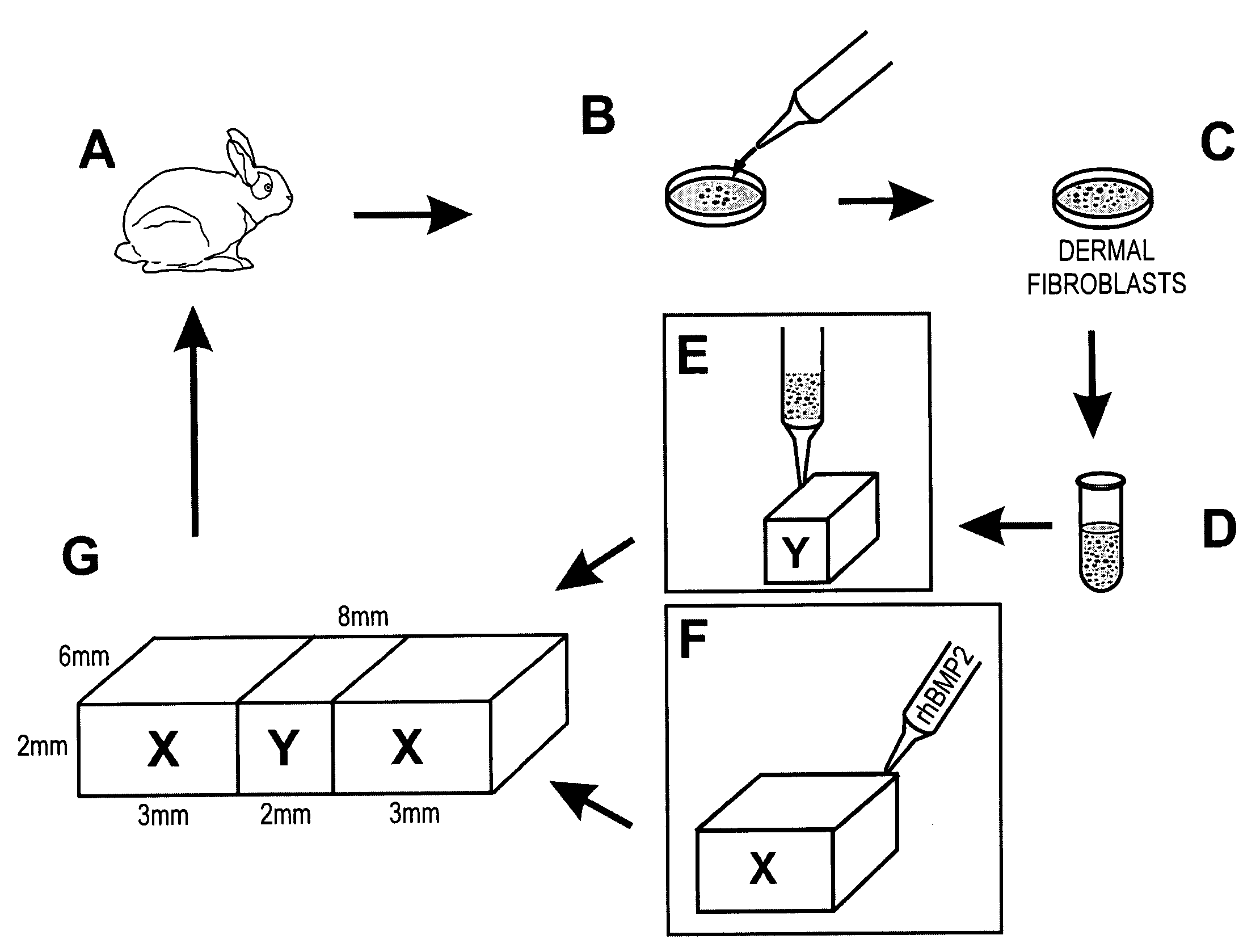
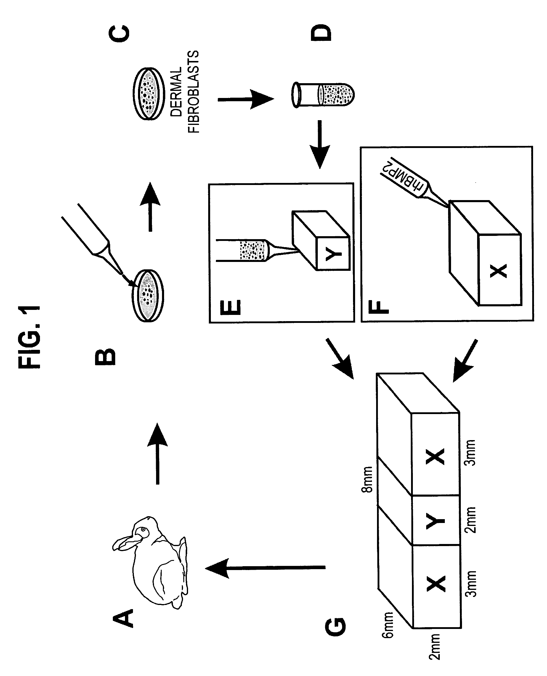
[0043]Eight, 8-week-old, male, New Zealand White rabbits were used in the isolation of dermal fibroblasts after approval by the Animal Care Committee of the University of Illinois at Chicago. Under general anesthesia and aseptic conditions, a 5-mm incision was made in the anterior tibial skin of the rabbit (FIG. 1A). A small piece of subcutaneous fibrous tissue (approx. 3×3 mm2) was removed, minced and digested with the enzyme Accutase under 37° C. for 1 hour, followed by neutralization with Dulbeccols Modified Eagle's Medium-Low Glucose (DMEM; Sigma, St. Louis, Mo.) supplemented with 10% fetal bovine serum (FBS) (Biocell, Rancho Dominguez, Calif.) (FIG. 1B).

[0044]The digested tissue solution was filtered through a cell restrainer (100 μm pore size). After centrifugation, the isolated dermal fibroblasts were plate-cultured (106 cells / 100 mm dish) and expanded in DM...


PUM

PropertyMeasurementUnit
pore sizeaaaaaaaaaa
temperatureaaaaaaaaaa
thickaaaaaaaaaa

Abstract

The in vivo synthesis of connective tissue by fibroblast or fibroblast precursor cells ensconced within a biocompatible scaffold is disclosed. The cells are preferably present in a biocompatible scaffold such as gelatin and placed between two other biocompatible scaffolds such as collagen sponges soaked with a collagenic amount of a member of the TGF-β family of proteins. This composition is then implanted in a host to produce cranial sutures, periodontal ligament or other fibrous tissue structures in vivo.

Description

CROSS-REFERENCE TO RELATED APPLICATION[0001]This application is based on Ser. No. 60 / 504,234 filed on Sep. 19, 2003.GOVERNMENTAL SUPPORT[0002]The present invention was made with governmental support pursuant to USPHS grants DE13964 and DE15391 from the National Institute of Dental and Craniofacial Research of the National Institutes of Health. The government has certain rights in the invention.TECHNICAL FIELD[0003]This invention relates to the formation of fibrous soft connective tissue in vivo. More particularly, this invention relates to the synthesis of cranial sutures by growth factors, fibroblast-like cells and stem cells encased within a biocompatible scaffold.BACKGROUND OF THE INVENTION[0004]Fibrous tissue consists of fibroblastic cells embedded in a hydrated extracellular matrix [Mow et al., Basic Orthopaedic Biomechanics, New York, Raven Press, 143-199 (1991)]. Fibroblasts are crucial to fibrous tissue histogenesis and maintenance [Hunziker (2000) Osteoarth. Cartil., 10:432...

Claims


Application Information

Patent Timeline
Patent Type & Authority Patents(United States)
IPC IPC(8): A61K45/00A61F2/02A61F2/08A61K38/18A61K38/19A61K47/36C12N
CPCA61L27/3804A61L27/3843C12N5/066A61K35/12C12N2533/54
Inventor MAO, JEREMY JIAN
Owner THE BOARD OF TRUSTEES OF THE UNIV OF ILLINOIS
Why Eureka
Try Eureka