Diagnostic test for analytes in a sample

a technology of analytes and visual tests, applied in the direction of instruments, analysis using chemical indicators, biomass after-treatment, etc., can solve the problems of large amount of conjugate, large sample volume, and long wait tim

Active Publication Date: 2008-07-01
ADVANTAGE DIAGNOSTICS
View PDF41 Cites 60 Cited by
  • Summary
  • Abstract
  • Description
  • Claims
  • Application Information

AI Technical Summary

Benefits of technology

[0013]In another embodiment, the test strip additionally comprises a control area located on the membrane downstream of the test area. The control area preferably comprises a control binding agent that is able to bind the conjugate. For example, if the analyte of interest is an antibody and the conjugate comprises an antibody binding agent such as protein A, the control binding agent may be rabbit IgG. If the analyte of interest is an antigen and the conjugate comprises a monoclonal antibody that is specific for the antigen, the control binding agent may be, for example, protein A.
[0014]The test strip may comprise one or more additional components. A buffer pad may be in fl

Problems solved by technology

However, such devices have typically required a relatively large sample volume and large amoun

Method used


Image

  • Diagnostic test for analytes in a sample
  • Diagnostic test for analytes in a sample
  • Diagnostic test for analytes in a sample

Examples


example 1

Construction of an HIV Test Card

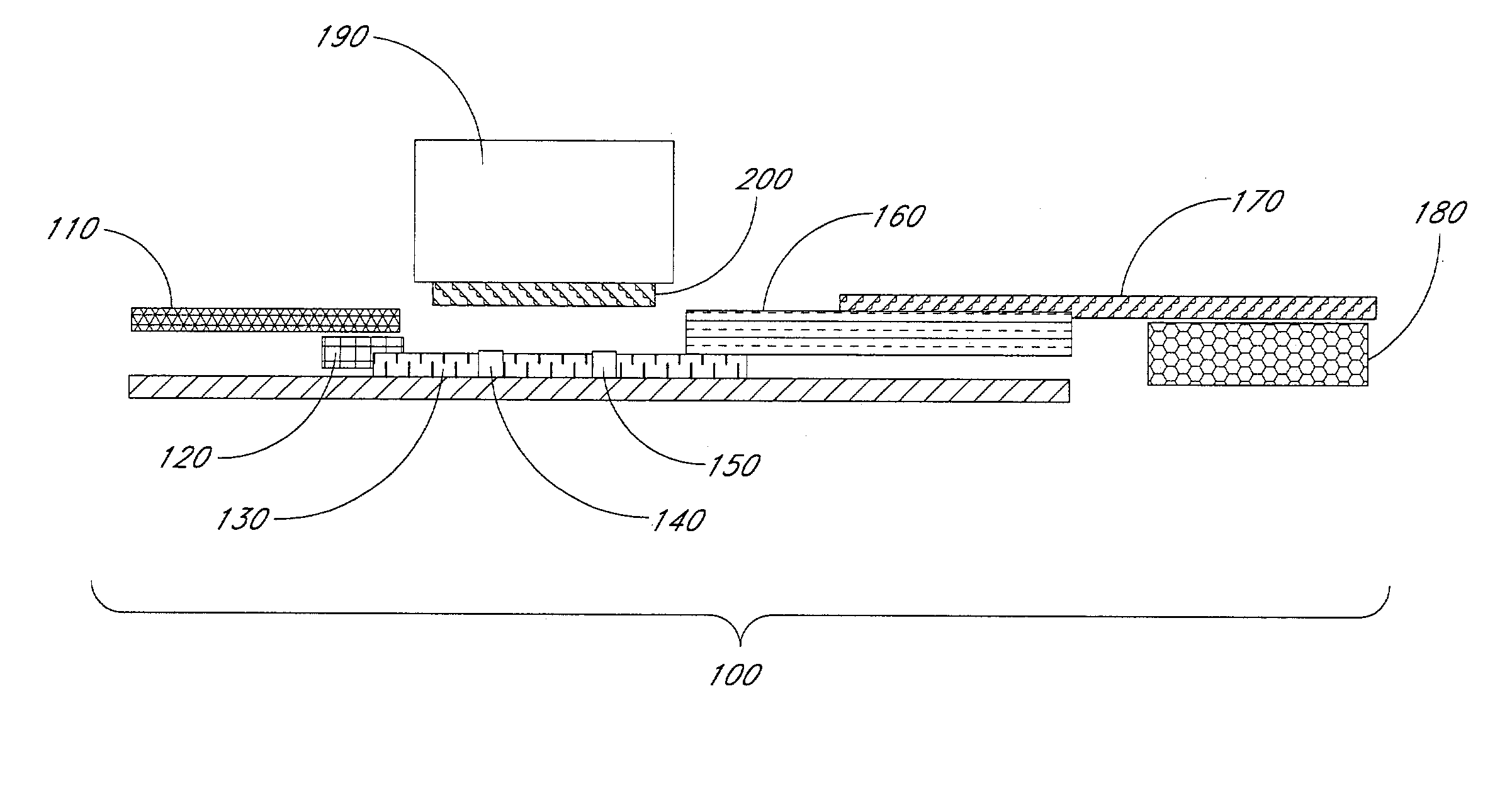
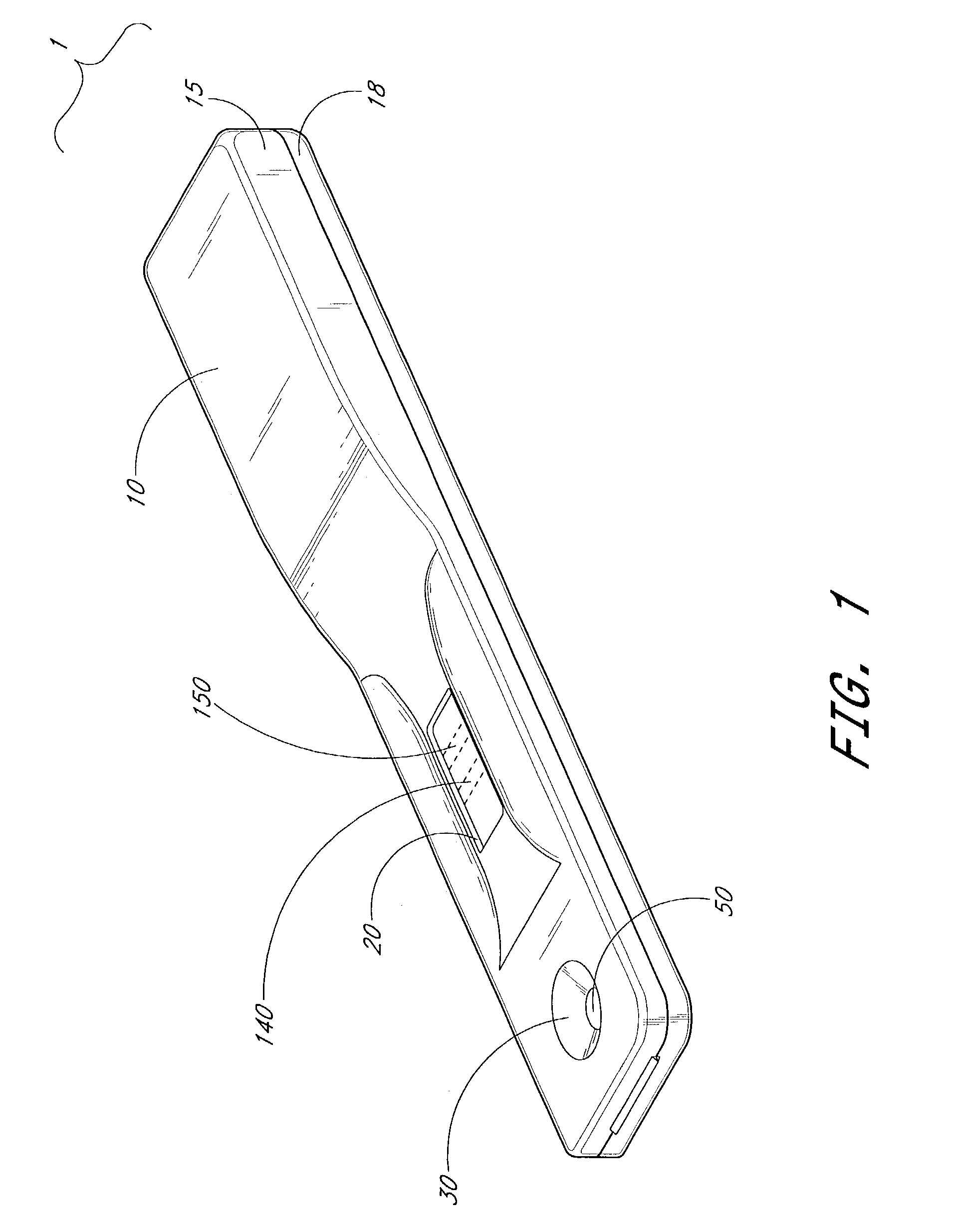
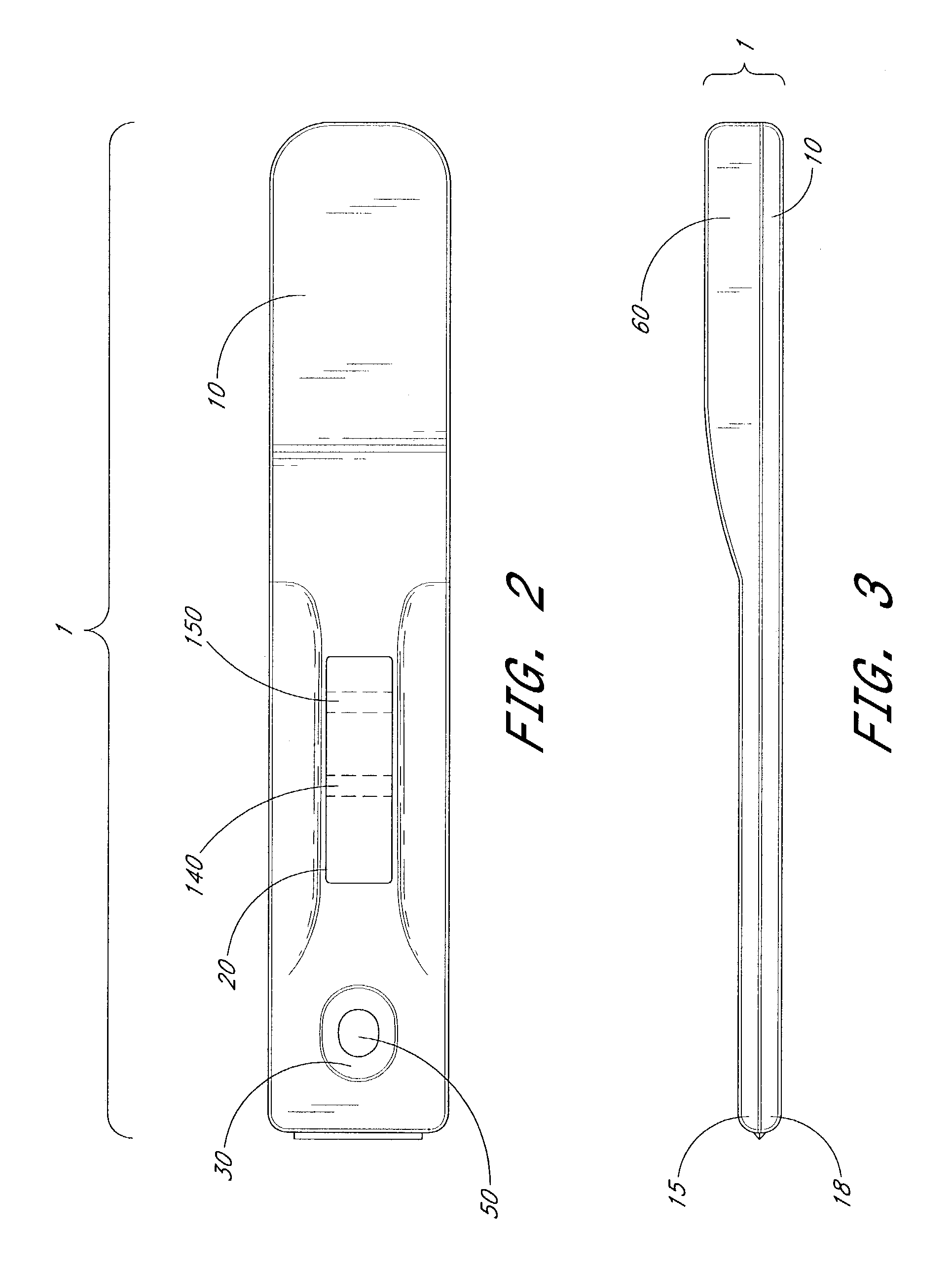
[0118]A lateral flow rapid visual test for the detection of antibodies to human immunodeficiency virus was prepared. The arrangement of the test strips was essentially as illustrated in FIG. 1.

[0119]The test strips comprised a plastic backing (6 cm×5 mm) with adhesive on both sides. The plastic backing was between about 2 mil and about 20 mil in thickness.

[0120]A test line and a control line were prepared on nitrocellulose membrane (Millipore HF09004) by spraying with a pump dispensing system (Kinematic Automation). The test line comprised a vertical line of HIV antigen. The HIV antigen was a mixture of HIV-1 recombinant glycoprotein antigens (GP120 (about 0.15 mg / ml) and P24 (about 0.1 mg / ml)) and HIV-2 recombinant glyco-protein antigen (GP36 (about 0.5 mg / ml), at a concentration of about 0.5 to about 1.0 mg / ml prepared in PBS comprising 5% trehalose. A total amount of from about 0.1 to about 0.5 μg of antigen was applied to the test area in a vertic...

example 2

Test for HIV

[0130]Serum or plasma was prepared from a whole blood sample obtained from a patient to be tested for HIV using proper venipuncture techniques. Samples were stored at 2° C. to 8° C. and used within 24 hours or frozen at −20° C. for use within 2 weeks. Frozen samples were thawed before use.

[0131]An HIV test card as described in Example 1, comprising a mixture of HIV-1 and HIV-2 antigens on the control area, was removed from the foil pack.

[0132]Approximately 5 μL of sample buffer comprising 0.025% HEPES and 0.2% sodium azide was applied to the membrane through the center of the test card window.

[0133]A sample pipette containing the serum or plasma to be tested was held in a vertical position over the test window on the test card and 5 μl of sample was placed onto the membrane in the center of the window.

[0134]Four drops of wash buffer solution (0.025% HEPES, 0.85% sodium chloride, 0.1% EDTA, 1% mannitol, 0.1% casein, 1% Triton X100, pH 7.2) were added to the buffer pad. Af...

example 3

HCV Test Card

[0137]A test strip for the diagnosis of hepatitis C virus was prepared as in Example 1. However, a hepatitis C antigen mixture comprising core (0.3 mg / ml), NS3 (0.4 mg / ml), and NS4 (0.1 mg / ml) was sprayed in a test line on the membrane to produce the test area, rather than HIV antigen. A total of about 0.4 μg of HCV antigen mixture was applied in the test line. Rabbit IgG diluted to 1 mg / ml was sprayed in a control line.


PUM


Abstract

Diagnostic devices and methods are provided. The diagnostic devices preferably comprise a test strip with a test area. The sample to be analyzed contacts the test area, which comprises a specific binding partner for the analyte of interest. Analyte, if present in the sample, binds to the immobilized binding partner in the test area and is subsequently contacted with a conjugate. The conjugate specifically binds the analyte and provides a visual indication of the presence of the analyte. The devices may be used for the diagnosis of particular diseases or disorders in a patient, such as HIV or hepatitis. They may also be used to determine if an individual is pregnant.

Description

BACKGROUND OF THE INVENTION[0001]1. Field of the Invention[0002]The present invention relates to lateral flow rapid visual test for the detection of analytes in a sample.[0003]2. Description of the Related Art[0004]Lateral flow testing devices are known in the art and may be used in clinical diagnosis to determine the presence of an analyte of interest in a sample, such as a bodily fluid. However, such devices have typically required a relatively large sample volume and large amounts of conjugate. They have also had a long wait time before the results of the test can be read.SUMMARY OF THE INVENTION[0005]In one aspect, an immunodiagnostic test strip is provided for the detection of an analyte of interest in a sample. In some embodiments, the analyte is an antibody, such as an anti-human immunodeficiency virus (HIV) antibody or an anti-hepatitis C virus (HCV) antibody. In other embodiments the analyte is an antigen, such as human chorionic gonadotropin (hCG).[0006]The diagnostic test...

Claims


Application Information

Patent Timeline
IPC IPC(8): G01N33/53G01N33/558
CPCG01N33/558Y10S435/973Y10S435/805Y10S435/971G01N2333/59G01N2333/16G01N2333/18Y10S436/828
Inventor CHARLTON, DAVID
Owner ADVANTAGE DIAGNOSTICS
Why Eureka
Try Eureka