Lambda phage display system and the process

a phage and display system technology, applied in the field of new phage display systems, can solve the problems of limiting the library size and the number of clones that can be screened, and the system has not gained much popularity, and achieves high efficiency cloning and high density display of proteins/peptides

Inactive Publication Date: 2008-08-12
UNIVERSITY OF DELHI
View PDF2 Cites 4 Cited by
  • Summary
  • Abstract
  • Description
  • Claims
  • Application Information

AI Technical Summary

Benefits of technology

[0010]Accordingly the instant invention describes a phage lambda display system which allows high efficiency cloning in phage lambda using lox-cre recombination system and high density display of protein / peptides fused to the head protein, ‘d’, of phage lambda. Further, the display of different molecules on phage lambda is compared with display of same molecules on phage M13.
[0011]The instant invention provides for a novel system for C-terminal display on phage lambda that avoids cloning into lambda phage DNA. This C-terminal display system achieves cloning efficiencies comparable to those obtained with any plasmid system and eliminates the step of in vitro packaging of phage lambda. The high-density display of foreign proteins is useful in studying low affinity protein-protein interactions, which is more efficient as compared to M13 display system.
[0012]The object of the instant invention is to provide a system for obtaining recombinant lambdoid bacteriophage with high-density display of functional peptides and proteins that has high efficiency cloning in phage lambda.

Problems solved by technology

However, the cumbersome technique of immobilizing expressed proteins on filters and screening large number of filters to obtain one specific binder is labour intensive and exhaustive, limiting the library size and number of clones that can be screened.
Therefore, it is not useful for studying interactions involving free-C-terminus of display partner and for making full-length cDNA libraries from polyA mRNA.
However, this system has not gained much popularity, which can be attributed to several reasons including the fact that(i) the lambda phage biology is more complex than of M13 phage.
Unlike M13 which grows by extruding from host cell, lambda can follow a lysogenic or lytic mode; therefore manipulation of lambda life cycle is more difficult,(ii) lambda genome is very large (50 kb), therefore isolation of viral DNA, insertion of user defined restriction sites, cloning of foreign fragments and then packaging of the ligated product in vitro to make lambda particles is difficult and the library sizes achieved are less than those obtained with phage / phagemid based M13 vectors.

Method used


Image

  • Lambda phage display system and the process
  • Lambda phage display system and the process
  • Lambda phage display system and the process

Examples


example 1

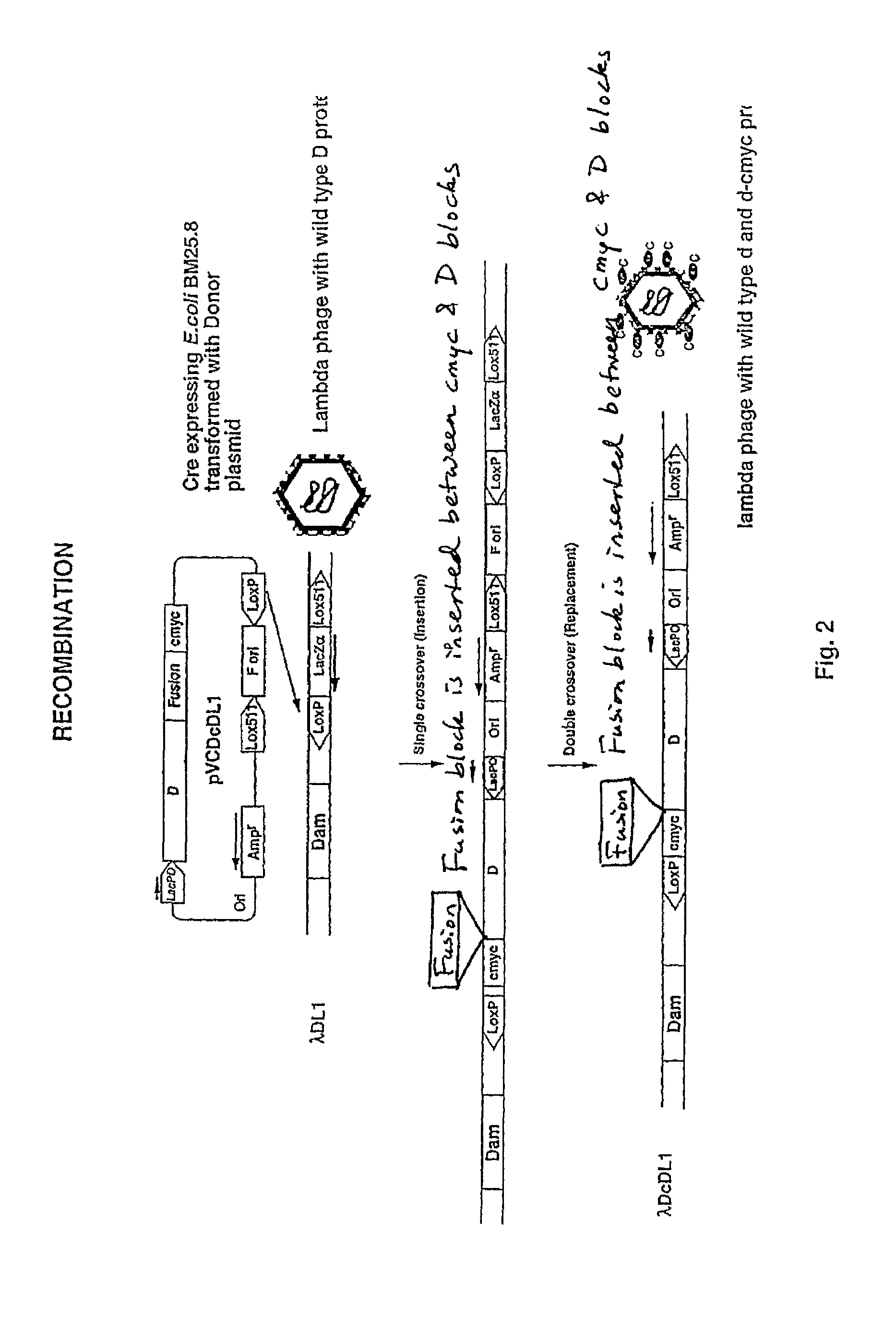
[0057]The system described above is optimized using donor plasmid pVCDcDL1, which expresses D protein fused to a ten amino acid tag, cmyc. pVCDcDL1 was used to transform BM25.8 (Cre+) cells and the transformants were infected with recipient lambda, λDL1. The lysate obtained after culture growth and lysis was plated to obtain ampicillin resistant transductants. These transductants were analysed by PCR to check for integration of plasmid into λ DNA.

[0058]Two kinds of cointegrates were obtained. Single crossover at one of the lox sites resulted in formation of SCO (single crossover cointegrates) which contained the entire donor plasmid and lacZα cassette (FIG. 2). A second crossover event resulted in excision off ori and lacZα cassette and this was called DCO (double crossover cointegrates). Both SCO and DCO produced same number of phages and the display of D-cmyc fusion protein was comparable.

[0059]DcDL1 phages were made and tested for display of D-cmyc in ELISA. Purified DcDL1 phages...

example 2

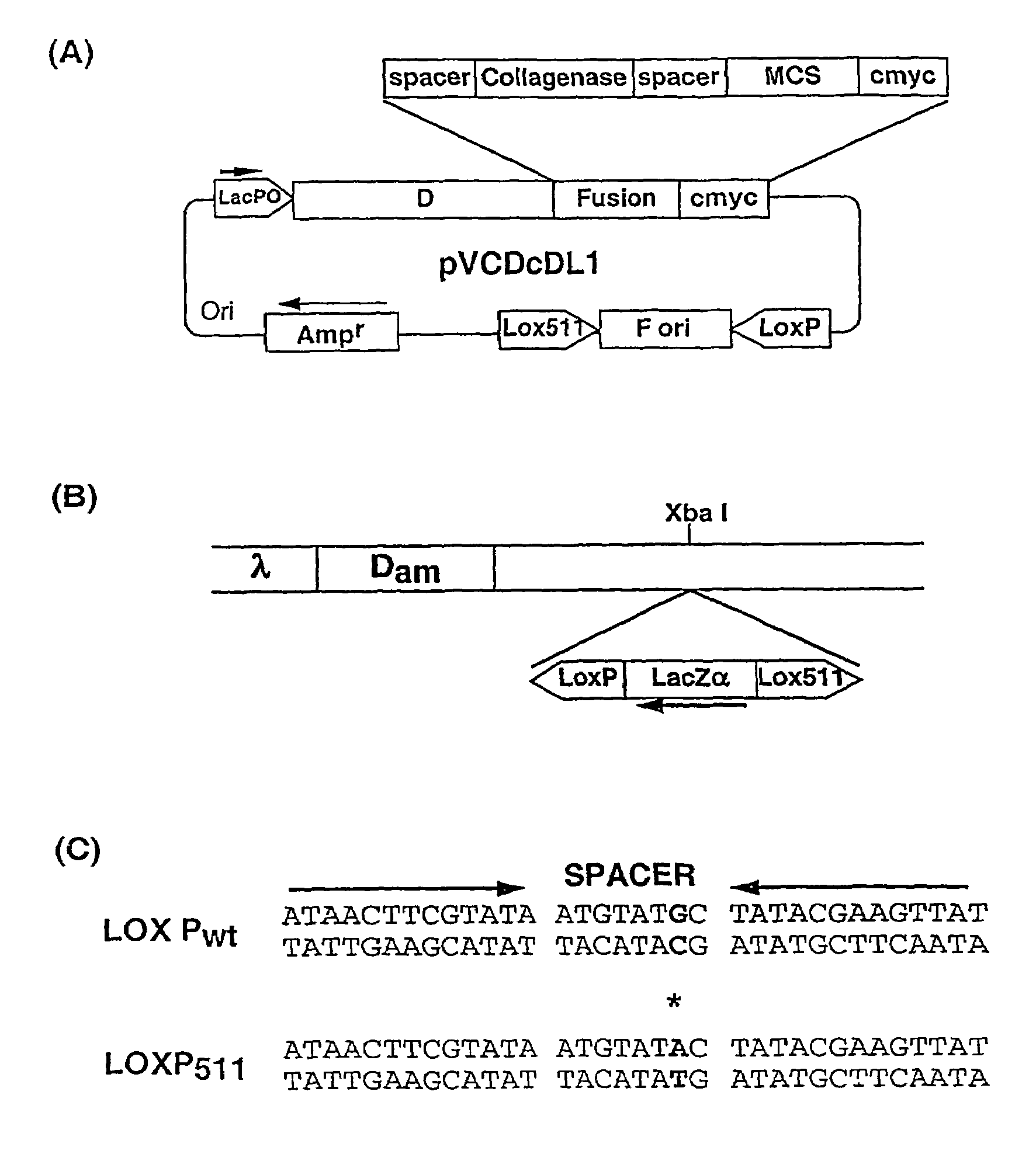
[0067]The ‘Donor’ plasmid vector pVCDcDL1, (FIG. 1A) a high copy number vector based on pUC119, was constructed for cloning sequences to be displayed as d-fusion protein on. This plasmid vector contains ‘d’ gene of under lacPO followed by a Gly-Ser spacer, collagenase cleavage site, multiple cloning site (MCS) and a decapeptide tag, cmyc. Cloning of DNA sequences in the MCS allows formation of d-fusion protein with the fusion protein present at C-terminus of d protein. The collagenase site allows cleavage of the fusion partner from d protein and is used for elution of bound phages after panning by collagenase treatment. The cmyc tag allows easy identification of the fusion protein. The vector contains M13 phage origin of replication (f ori), flanked by Lox Pwt and Lox P511 sites (Hoess et al., 1982).

[0068]This cassette is extremely stable and there is no loss of ‘f ori’ sequence upon repeated cycles of growth (more than five times) in Cre+ / Cre−host (data not shown).

[0069]pVCDcDL1 wa...

example 3

Comparison of Lambda Display System with M13 Display System

[0075]DNA sequences encoding different length fragments of HIV capsid protein p24 were amplified from pVCp24210 (Gupta et al., 2000) using 5′ primer which annealed to first eight codons of the fragment and contained Nhe I site and 3′ primer which annealed to last eight codons of the fragment and contained Mlu I site. The amplified products were digested with Nhe I and Mlu I and cloned in Nhe I-Mlu I digested, dephosphorylated vector, pVCDcDL1 to create pVCDc(p241)DL1, pVCDc(p246)DL1 and pVCDc(p24)DL1.

[0076]The three constructs were then used to transform Cre+ host BM25.8 and recombination was carried out to obtain Dc(p241)DL1, Dc(p246)DL1 and Dc(p24)DL1.

[0077]DNA encoding different p24 fragments were cloned as Nhe I-Mlu I inserts into gIII display vector, pVC3TA726 (Sampath et al., 1997) and similar gVIII display vector, pVCp2418426 (VKC, personal communication). The recombinants pVC(p241)3426, pVC(p246)3426 and pVC(p24)3426...


PUM

PropertyMeasurementUnit
densityaaaaaaaaaa
sizeaaaaaaaaaa
sizesaaaaaaaaaa

Abstract

The present invention relates to a process of obtaining recombinant lambdoid bacteriophage with high density display of functional peptides and proteins on surface of said phage comprising of: constructing a donor plasmid having a nucleotide sequence that defines the elements for replication of the vector in bacteria, a selectable marker, a nucleotide sequence flanked by two non-compatible recombination sequences, and an inducible cistron for expression of a capsid protein and a fusion protein; constructing a recipient phage having a nucleotide sequence that defines the lambdoid elements for replication and packaging of the vector into an assembled bacteriophage and encodes an inducible cistron for expression of a selectable marker flanked by two non-compatible recombination sequences; transferring the said donor plasmid to said recipient plasmid to obtain cointegrates; growing said cointegrates in selective liquid medium; harvesting phages displaying protein encoded by the foreign DNA encapsulated in said harvested phage particle.

Description

FIELD OF INVENTON[0001]This invention relates to a process of obtaining recombinant lambdoid bacteriophage and the resultant novel phage display system.BACKGROUND OF INVENTION[0002]Identification of specific binding sequences is an integral part of research; delineation of antibody recognition sites, study of host-pathogen interaction, elucidation of signal transduction pathways, understanding of cellular responses, all these involve interaction of various macromolecules with one another; most of these interactions occurring between proteins or of a protein with another macromolecule.[0003]For several years, expression libraries have been used for isolating ligands of desired specificity. However, the cumbersome technique of immobilizing expressed proteins on filters and screening large number of filters to obtain one specific binder is labour intensive and exhaustive, limiting the library size and number of clones that can be screened.[0004]It was the concept of phage display intro...

Claims


Application Information

Patent Timeline
Patent Type & Authority Patents(United States)
IPC IPC(8): C12N15/65C12N15/74A61KC07H21/04C12N1/20C12N7/00C12N15/00C12N15/10C12N15/86C12P21/04C12P21/06C12Q1/68C40B40/02
CPCC07H21/04C40B40/02C12N15/1037
Inventor CHAUDHARY, VIJAY KUMARGUPTA, AMITAADHYA, SANKARPASTAN, IRA
Owner UNIVERSITY OF DELHI
Why Eureka
Try Eureka