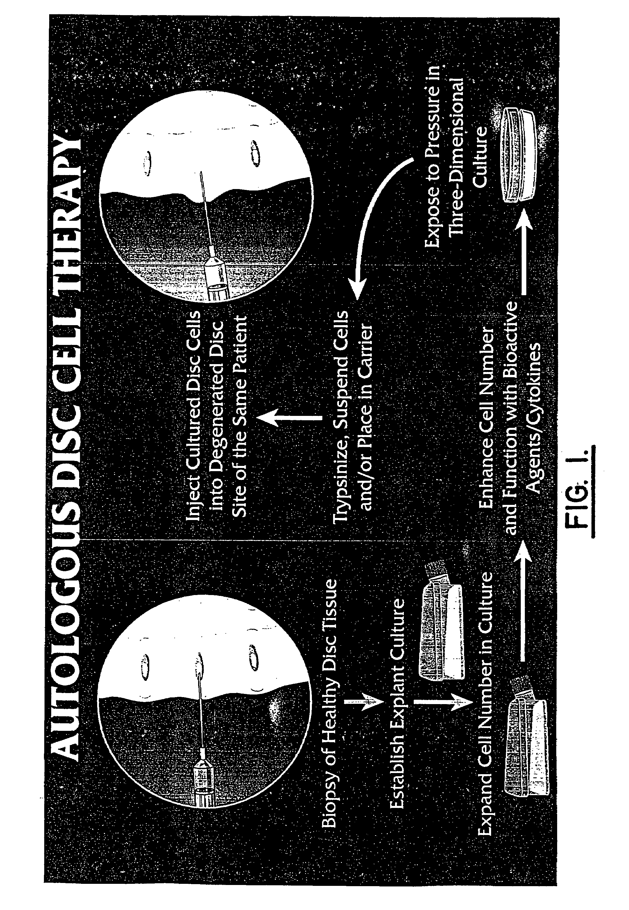
Method for producing human intervertebral disc cells for implantation
a technology of intervertebral discs and implantation, which is applied in the direction of skeletal/connective tissue cells, drug compositions, peptide/protein ingredients, etc., can solve the problems that the cell biology of human intervertebral disc cells has been neglected
- Summary
- Abstract
- Description
- Claims
- Application Information
AI Technical Summary
Benefits of technology
Problems solved by technology
Method used
Image

Examples
example 1
[0048]This example illustrates the growth of healthy disc cultures. Healthy disc tissue was obtained and rinsed with phosphate buffered saline at pH 7.4. The tissue was placed in modified Minimal Essential Medium (MEM) with Earle's salts (MEM, Gilco, Grand Island, N.Y.) with 1% (v / v) L-glutamine (Irvine Scientific, Santa Ana, Calif.), 1% (v / v) penicillin-streptomycin (Irvine Scientific, Santa Ana, Calif.), but without added serum. The disc tissue was incubated in three 15-minute rinses of MEM with 1% fungizone (Irvine Scientific) as a precaution against contamination of the cells during removal.
[0049]Regions of annulus and nucleus were visually identified and representative pieces of annulus and nucleus were dissected. Cartilaginous and vascular regions removed from tissue designated for culturing. The disc tissue to be cultured was minced with a scalpel into 1-2 mm square pieces, again rinsed twice with saline to remove clots or residual debris, placed into 35 mm culture dishes and...
example 2
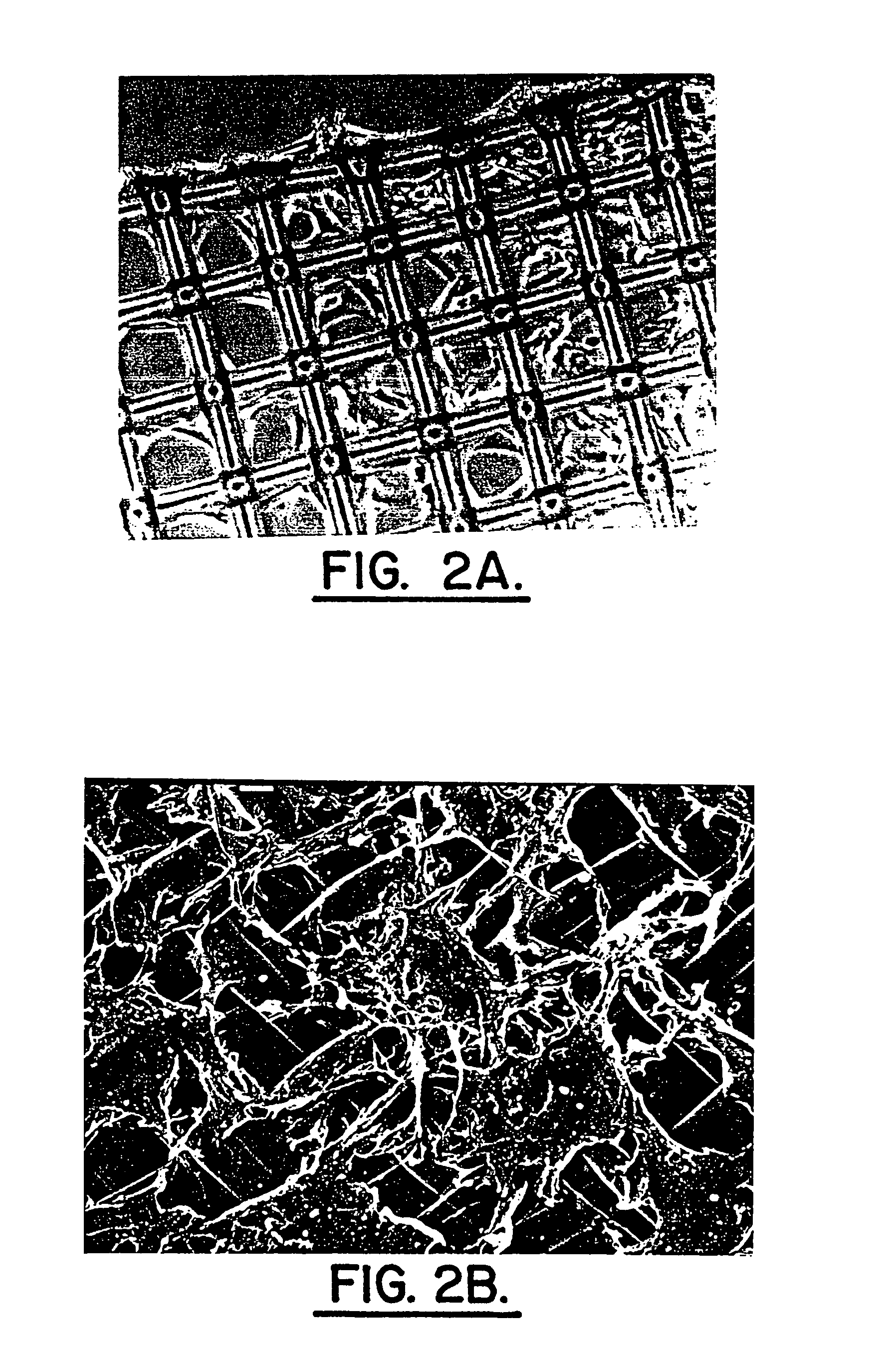
[0053]In this example, primary cell cultures were grown in alginate. Cells established in monolayer primary culture were seeded either into alginate layers on inserts (P1 cultures) or continued as monolayer P1 cultures which were subsequently split and used as the cell source for sequential P2, P3 or P4 passages seeded into alginate. Several cryofrozen cultures were thawed and expanded as P1 passages on monolayer with subsequent P2 and later passages seeded into alginate.
[0054]The alginate solution was prepared from a 1.2% solution of Keltone LV alginate (Kelco, San Diego, Calif.) in 0.9% physiological saline with stirring for one hour. The alginate solution was sterilized by filtering through a 0.2 μm bottle top filter fitted onto a 100 ml sterile bottle.
[0055]Trypsinized primary cell cultures were assayed for cell viability and the required volume of cell suspension centrifuged at 500 rpm for five minutes in an IEC MP4R centrifuge and the medium subsequently aspirated off. An appr...
example 3
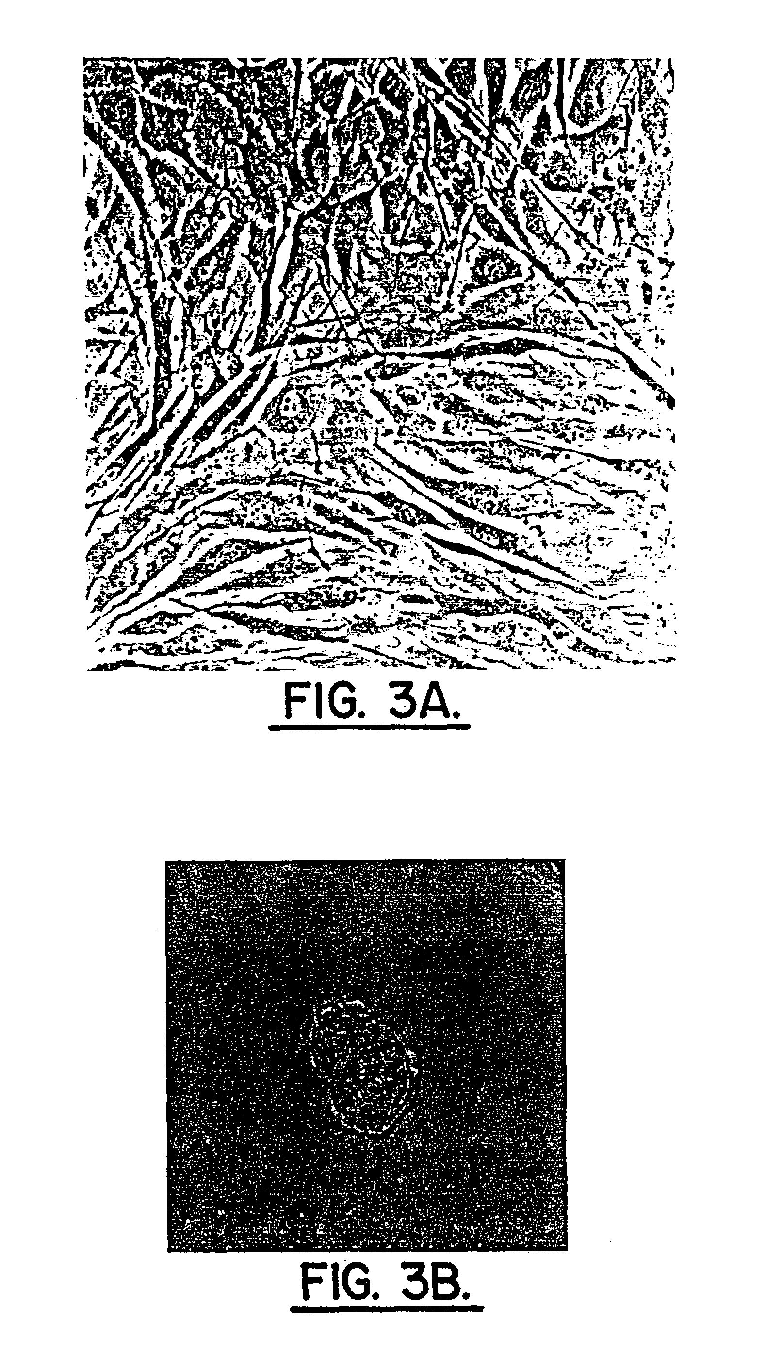
[0060]Adjacent sections of three-dimensional cultures from Example 2 cut en face were collected and utilized for immunohistochemical localization of rabbit anti-human collagen Type I or rabbit anti-human collagen Type II (Biodesign International, Kennebunk, Me.), monoclonal anti-proteoglycan delta DI-4S (ICN, Costa Mesa, Calif.) or monoclonal anti-keratin sulfate (Seikagaku Corp., Tokyo, Japan).
[0061]Negative controls were incubated minus primary antibodies. Localization utilized DAB calorimetric visualization of extracellular matrix constituents. Cells or colonies were scored for positive immunohistochemical localizations using a Nikon microscope (20× objective) and OsteoMeasure computer software (OsteoMetrics, Inc., Atlanta, Ga.). The mean number of cells or colonies assessed for each localization was: Type I collagen: 272; Type II collagen: 260; proteoglycan delta DI-4S, 241, and keratin sulfate, 201. Nine surgical and two normal series of cells were studied in this experiment. P...
PUM
| Property | Measurement | Unit |
|---|---|---|
| temperature | aaaaa | aaaaa |
| humidity | aaaaa | aaaaa |
| humidity | aaaaa | aaaaa |
Abstract
Description
Claims
Application Information