Radiopaque marker for a surgical sponge

a radiopaque marker and sponge technology, applied in the field of sponges, can solve the problems of disturbing regularity, pain, infection, death of patients, accidental retention of surgical implements, sponges and the like, etc., and achieve the effects of high radiographic density, high contrast, and easy recognition and differentiation

Inactive Publication Date: 2008-12-16
FABIAN CARL E
View PDF23 Cites 129 Cited by
  • Summary
  • Abstract
  • Description
  • Claims
  • Application Information

AI Technical Summary

Benefits of technology

[0009]The present invention provides a radiopaque marker suitable for association with a surgical sponge or other surgical implement. The marker has a high radiographic density and a distinctive shape, whereby the marker produces an x-ray image with high contrast and a shape that is readily recognizable and differentiated from the images produced by other items and structures commonly seen in x-rays of post-operative patients. The marker is suitable for association with a surgical sponge and has an x-ray density equivalent to at least about 0.1 g / cm2 of BaSO4.
[0012]The combination of the distinctive shape of the marker and its high x-ray density causes it to produce an x-ray image that much better suited to be readily and unambiguously detected than images produced by previous radiopaque elements. As a result, a sponge comprising a marker constructed in accordance with the present invention will not likely remain undetected on an x-ray if inadvertently left within a patient following a surgical procedure.
[0013]Advantageously, the presence of the EAS tag in the surgical sponge of the invention further reduces the likelihood of a sponge being retained inadvertently, since remote scanning can be carried out simply without the need of an x-ray and without risk to the patient before completion of a surgical procedure in which the sponge is used.

Problems solved by technology

When this occurs, serious consequences often ensue.
The patient may suffer pain, infection, intestinal obstruction, and even death.
Notwithstanding these precautionary measures the accidental retention of surgical implements, especially sponges and the like, continues to occur to this day with disturbing regularity, even in highly respected institutions.
Surgeons and related medical professionals regard this eventuality as a major unsolved problem.
A number of difficulties compromise the efficacy of visual searching.
Sponges also tend to become compacted and wadded after being moistened.
In addition, some approaches have been proposed that would allow overlooked sponges and other surgical items to be located by remote electronic techniques and removed before completion of surgery, but these methods have not yet come into widespread use.
However, intraoperative x-rays are not routinely performed before closure of the incision for several reasons: They entail the risks that inevitably arise with the extension of operative time and anesthesia, as well as undesirable expense, inconvenience, and radiation exposure.
These devices generally have a lower output than fixed, standard machines, necessitating longer exposures and resulting in inferior resolution, for example due to motion of the patient which causes blurring of the image.
Postoperative x-rays, even if done with a fixed machine, are still subject to some of the same disadvantages and are not routinely done unless there is a specific question or suspicion of a retained implement in a given case.
Moreover, even when postoperative x-rays are obtained, retained surgical implements are still overlooked in many cases, owing to the presence of artifacts or other competing shadows on the film or the unfavorable orientation of the object relative to the x-ray incidence direction and the position of the x-ray film.

Method used


Image

  • Radiopaque marker for a surgical sponge
  • Radiopaque marker for a surgical sponge
  • Radiopaque marker for a surgical sponge

Examples


Embodiment Construction

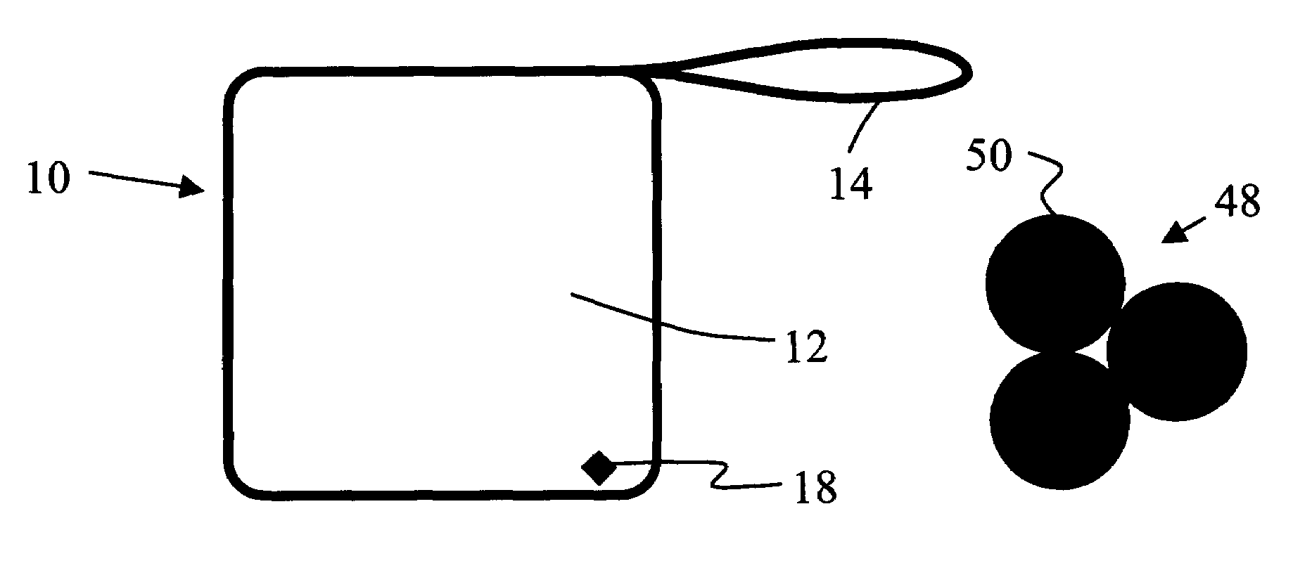
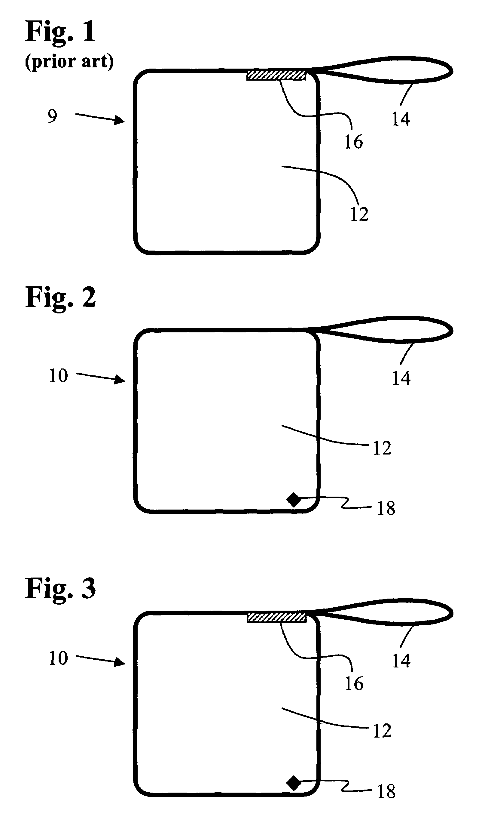
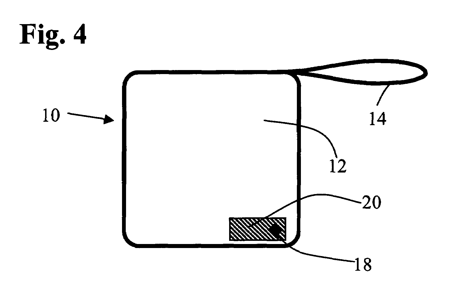
[0027]The present invention provides a radiopaque marker that is adapted to be attached to a surgical sponge or surgical implement. As a result of the high radiographic density and distinctive shape of the marker, an x-ray image thereof is easily recognized. If a sponge or implement bearing such a marker is inadvertently allowed to remain within the patient after the conclusion of surgery, it may still be detected and localized by a routine postoperative x-ray and subsequently removed to prevent serious medical consequences.

[0028]Referring now to FIG. 1 there is depicted a prior art surgical sponge 9 composed of gauze 12 and having a fabric loop 14 to facilitate identification and location of the sponge. The sponge bears a generally rectangular, sheet-form radiopaque element 16. Each of FIGS. 2-4 depicts an aspect of a surgical sponge 10 of the invention. As depicted, each sponge includes an optional fabric or thread loop 14 to facilitate identification and location of the sponge. S...


PUM


Abstract

A surgical sponge comprises a plurality of radiopaque markers having a high radiographic density and a distinctive, visually recognizable shape. The markers have an x-ray density equivalent to at least about 0.1 g / cm2 of BaSO4. The markers produce an x-ray image with high contrast and a shape that is readily recognizable and differentiated from the images produced by other items and structures commonly seen in x-rays of post-operative patients. Owing to the distinctive, high contrast image produced by the markers, the sponge is reliably and unambiguously detected. This is so even in situations where the sponge is inadvertently left in the surgical wound. Discomfort, trauma, and possibly fatal consequences that might otherwise occur are virtually eliminated. The surgical procedure is carried out with decreased likelihood of a sponge being retained inadvertently.

Description

BACKGROUND OF THE INVENTION[0001]1. Field of the Invention[0002]The present invention relates to a surgical sponge, and more particularly, to a surgical sponge or similar absorbent article having associated therewith a radiopaque marker that produces a visually distinctive shadow on an x-ray image of the sponge, rendering the sponge easily detectable and locatable within a surgical patient.[0003]2. Description of the Prior Art[0004]During the course of a surgical operation it is generally necessary for articles, such as surgical sponges, gauzes, instruments and the like, to be placed into a wound cavity. Sponges are often used to protect and isolate organs and tissues not directly involved in the surgical procedure; to absorb incidental blood and other bodily fluids; and to serve as aids in grasping and displacing structures to facilitate access needed to various internal regions of the patient's body.[0005]Despite rigorous attention given to locating and removing all these items pr...

Claims


Application Information

Patent Timeline
Patent Type & Authority Patents(United States)
IPC IPC(8): A61F13/15A61B19/00A61N5/00A61F13/20A61F13/44
CPCA61F13/44A61F2013/8497
Inventor FABIAN, CARL E.
Owner FABIAN CARL E
Why Eureka
Try Eureka