Tissue graft scaffold made from cholecyst-derived extracellular matrix

a tissue graft and extracellular matrix technology, applied in the field of bioengineered tissue graft scaffolds, can solve the problems of complex manufacturing, inability to modify as easily, and inability to replace or repair the tissue satisfactorily or within an appropriate time scale, so as to reduce the likelihood of inducing an inflammatory response and improve tensile strength

Inactive Publication Date: 2009-06-23
THE NAT UNIV OF IRELAND GALWAY
View PDF2 Cites 36 Cited by
  • Summary
  • Abstract
  • Description
  • Claims
  • Application Information

AI Technical Summary

Benefits of technology

[0014]One object of the present invention is to provide an alternative tissue scaffold. A further object is to provide a tissue scaffold that has a reduced likelihood of inducing an inflammatory response on implantation in a patient. It is also an object to provide a tissue scaffold that harbors fewer cellular remnants than currently available tissue scaffolds. Another object is to provide a tissue scaffold with improved tensile strength. A still further object is to provide an improved method of producing a tissue scaffold.SUMMARY OF THE INVENTION
[0032]Suitably the tissue is delaminated. Delamination may be achieved by mechanical means, such as by scraping. Preferably, the tissue is immersed in 10% neutral buffered formalin for at least 1 min and up to 12 hours prior to delamination. Preferably the immersion is for 2-4 hours. This helps to prevent tearing during delamination.
[0037]Furthermore the submucosa of the small intestine is highly cellular due to the nature fibroblasts and submucosal glands, whereas the extracellular matrix of the cholecyst has relatively few cellular components, esp. devoid of vasculature, submucosal glands, lymphatic and nervous networks. Cellular components have been shown to generate antigenicity in the scaffold rendering it to be ineffective in long term applications because of the potential immunogencicity. The scaffold of the present invention is about 130 μm giving the scaffold a more elastic nature. Additionally the proportion of collagen in SIS is less than that in the cholecyst extracellular matrix which means the scaffold of the present invention is stronger.

Problems solved by technology

Many disease conditions or injuries of the body require the repair or replacement of damaged tissues, but the body itself may not be able to replace or repair the tissue satisfactorily or within an appropriate time scale.
On the other hand, these scaffolds exhibit great variability in terms of their morphological and biological properties, they are complex to manufacture and susceptible to contamination.
Additionally, modifications are not as easy compared to synthetic scaffolds.
There are limitations such as size, shape and the properties that can be imparted to a biological scaffold.
These processing procedures may alter the innate characteristics of the scaffold.
Synthetic scaffolds have the disadvantage that cell recognition is difficult, surface alterations such as mobilisation of growth factors to the scaffold (to permit cell migration, proliferation and adhesion) might be needed in some applications, they are subject to hydrolysis / enzymatic degradation and generate no biological signals.
Although scaffolds derived from small intestine and urinary bladder can be used for certain applications, they are not suitable for universal use because of their defined range of mechanical and biological properties.
Non-biodegradable membranes are prone to rejection in the short term and failure in the long term.
This healed area may be very different from the original tissue, because the prosthetic membrane does not elicit a proper healing response.

Method used


Image

  • Tissue graft scaffold made from cholecyst-derived extracellular matrix
  • Tissue graft scaffold made from cholecyst-derived extracellular matrix
  • Tissue graft scaffold made from cholecyst-derived extracellular matrix

Examples


Embodiment Construction

Materials and Methods

Isolation of Cholecyst Extracellular Matrix

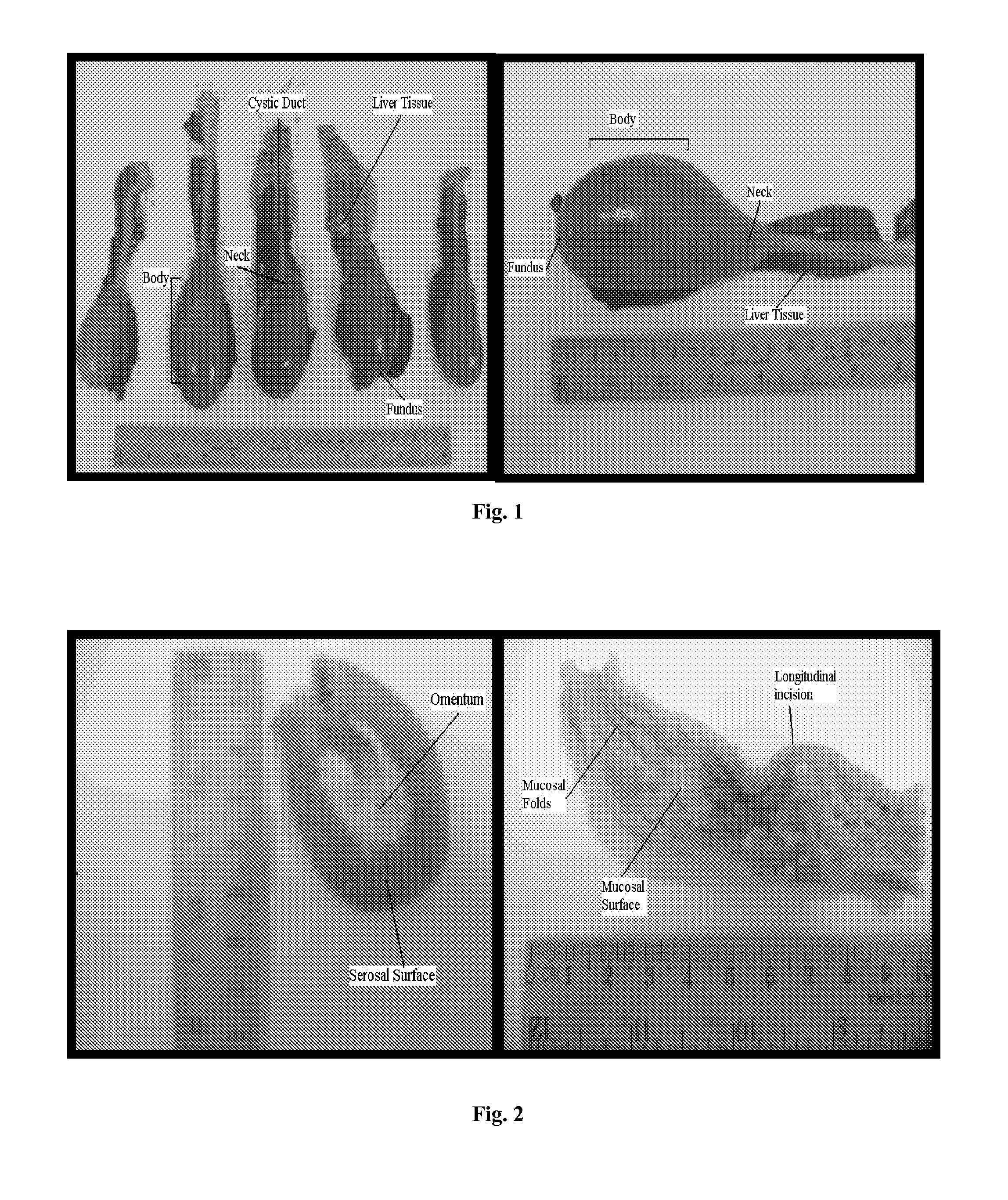
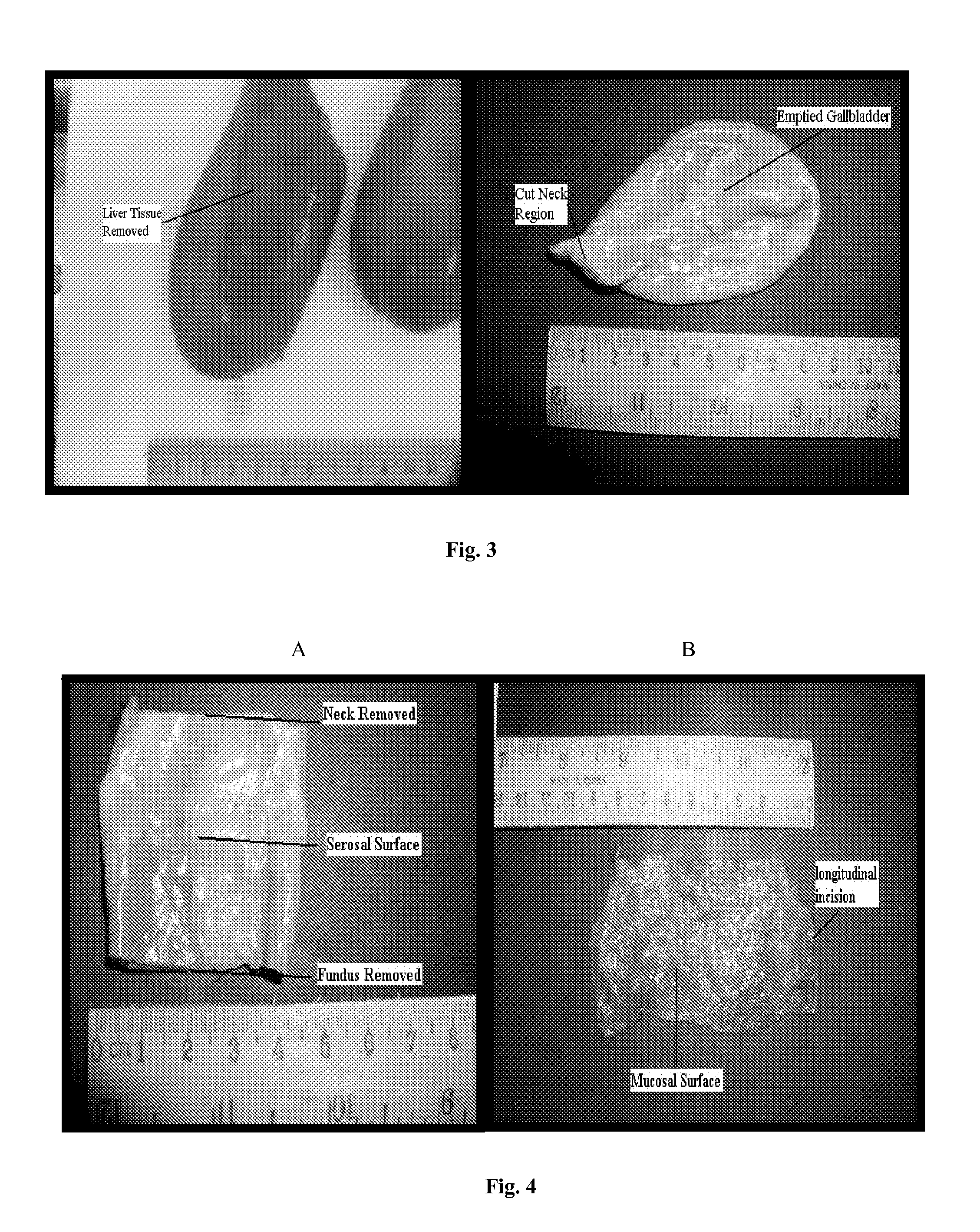
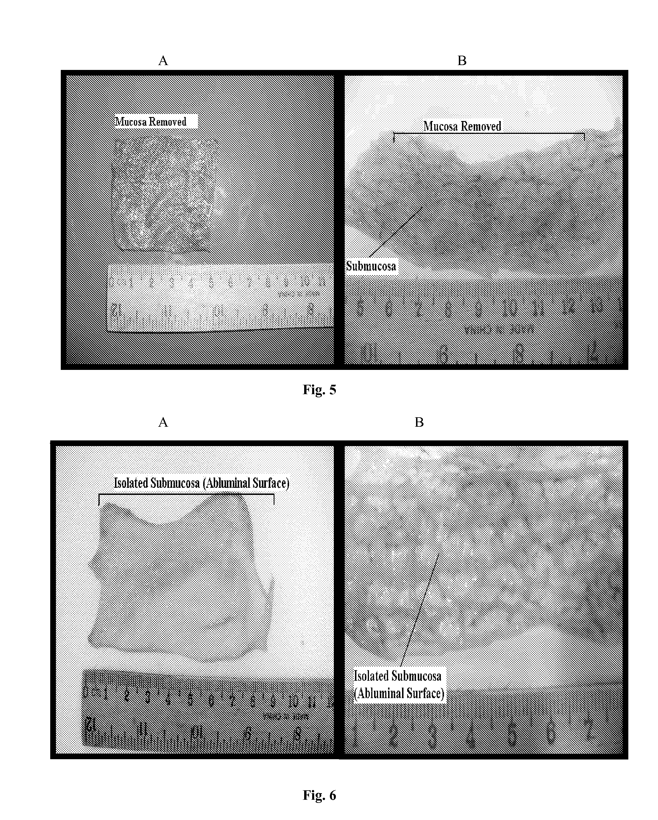
[0073]Cholecyst and small intestine tissue was obtained from market weight pigs, slaughtered at Duffy's Meats abattoir, Gort. Co. Galway. Porcine specimens were fixed immediately post mortem by total immersion in 10% neutral buffered formalin (NBF) for light microscopy and 3% Glutaraldehyde Fixative for electron microscopy. Whole cholecysts were punctured and drained of bile before immersion in fixatives, while small intestinal samples were cut into approximately 10 cm long sections before immersion in fixative. Samples were subsequently transported to a suitable laboratory environment for further tissue processing. Before fixation had fully occurred (2 hours), samples were further processed to allow for total fixation and for the isolation of extra cellular matrix. This process resulted in the procurement of whole tissue and delaminated extra cellular matrix, for light and electron microscopy. Before delamination occur...


PUM


Abstract

Bioengineered tissue graft scaffolds and method for producing such scaffolds are provided. The scaffolds are useful to replace or repair damaged mammalian tissues and organs.

Description

FIELD OF THE INVENTION[0001]The present invention relates to bioengineered tissue graft scaffolds and to methods of producing such scaffolds. The scaffolds find uses to replace or repair damaged mammalian tissues and organs.[0002]In this context a scaffold can be defined as a biodegradable material that supports the growth of cells and will eventually degrade leaving behind regenerated tissue structure. The scaffold provides a support upon which cells can organise and develop.BACKGROUND OF THE INVENTION[0003]Tissue engineering attempts to create three-dimensional tissue structures on which cells and other biomolecules may be incorporated. These structures or scaffolds guide the organization, growth and differentiation of cells in the process of forming functional tissue by providing physico-chemical cues. To successfully incorporate a scaffold within the host body depends on efficient communication between cells, tissues and the host system as a whole. The scaffolds and cells to be ...

Claims


Application Information

Patent Timeline
Patent Type & Authority Patents(United States)
IPC IPC(8): A61F2/00
CPCA61L27/3633A61L27/3683A61L27/38A61L27/3687
Inventor PANDIT, ABHAYANILKUMAR, THAPASIMUTHU VIJAYAMMA
Owner THE NAT UNIV OF IRELAND GALWAY
Why Eureka
Try Eureka