

Method and apparatus for detecting and quantifying bacterial spores on a surface
a technology of bacterial spores and methods, applied in the field of chemical detection, can solve the problems of large underestimation of bioburden, process takes 3-5 days to complete, and limits of culture-dependent methods
- Summary
- Abstract
- Description
- Claims
- Application Information
AI Technical Summary
Benefits of technology
Problems solved by technology
Method used
Image

Examples
example 1
Bacterial Spore Capture / Transfer Methods
[0064]Capture from solid surfaces: (FIG. 2A,B) For capture of bacterial spores from solid surfaces, adhesive polymer polydimethylsiloxan (PDMS) was used, purchased from Dow Corning. D2A agar (Difco) was also used in the capture of bacterial spores from solid surfaces. Cotton swabbing of solid surfaces: cotton swabs with bacterial spores were either suspended into water and plated onto testing surface, or water suspension of spores was filtered onto 0.2-μm membrane filter and then transferred onto test surface by “streaking” (Example 2) (See Table 1).
[0065]Capture from water: (FIG. 2C) For capture of bacterial spores from water a 0.2-μm membrane filter was used (Millipore). One of skill in the art can envision several mechanisms for separating and collecting bacterial spores from water using variations on the disclosed membrane water filter discussed above. Transfer of spores from filter to a testing surface is done by “streaking” (Example 2) (...
example 2
Test Surface
[0068]“Streaking”: Spores on the surface of a membrane filter are transferred to a test surface by contacting the two surfaces at one end and dragging across to the other end to effect the transfer of spores from a membrane onto a test surface. This process is referred to as “streaking”. Alternatively spores on an adhesive polymer such as PDMS can also be streaked from the polymer onto a test surface.
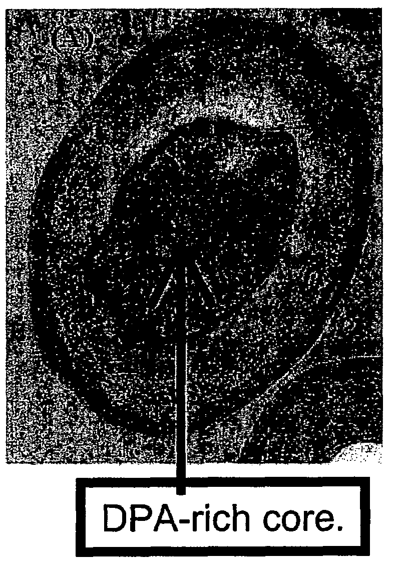
[0069]Bacteria spores were immobilized onto a test sample surface of thin, flexible, clear, adhesive polymer polydimethylsiloxan (PDMS) (Dow Corning). PDMS was doped with L-alanine (Aldrich) to induce germination and generate local high concentration of DPA / DP. TbCl3 (Aldrich) was also doped into the PDMS sample. The bacterial spores immobilized on the L-alanine and TbCl3-containing polymer were physically lysed by microwave irradiation (Vaid and Bishop, 1998?), wherein DPA / DP was released and luminescence was turned on.
[0070]The test surface in FIG. 3 was prepared by adding...
example 3
Detection and Quantifying, apparatus
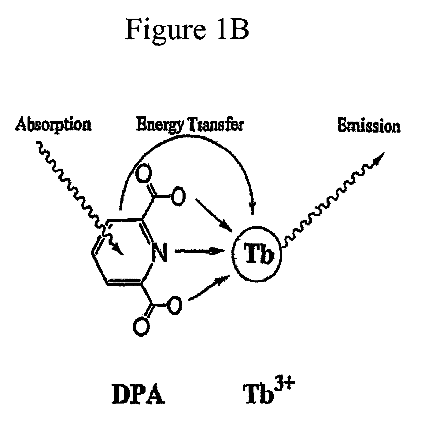
[0072]An apparatus for detecting and quantifying bacterial spores on a surface including lanthanide ions and aromatic molecules released from the bacterial spores on the surface. The apparatus in FIG. 5 comprises a UV-light radiation device for exciting a complex of a Tb3+ ion and DPA / DP to generate a characteristic luminescence of the complex on a surface. The source for the UV-light was a Xenon flash lamp, which was approximately 5 cm away the test surface. Between the Xenon flash lamp and the test surface were two C-amount elliptical lenses. The Xenon flash lamp and the test substrate were positioned at an angle of 45 degrees to each other. The area of irradiation by the Xenon flash lamp was observed by a microscope objective with a red bandpass filter suitable for Eu3+ for detecting and quantifying bacterial spores exhibiting the luminescence of the complex on the surface. The image was transferred from the microscope to the imaging device for...
PUM
| Property | Measurement | Unit |
|---|---|---|
| Melting Point | aaaaa | aaaaa |
| diameter | aaaaa | aaaaa |
| delay time | aaaaa | aaaaa |
Abstract
Description
Claims
Application Information