Systems and methods for spectroscopy of biological tissue

a biological tissue and spectroscopy technology, applied in the field of biological tissue spectroscopy systems and methods, can solve the problems of unsatisfactory current systems, unreasonably long collection time, and strict application limitations of probe and fiber bundle size, and achieve accurate assessment and rapid and non-destructive nature.

Inactive Publication Date: 2010-01-12
MASSACHUSETTS INST OF TECH
View PDF34 Cites 89 Cited by
  • Summary
  • Abstract
  • Description
  • Claims
  • Application Information

AI Technical Summary

Benefits of technology

[0014]The present invention includes the diagnostic classification of atherosclerotic plaques in human coronary arteries by quantitative assessment of their morphologic composition using Raman spectroscopy. The rapid and nondestructive nature of Raman spectroscopy provides the opportunity to diagnose coronary artery plaques in-vivo, when applied in a clinical setting using optical fiber technology. So used, the preferred embodiments of the present invention classify an atherosclerotic lesion, and can provide in-vivo quantitative assessment of its morphologic features, such as the presence of foam cells (FC), necrotic core (NC), and cholesterol crystals (CC), which may be used to assess plaque instability and the extent of disease progression, and thereby, the risk of life-threatening complicatio...

Problems solved by technology

More recently there have been reports of in-vivo work that however have either been confined to studies of skin or other easily accessible organs, or have used optical fiber configurations that require collection times that are unreasonably long for practical clinical use.
The majority of applications require remote sampling via optical fibers, and the size of the probe and fiber bundle is strictly limited by the application.
A particular example that current commercial systems cannot provide is the ability to evaluate atherosclerotic lesions in-vivo in real-time, through an angiographic catheter, thus aiding cardiologists in dir...

Method used


Image

  • Systems and methods for spectroscopy of biological tissue
  • Systems and methods for spectroscopy of biological tissue
  • Systems and methods for spectroscopy of biological tissue

Examples


Embodiment Construction

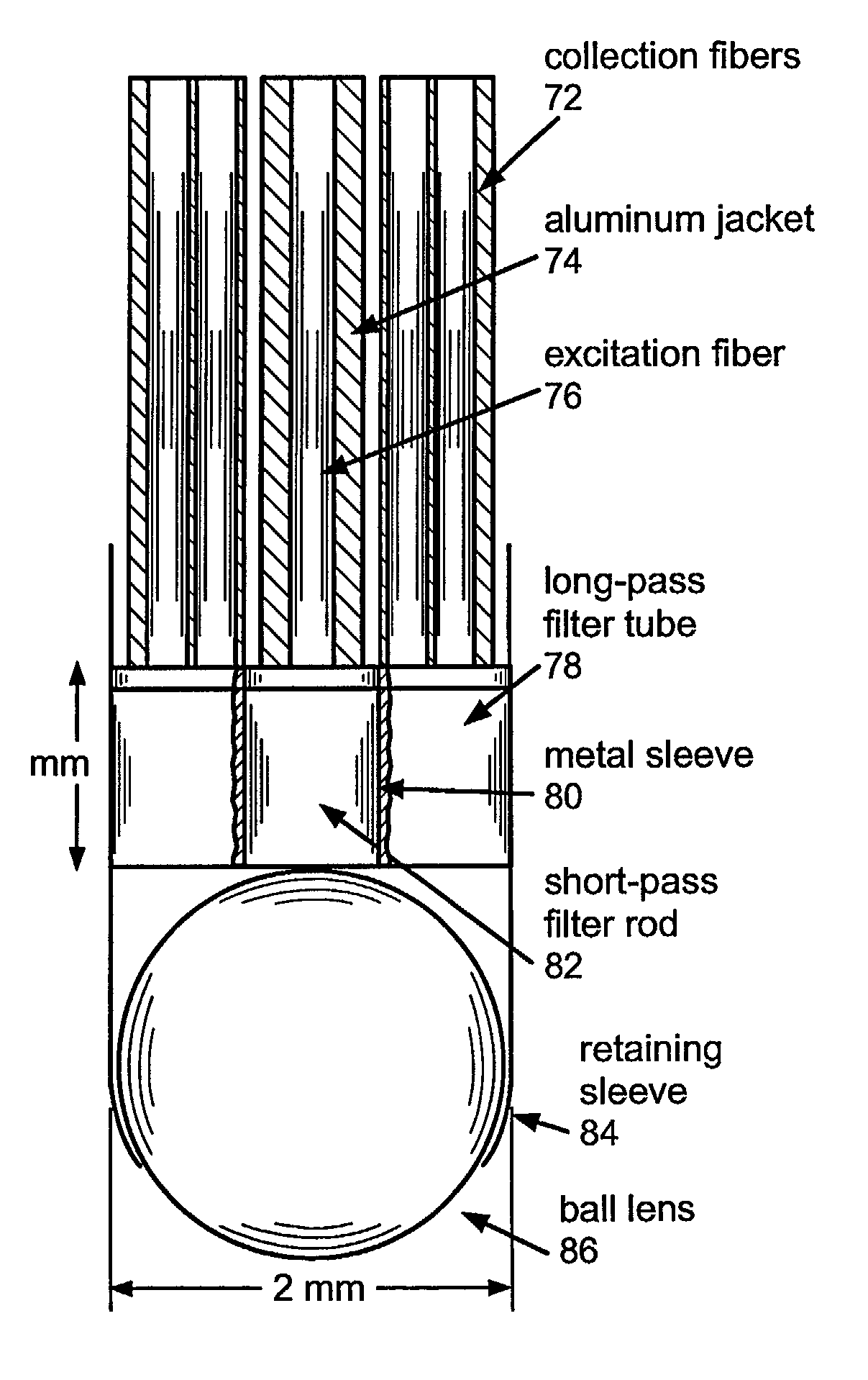
[0064]The present invention is directed to systems and methods for using Raman spectroscopy of tissue. A predicate for developing systems and methods for in-vivo applications using angiographic catheters to aid cardiologists in directing the appropriate treatment is the development of optical fiber probes for Raman spectroscopy capable of delivering low energy laser light to, and efficiently collecting the resulting Raman spectral signature from, in-vivo tissue. The probes in preferred embodiments are small, and use micro-optical design principles.

[0065]Methods for performing Raman spectroscopy for diagnosis and treatment of tissue are described in U.S. Pat. No. 5,615,673 issued on Apr. 1, 1997, in U.S. Pat. No. 5,304,173 issued on Apr. 19, 1994, in International Publication No. WO 92 / 15008, published on Sep. 3, 1992 and in International Publication No. WO 96 / 29925 published on Oct. 3, 1996, the entire teachings of all the references are incorporated herein by reference.

[0066]Th...


PUM


Abstract

The system and method of the present invention relates to using spectroscopy, for example, Raman spectroscopic methods for diagnosis of tissue conditions such as vascular disease or cancer. In accordance with a preferred embodiment of the present invention, a system for measuring tissue includes a fiber optic probe having a proximal end, a distal end, and a diameter of 2 mm or less. This small diameter allows the system to be used for the diagnosis of coronary artery disease or other small lumens or soft tissue with minimal trauma. A delivery optical fiber is included in the probe coupled at the proximal end to a light source. A filter for the delivery fibers is included at the distal end. The system includes a collection optical fiber (or fibers) in the probe that collects Raman scattered radiation from tissue, the collection optical fiber is coupled at the proximal end to a detector. A second filter is disposed at the distal end of the collection fibers. An optical lens system is disposed at the distal end of the probe including a delivery waveguide coupled to the delivery fiber, a collection waveguide coupled to the collection fiber and a lens.

Description

CROSS REFERENCES TO RELATED APPLICATIONS[0001]The present application claims the benefit of the U.S. Provisional Patent Application No. 60 / 370,197, filed Apr. 5, 2002. The entire contents of the above application is incorporated herein by reference in its entirety.GOVERNMENT SUPPORT[0002]This invention was supported, in whole or in part, by grants P41-RR-02594 and NIH-HL-64675 from the National Institute of Health. The Government has certain rights in the invention.BACKGROUND OF THE INVENTION[0003]Optical methods are increasingly being used for the detection of disease. Near-infrared Raman spectroscopy in particular, because of its chemical specificity, is proving to be a useful tool for both disease diagnosis and the study of disease progression. Over the past decade Raman spectroscopy has been applied to many diseases and biological problems and there have been many advances in-vitro. More recently there have been reports of in-vivo work that however have either been confined to s...

Claims


Application Information

Patent Timeline
IPC IPC(8): A61B6/06A61B1/00A61B5/00G01N21/47G01N21/65
CPCA61B5/0075A61B5/0084A61B5/0086A61B5/415G01N21/474G01N21/65A61B5/0071A61B5/0091A61B5/4519G01N2021/4745G01N2021/4759G01N2021/656A61B5/02007A61B5/4312A61B5/7232
Inventor MOTZ, JASON T.GALINDO, LUIS H.HUNTER, MARTINDASARI, RAMACHANDRAFELD, MICHAEL S.
Owner MASSACHUSETTS INST OF TECH
Why Eureka
Try Eureka