One
disadvantage of these previously known resonators, however, is that they typically use lumped element circuits (with discrete inductors and capacitors) to achieve resonance at the selected
radio frequency.
Because lumped element circuits lose more energy to
radiation at high frequencies where the circuit is greater than 1 / 10
wavelength, the lumped element resonator is less efficient for
high field MR imaging applications compared to
lower field strengths.
Because lumped element circuits are more radiative, they are electrically less efficient and have a lower
Q factor.
Especially at higher frequencies and for larger (clinical) volumes, lumped element resonators may achieve self-resonance below the desired frequency of operation as well as increased
electromagnetic radiation losses.
However, there are still some problems associated with both the resonator disclosed by Vaughan and the previously known lumped element resonators.
However, it is difficult to provide comfort and
accessibility for the subject and ease of use for the
technician and maintain a high coil performance.
Furthermore, some commercial systems don't provide the space for a coil that slides over a subject's head.
However, these commercial coils that separate into halves are not popular with some research applications such as fMR.
Apparently the
electrical contacts that are broken and remade to open and close the coil each time a new subject is loaded, become unstable over time due to wear and oxidation, resulting in
noise “spikes” and temporal instabilities often seen in EPI images for example.
While commercial coils must meet rigorous FDA
safety criteria, it could be imagined that
electrical contacts in a coil might possibly
pose safety risks in certain situations, especially where electrolytic bodily fluids were present.
The present body coils are built very large to allow for access and comfort, but as a consequence are very inefficient and are poorly coupled to the MR
region of interest in a body.
Still, these coils provide little shoulder room for the largest human subjects.
Limb coils, especially leg coils, are similarly limited by conventional practice.
This latter coil however by conventional designs requires the problematic
electrical contacts described for the head coil above.
A significant problem with this structure is that many medical subjects do not possess the
physical ability to manipulate their heads and bodies into the positions required for the MR without significant difficultly or pain.
This can be painful or impossible for most medical subjects who are asked to do this while
lying on their back.
Movement will result in lower resolution images and in image “
ghosting”, thereby limiting the
diagnostic quality of an image and often requiring a repeated imaging session.
While this has the effect of reducing the subject's head movement, it does not eliminate all of the subject's head movement.
Further, all of the padding placed around the subject's head can apply uncomfortable pressure and can intensify the subject's feelings of
claustrophobia.
Another, problem associated with many MR protocols, is they can be painfully loud.
The acoustic
noise is attributed to the electro motive forces generated by switched electrical currents in the wires of the
magnet's gradient coils.
While the methods mentioned above are generally effective for
gradient noise reduction, coils must be built to larger and less efficient dimensions to accommodate the
head restraint and
hearing protection devices placed about the head.
A problem with existing systems of this sort is that 1) they are often fixed in position which requires that a subject be adjusted to the device, and 2) they typically protrude above the head coil so as to preclude their use in close fitting head only MR systems, and in head gradient inserts used in
whole body MR systems.
This increased length of the birdcage has many problems.
It creates a head coil, which can increase feelings of
claustrophobia for the subject.
A subjects breath pushed back into their face by the inside coil wall creates a very uncomfortable / claustrophobic feeling for the subject.
This is an undesirable result since the MR exam may take 20 to 90 minutes.
Additionally, general medical access and vocal communications are impeded with the coil extending over the subject's mouth and
chin.
Further, if the subject has a
large head,
nose, and / or
chin, it becomes increasingly difficult to fit the subject's head inside of the coil.
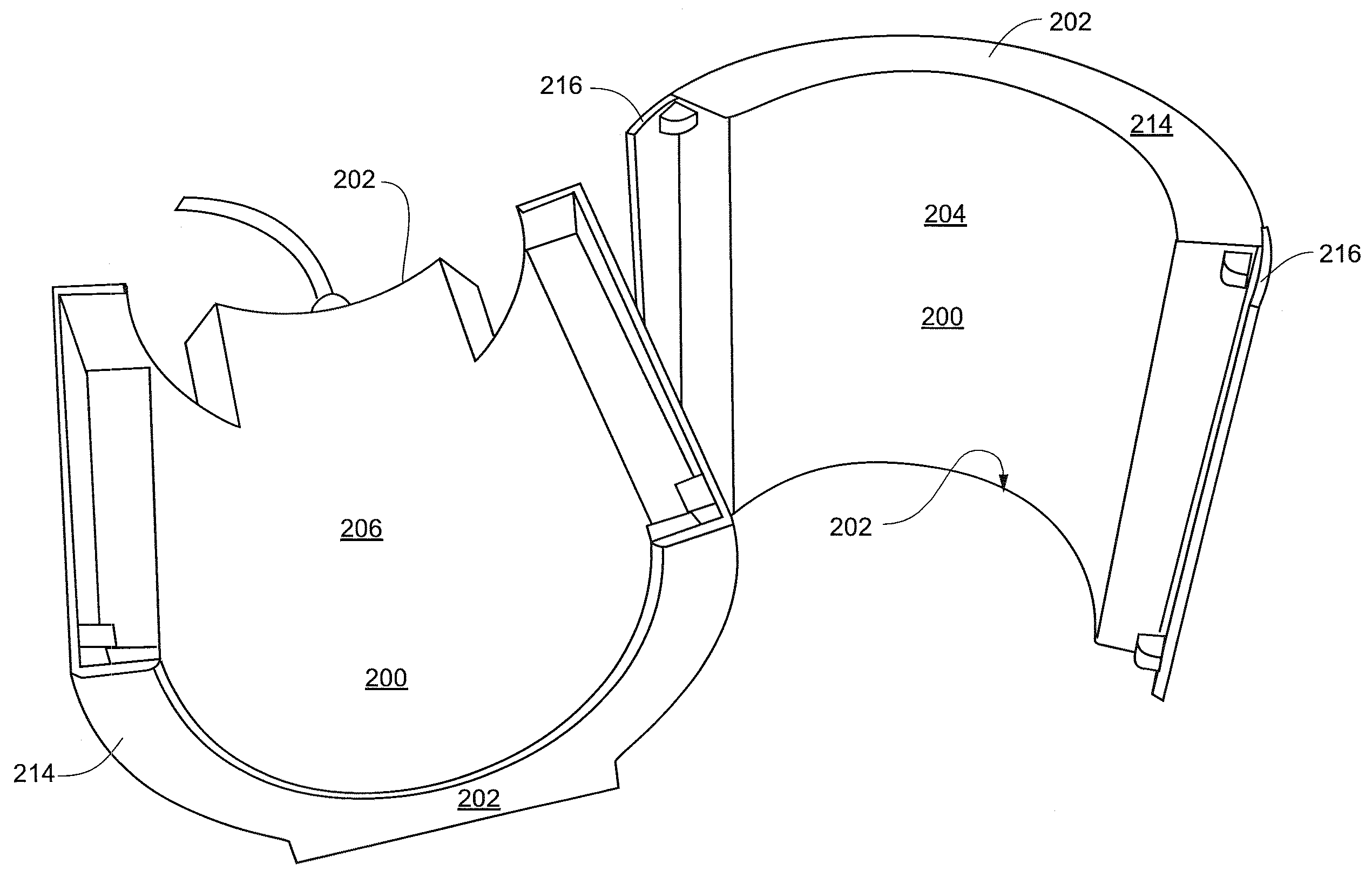
Another additional disadvantage for coils not having end caps is additional electromagnetic energy is lost from the top of the coil and thus the coil is less efficient at high frequencies.
While a back wall in a head coil is more desirable for coil performance and ergonomics in relation to the subject's mouth and
chin (i.e., with a back wall the head coil body can be shorter and thus the head coil does not have to extend over the chin), a back wall is undesirable for a couple of reasons.
Second, the back plane limits access to the subject from the back of the
magnet.
As stated above, a problem associated with head coils is the amount of space provided inside of the coil.
However, the padding,
hearing protection and communication equipment can not only make the MR experience uncomfortable for the subject but this equipment also occupies limited space within the head coil.
All of these problems listed above, individually and collectively, degrade the overall quality of NMR images and spectra, add to the discomfort of the subject, and limit subject access for the physician or researcher.

