Compositions and methods of treating disease with FGFR fusion proteins

a technology of fusion proteins and fibroblasts, applied in the direction of fusion polypeptides, peptide/protein ingredients, depsipeptides, etc., can solve the problems of patient safety, serum half-life, intracellular delivery, and the ability of specificity to be blocked by specific antibodies, so as to achieve less susceptible to cleavage

Inactive Publication Date: 2010-03-16
FIVE PRIME THERAPEUTICS
View PDF30 Cites 98 Cited by
  • Summary
  • Abstract
  • Description
  • Claims
  • Application Information

AI Technical Summary

Benefits of technology

[0007]The invention provides an FGFR fusion protein comprising a first polypeptide that comprises an extracellular domain of an FGFR polypeptide and a fusion partner, wherein the extracellular domain comprises a C-terminus, wherein the C-terminus comprises a variant of a wildtype FGFR extracellular domain C-terminus, wherein the variant comprises a deletion of 1-22 amino acid residues present in a wildtype FGFR1, FGFR2, FGFR3, or FGFR4 extracellular domain C-terminus, and wherein the FGFR fusion protein binds at least one FGF ligand or a biologically active fragment thereof. In an embodiment, the deletion is C-terminal to a valine residue situated at the C-terminus of the IgIII domain and commonly aligned among the wildtype FGFR1, FGFR2, FGFR3, and FGFR4 extracellular domain C-termini. In an embodiment, the FGFR fusion protein is less susceptible to cleavage.

Problems solved by technology

Previously proposed therapeutic approaches using specific antibodies to block FGF function do not address the issue of redundancy within the FGF family in activating multiple FGFRs, since cancers or other proliferative cells may express upregulated levels of more than one FGF or FGFR.
Antisense oligonucleotide or related siRNA therapies have potential problems with specificity, serum half-life, and intracellular delivery.
Gene transfer therapies, including those using adenovirus, have raised issues of patient safety and a number of clinical gene therapy studies have been halted due to patient death.
Small molecule tyrosine kinase inhibitor therapies suffer from issues of target specificity, toxicity, and manifestations of drug resistance.
To date, no drug which targets an FGFR signaling pathway has been approved for treating any human disease.

Method used


Image

  • Compositions and methods of treating disease with FGFR fusion proteins
  • Compositions and methods of treating disease with FGFR fusion proteins
  • Compositions and methods of treating disease with FGFR fusion proteins

Examples


example 1

Sequence Alignment of Partial IgIII Domains and the Membrane Proximal Regions of FGFRs and FGFR Variants

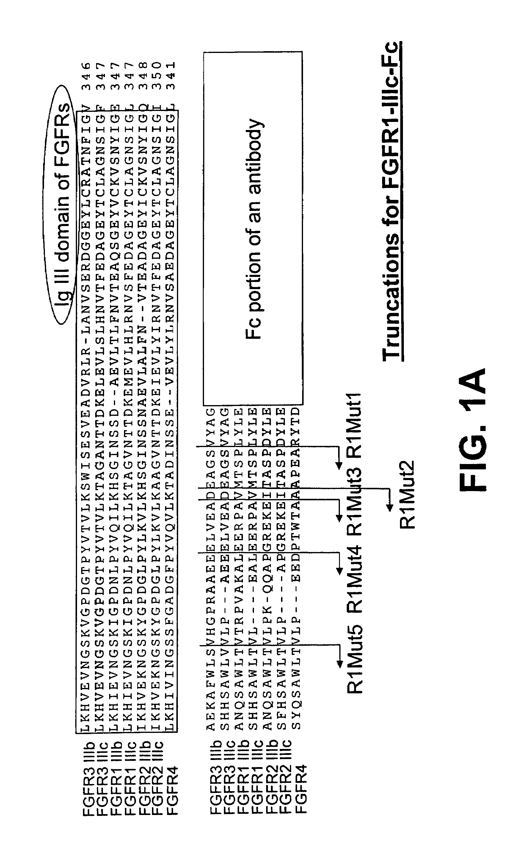
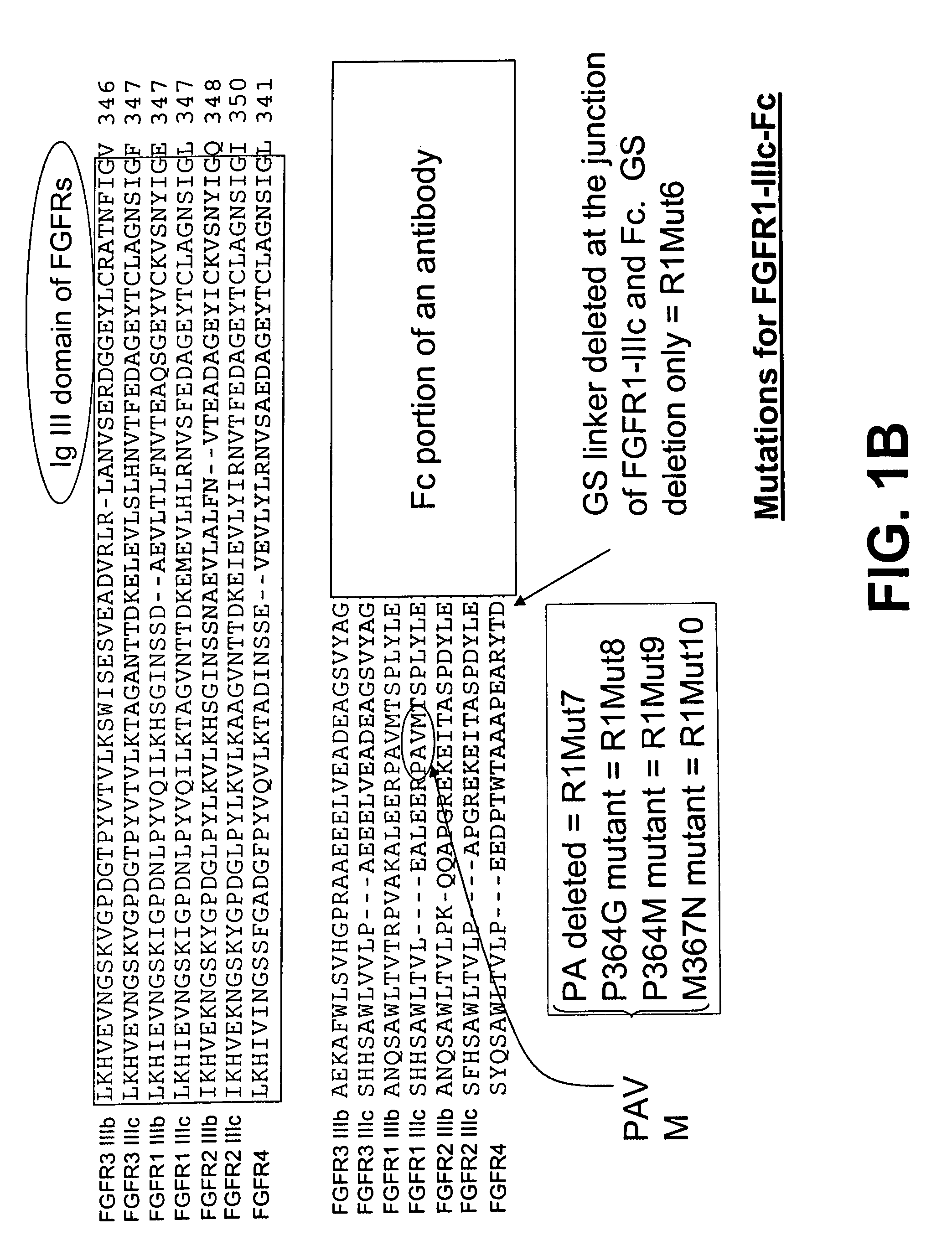
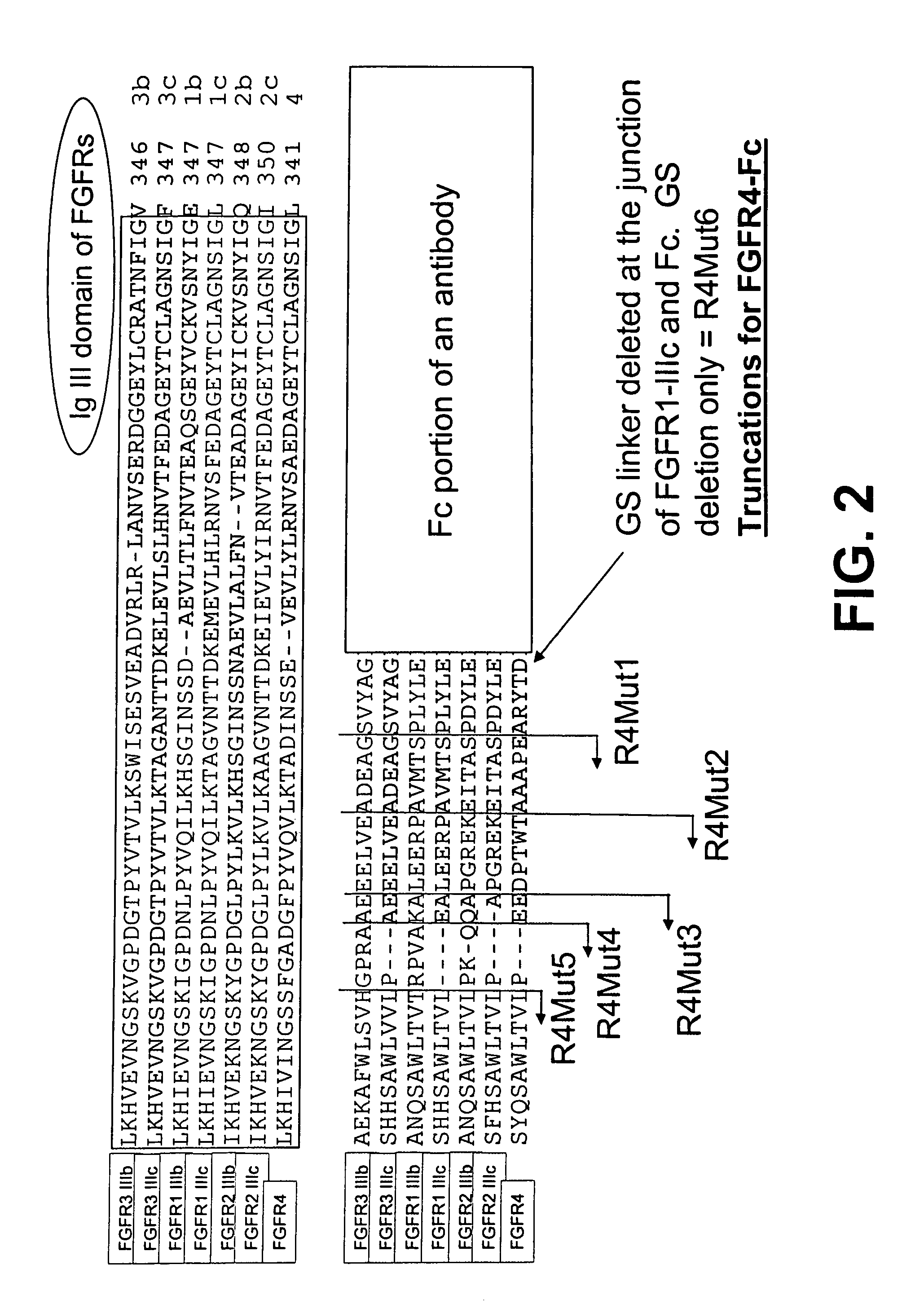
[0245]FIG. 1A shows an alignment of the amino acid sequences of a part of the IgIII domain of each of the seven FGFR family members, FGFR1-IIIb, FGFR1-IIIc, FGFR2-IIIb, FGFR2-IIIc, FGFR3-IIIb, FGFR3-IIIc, and FGFR4, using the Clustal W version 1.8 program of the European Molecular Biology Laboratory (EMBL) bioinformatics search site.

[0246]FIG. 1A also illustrates the organization of the parental FGFR1-IIIc-Fc fusion protein (nucleotide SEQ ID NO: 4, protein SEQ ID NO: 95) and corresponding mutants. The IgIII domain is followed by the C-terminal portion of the ECD, which is followed by the Fc portion of an antibody. The alignment marks the truncation locations of the R1Mut1 (nucleotide SEQ ID NO: 6, protein SEQ ID NO: 97), R1Mut2 (nucleotide SEQ ID NO: 7, protein SEQ ID NO: 98), R1Mut3 (nucleotide SEQ ID NO: 8, protein SEQ ID NO: 99), R1Mut4 (nucleotide SEQ ID NO: 9, protein SEQ ID...

example 2

Expression of FGFR Fusion Proteins

[0249]The fusion proteins of the invention were expressed in 293-6E host cells using the pTT5 vector (Biotechnology Research Institute; Montreal, Canada) transfected into 293-6E cells (Biotechnology Research Institute; Montreal, Canada), which were then cultured to produce the fusion proteins. An expression vector that comprised the cDNA of FGFR1-IIIc-Fc (SEQ ID NO: 4), encoding the extracellular domain of human FGFR1-IIIc (SEQ ID NO: 1) was constructed from an open-reading frame cDNA library prepared internally. This cDNA was linked at its C-terminus through a linker encoding the amino acids GS to cDNA encoding an Fc fragment of human IgG1 heavy chain (SEQ ID NO: 80) to produce a fusion construct hereafter referenced as “FGFR1-IIIc-Fc cDNA” and the expression product thereof as “FGFR1-IIIc-Fc protein.” The Fc fragment was also obtained from an open-reading frame cDNA library prepared internally. This cDNA fusion construct was inserted into a pTT5 v...

example 3

Transient Expression of Fusion Proteins in 293-6E Cells and CHO-S Host Cells

[0258]The FGFR1-IIIc-Fc / pTT5 expression vector was designed to provide transient expression in 293-6E host cells. The 293-6E cells were previously adapted to serum-free suspension culture in Free-Style medium (Invitrogen; Carlsbad, Calif.). The cells were transfected with the expression vector while in logarithmic growth phase (log phase growth) at a cell density of between 9×10⁵ / ml and 1.2×10⁶ / ml.

[0259]In order to transfect 500 ml of cell suspension, a transfection mixture was first made by mixing 500 micrograms (ug) of the expression vector DNA in 25 milliliters (ml) of sterile phosphate buffered saline (PBS) with 1 milligram (mg) of polyethylenimine (at a concentration of about 1 mg / ml solution in sterile water) in 25 ml of sterile PBS. This transfection mixture was incubated for 15 min at room temperature. Following incubation, the transfection mixture was added to the 293-6E cells in log phase growth to...


PUM

PropertyMeasurementUnit
concentrationaaaaaaaaaa

Abstract

The invention provides FGFR fusion proteins, methods of making them, and methods of using them to treat proliferative disorders, including cancers and disorders of angiogenesis. The FGFR fusion molecules can be made in CHO cells and may comprise deletion mutations in the extracellular domains of the FGFRs which improve their stability. These fusion proteins inhibit the growth and viability of cancer cells in vitro and in vivo. The combination of the relatively high affinity of these receptors for their ligand FGFs and the demonstrated ability of these decoy receptors to inhibit tumor growth is an indication of the clinical value of the compositions and methods provided herein.

Description

RELATED APPLICATIONS[0001]The invention relates to applications 60 / 701,479, filed Jul. 22, 2005; 60 / 729,401, filed Oct. 21, 2005; 60 / 757,398, filed Jan. 10, 2006; and 60 / 800,005, filed May 15, 2006, all of which are incorporated by reference in their entireties.TECHNICAL FIELD[0002]The present invention relates to fusion molecules comprising an extracellular domain of a fibroblast growth factor receptor (FGFR). It relates to polypeptide and polynucleotide sequences, vectors, host cells, compositions, kits, and animals comprising FGFR fusion proteins. The invention also relates to methods of making and using FGFR fusion molecules and variants and fragments thereof to diagnose, prevent, determine the prognosis for, and treat proliferative diseases, including cancer and disorders of angiogenesis.BACKGROUND ART[0003]Fibroblast growth factors (FGFs) and their receptors (FGFR) are a highly conserved group of proteins with instrumental roles in angiogenesis, vasculogenesis, and wound heali...

Claims


Application Information

Patent Timeline
Patent Type & Authority Patents(United States)
IPC IPC(8): A61K38/00A61K38/16A61K39/00
CPCC07K14/71A61K45/06C07K17/08A61K47/48415C07K14/765C07K16/2863A61K47/48369C07K2319/30C07K2319/32A61P1/02A61P1/04A61P11/00A61P13/10A61P13/12A61P15/00A61P17/02A61P25/00A61P27/02A61P35/00A61P35/02A61P35/04A61P43/00
Inventor BOSCH, ELIZABETHHOLLENBAUGH, DIANELEE, ERNESTINEQIN, MINMINSADRA, ALI
Owner FIVE PRIME THERAPEUTICS
Why Eureka
Try Eureka