
Mass spectrometric imaging method under ambient conditions using electrospray-assisted laser desorption ionization mass spectrometry

a mass spectrometry and electrospray technology, applied in the field of molecular imaging, can solve the problems of inability to detect macromolecules such as peptides or proteins, tedious preparation work, and inability to achieve satisfactory results

Active Publication Date: 2010-03-30
NAT SUN YAT SEN UNIV
View PDF11 Cites 22 Cited by
  • Summary
  • Abstract
  • Description
  • Claims
  • Application Information

AI Technical Summary

Benefits of technology

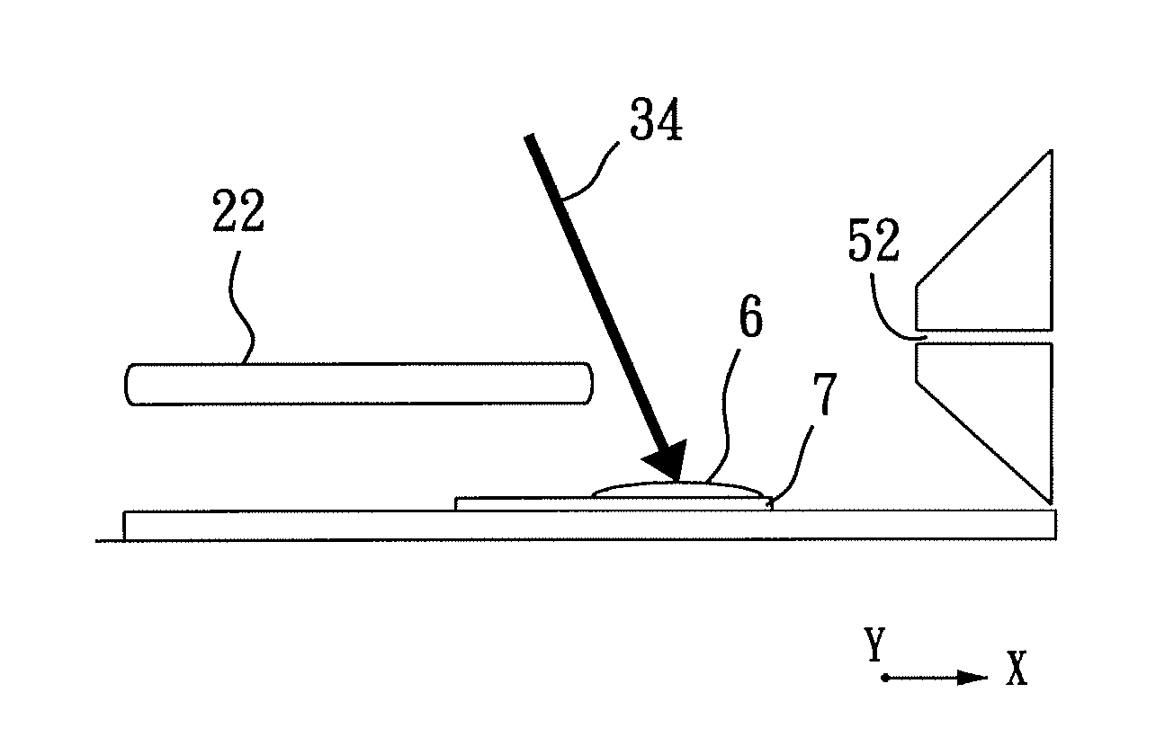
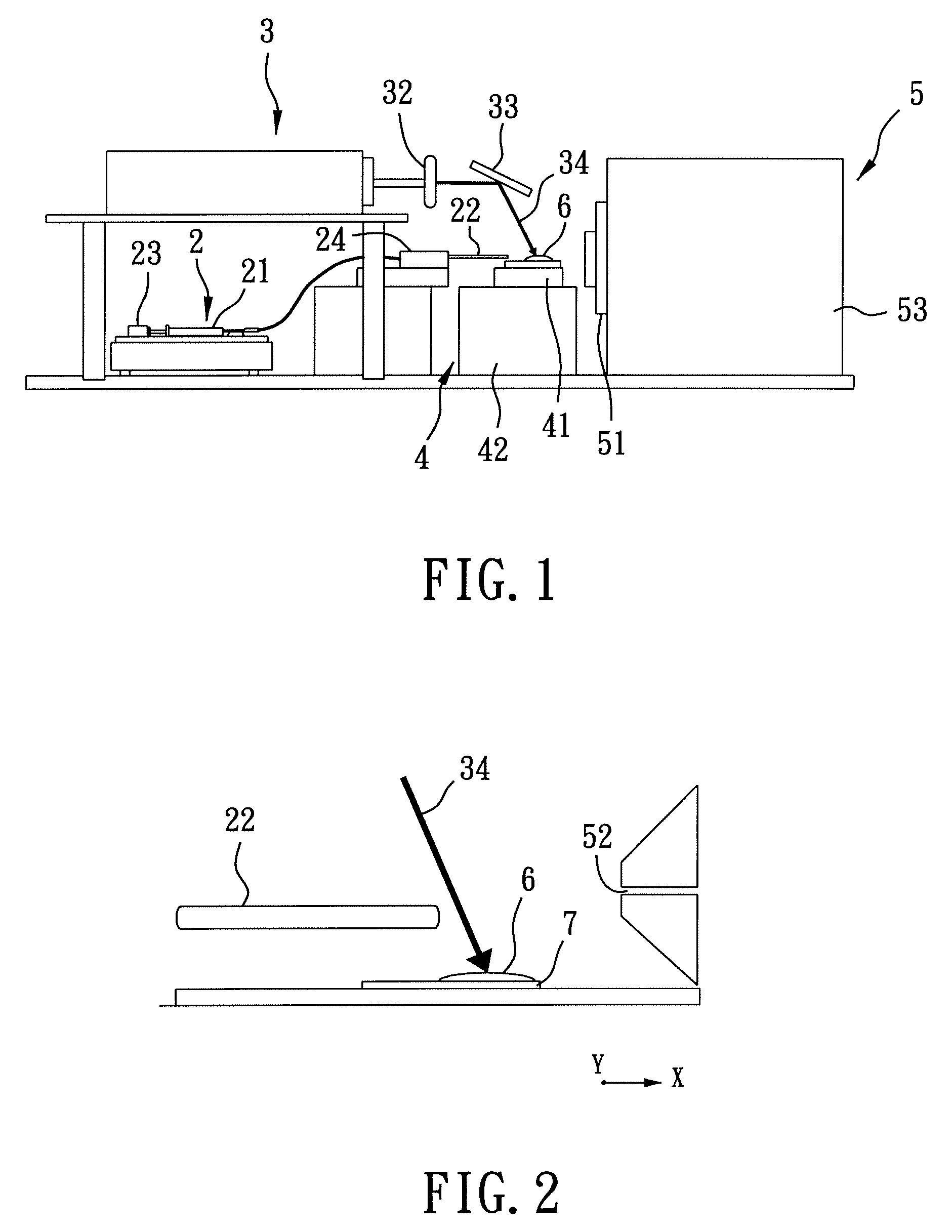
[0011]Therefore, the object of the present invention is to provide a mass spectrometric imaging method that can be conducted under ambient conditions, and that can be used to obtain an imaging profile of a sample that has a self-sustained shape with speed and accuracy. A further object of the present invention is to provide a mass spectrometric imaging method that is capable of swiftly and un-obstructively maneuvering a sample to move relative to a desorption mechanism such that mass spectrometric results for substantially continuous areas of the sample can be obtained in a desirable short period of time.

Problems solved by technology

However, due to the biological nature of target protein, e.g., being more labile to ionization energy and being in a state of flux, such efforts did not prove to be satisfactory.
However, SIMS is only capable of detecting analytes such as metal ions or small organic molecules, and is unable to detect macromolecules such as peptide or proteins because the macromolecules are either spoiled during ionization or unable to be effectively desorbed from the surface of the sample.
Although MALDI-MS is capable of successfully desorbing peptide or protein molecules from a solid biological sample, and the result thereof is used to distinguish abnormal or cancerous tissues from normal tissues, several drawbacks still exist for MALDI, such as involving a tedious preparation work and requiring to be conducted in vacuum, etc.
However, there are several disadvantages involved in DESI-MS, including the difficulty in controlling the precision of striking electron-carrying spray droplets onto the tissue slice, and the inability in desorbing protein molecules from the tissue slice.
It can be seen from the above that a variety of difficulties and inconveniences are encountered when obtaining molecular images through the methods of mass spectrometry.

Method used


Image

  • Mass spectrometric imaging method under ambient conditions using electrospray-assisted laser desorption ionization mass spectrometry
  • Mass spectrometric imaging method under ambient conditions using electrospray-assisted laser desorption ionization mass spectrometry
  • Mass spectrometric imaging method under ambient conditions using electrospray-assisted laser desorption ionization mass spectrometry

Examples


exemplary example 1

Imaging Mass Spectrometric Analysis using Electrospray-assisted Laser Desorption Ionization Mass Spectrometry (ELDI-MS) on Glossy Ganoderma (Ganoderma Lucidum)

[0068]As shown in FIG. 4(a) and FIG. 5(a), a slice of glossy ganoderma, a genus of polypores, was obtained for exemplary example 1 using a razor blade, where FIG. 4(a) shows a photograph of the glossy ganoderma slice and FIG. 5(a) is a negative image of FIG. 4(a). The glossy ganoderma slice was measured 10 mm in length, 35 mm in width, and 3 mm in thickness. The glossy ganoderma slice was placed on the sample stage 41 of the sample stage unit 4 (refer to FIG. 1) to be irradiated by the laser beam 34 (refer to FIG. 1 and FIG. 2) for conducting imaging mass spectrometric analysis using ELDI-MS.

[0069]While the laser beam 34 irradiates the glossy ganoderma slice to form a laser spot of 100×150 μm2 thereon at an operating frequency of 10 Hz, i.e., 10 laser shots per second, the sample stage 41 is moved relative to the laser beam 34...

exemplary example 2

Imaging Mass Spectrometric Analysis using ELDI-MS on Antrodia Camphorata

[0073]As shown in FIG. 7(a), a slice of antrodia camphorata, a special Taiwanese fungus species that only grows on cinnamomum kanehirae, was obtained for exemplary example 2 using a razor blade, where FIG. 7(a) shows a photograph of the antrodia camphorata slice. The antrodia camphorata slice was measured 21 mm in length, 3 mm in width, and 1 mm in thickness.

[0074]The sample stage 41 was moved relative to the laser beam 34 in the longitudinal direction (X) in the same manner as described above for exemplary example 1, such that two subsequent laser spots formed on the antrodia camphorata slice in the longitudinal direction (X) are spaced apart from each other for 0.02 mm. The sample stage 41 was further moved in the transverse direction (Y) in consecutive increments of 3 / 26 mm upon control by the computer-controlled positioning mechanism 42. In other words, the laser beam 34 scans across the antrodia camphorata ...

exemplary example 3

Imaging Mass Spectrometric Analysis using ELDI-MS on Angelica Sinensis Diels

[0077]As shown in FIG. 10(a) and FIG. 11(a), two slices of angelica sinensis diels, a traditional Chinese medicine, were obtained for exemplary example 3, where FIG. 10(a) shows a photograph of the angelica sinensis diels slices, and FIG. 11(a) shows a negative image of FIG. 10(a). The angelica sinensis diels slices were respectively measured 2 cm and 2 cm in length, 2 cm and 1 cm in width, and 2 mm and 2 mm in thickness.

[0078]The sample stage 41 was moved relative to the laser beam 34 in the longitudinal direction (X) in the same manner as described above for exemplary example 1, such that two subsequent laser spots formed on each of the angelica sinensis diels slices in the longitudinal direction (X) are spaced apart from each other for 0.02 mm. The sample stage 41 was further moved in the transverse direction (Y) in consecutive increments of 1 / 15 cm for analyzing the angelica sinensis diels slices simulta...


PUM


Abstract

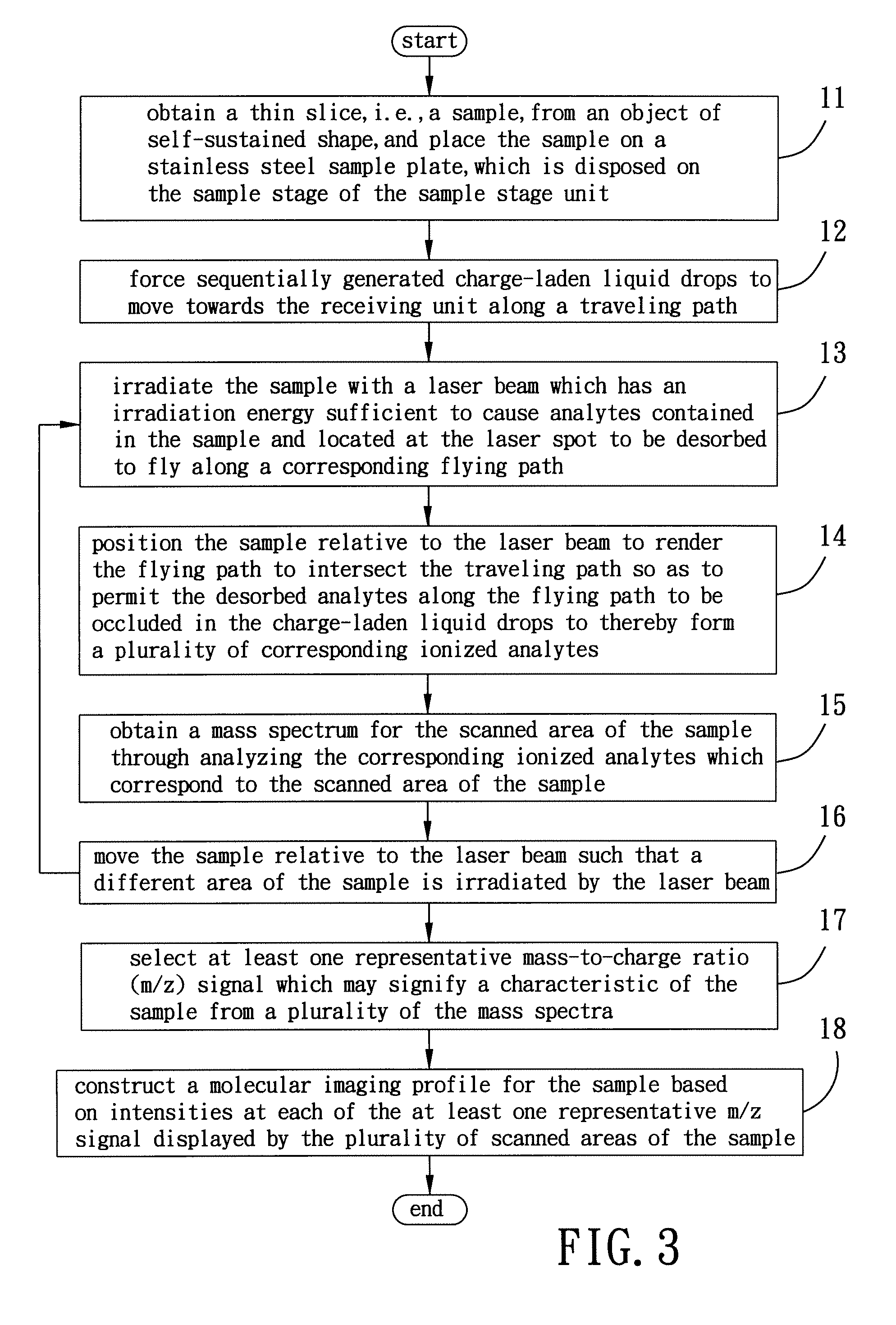
A mass spectrometric imaging method includes the steps of: forcing sequentially generated charge-laden liquid drops to move towards a receiving unit of a mass spectrometer along a traveling path; scanning a sample with a laser beam which has an irradiation energy sufficient to cause analytes contained in the sample to be desorbed to fly along a plurality of flying paths respectively; and positioning the sample relative to the laser beam to render the plurality of flying paths intersecting the traveling path so as to permit a plurality of the analytes respectively along the plurality of flying paths to be occluded in a plurality of the charge-laden liquid drops respectively to thereby form a plurality of corresponding ionized analytes.

Description

CROSS-REFERENCE TO RELATED APPLICATION[0001]This application is a continuation-in-part (CIP) of U.S. patent application Ser. No. 11 / 561,131, entitled “ELECTROSPRAY-ASSISTED LASER DESORPTION IONIZATION DEVICE, MASS SPECTROMETER, AND METHOD FOR MASS SPECTROMETRY”, filed on Nov. 17, 2006, which claims priority from Taiwan 095103439 filed Jan. 27, 2006.BACKGROUND OF THE INVENTION[0002]1. Field of the Invention[0003]The invention relates to molecular imaging, more particularly to mass spectrometric imaging under ambient conditions using electrospray-assisted laser desorption ionization mass spectrometry.[0004]2. Description of the Related Art[0005]Imaging mass spectrometry (IMS) is widely used in the investigation of chemical or molecular distributions of solid samples, such as metals, polymers, semiconductors, and geological substances.[0006]Many attempts have been made to explore the feasibility of using imaging mass spectrometry in studying spatial distribution of proteins in various ...

Claims


Application Information

Patent Timeline
Patent Type & Authority Patents(United States)
IPC IPC(8): H01J49/10
CPCH01J49/0004H01J49/0463H01J49/165
Inventor SHIEA, JENTAIE
Owner NAT SUN YAT SEN UNIV
Learn More