Cardiac display methods and apparatus

a display method and heart technology, applied in the field of ct, pet, and mr examination of the heart, can solve the problems of high operator dependence, depressed ventricular function, poor prognosis,

Active Publication Date: 2010-03-30
GENERAL ELECTRIC CO
View PDF21 Cites 19 Cited by
  • Summary
  • Abstract
  • Description
  • Claims
  • Application Information

AI Technical Summary

Problems solved by technology

On the other hand, even with good coronaries and normal perfusion, depressed ventricular function indicates a poor prognosis.
One disadvantage of the echocardiographic methods is that they are highly operator dependent.
Creating several views in different orientations can be very time consuming.

Method used


Image

  • Cardiac display methods and apparatus
  • Cardiac display methods and apparatus
  • Cardiac display methods and apparatus

Examples


Embodiment Construction

[0024]In the PET radionuclide imaging methods (also known as radionuclide angiography), a blood pool is labeled with a radioisotope (such as technetium-99m) and scanned using a gamma camera with R wave (ECG) gating. Volume changes within the ventricular cavity are computed from the quantification of the count rate changes. Although the radionuclide method is considered the ‘gold standard’ for quantitative assessment of global ventricular function and is relatively operator independent, it provides limited assessment of regional ventricular wall motion.

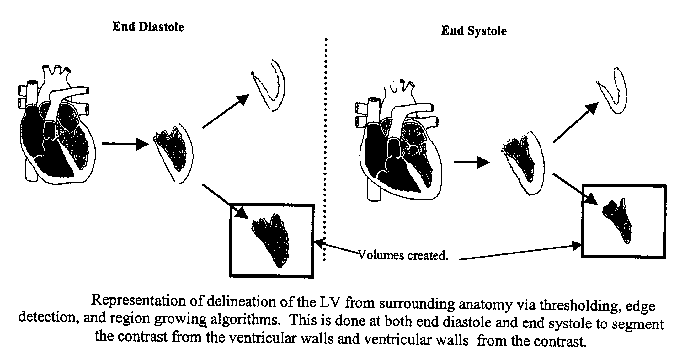
[0025]In the MR imaging methods, tomographic sections of the heart are combined to reconstruct a 3D image. Due to intrinsic contrast between blood and myocardial tissue, a blood pool can be segmented from the adjacent tissue in sectional views and the total volumes are calculated in systole & diastole. Although no contrast is injected and no radiation is administered to the patient, scanning times are typically relatively long (e.g., 1...


PUM


Abstract

A method includes receiving a multi-phase axial cardiac dataset, receiving a selection of a phase from a user, when the received selection is systole, generating an endocardial volume of a left ventricle at an end systole phase without further user intervention, and when the received selection is diastole, generating an endocardial volume of the left ventricle at an end diastole phase without further user intervention.

Description

BACKGROUND OF THE INVENTION[0001]This invention relates generally to CT, PET, and MR examinations of the heart, and more specifically to methods and apparatus for automating image generation and review.[0002]Accurate evaluation of the cardiac function, particularly left ventricular (LV) function (e.g., stroke volume, ventricular ejection fraction, regional wall motion) is useful in cardiac diagnosis, guiding patient therapy, determining prognosis, and following the course of a disease. LV function is also a useful component of any comprehensive cardiac diagnostic examination for stable coronary artery disease (e.g., stable angina) and acute cardiac syndromes (e.g., acute myocardial infarction and unstable angina). Measurement of ventricular function complements and supplements other cardiac diagnostic procedures such as cardiac catheterization and coronary artery imaging for vessel patency and myocardial perfusion. In patients with a variety of heart diseases, cardiac function has a...

Claims


Application Information

Patent Timeline
Patent Type & Authority Patents(United States)
IPC IPC(8): A61B5/05A61B5/055A61B6/03G06T7/60G06T17/00
CPCA61B6/032G06T17/00A61B6/503G06T7/60A61B6/037A61B5/055A61B5/7289
Inventor MOHR, KELLY ANNSTEFANI, LAURENTLAUNAY, LAURENTTHOMSON, HELEN JANE
Owner GENERAL ELECTRIC CO
Why Eureka
Try Eureka