Methods and kits for separation and detection of proteins in biological samples

a technology for biological samples and proteins, applied in the field of methods and kits for separating a mixture of proteins in a biological sample, can solve the problems of affecting the separation/characterization of proteins in serum and plasma, affecting the effect of serum analysis, and non-specific binding,

Inactive Publication Date: 2010-05-04
QUEENS UNIV OF KINGSTON
View PDF11 Cites 5 Cited by
  • Summary
  • Abstract
  • Description
  • Claims
  • Application Information

AI Technical Summary

Benefits of technology

[0013]Another object of the present invention is to provide kits for producing a profile of proteins in a biological sample. Kits of the present invention comprise a solution containing a sulfhydryl reducing agent, an anionic detergent, and at least one detergent selected from the group consisting of another ionic detergent, a non-ionic detergent and a zwitterionic detergent, and instructions for separating proteins in said biological sample. Kits of the present invention further comprise a means for characterization of the separated proteins so that a profile of proteins in the biological sample can be generated.

Problems solved by technology

However, separation / characterization of proteins in serum and plasma is hampered by the presence at high levels of such proteins as albumin and immunoglobulin, which non-specifically bind (“stick”) to other proteins.
The effectiveness of analysis of serum is also limited because such serum proteins often bind to marker proteins, and hence interfere with the migration of the marker proteins on a gel.
In cases with low serum level of marker protein, such as mild or chronic disease states, where the amount of target epitope is low, the binding by other serum proteins may reduce access to target epitope enough to cause false negative results.
However, problems exist with many of the commercially available detection kits currently in use for these biomarkers.
Further, commercial kits can not differentiate whether or not modified forms of cTnI are present.
Nor can these kits identify which forms of cTnI are in the sample.
Another problem is that proteins in serum are bound to each other as well as to other proteins which can mask or hide epitopes, thus rendering the proteins undetectable by the antibodies of the kit.
Thus, for some patients experiencing an acute myocardial infarction (AMI), the current cTnI kits do not detect all of the intact cTnI and modification products thereof that are released into the serum.
In addition, current cTnI kits are not always capable of detecting intact cTnI and modification products thereof in cardiac patients who are not experiencing an AMI, as the commercially available assays appear to have low analytical and diagnostic sensitivity.
Unfortunately, known methods for removing native serum proteins from serum / plasma under native conditions (e.g., non-denaturing, non-reducing conditions, such as immunoprecipitation, acid extraction, gel filtration, ion exchange chromatography) typically lead to substantial loss of the marker protein or proteins, due in large part to the above-mentioned non-specific sticking.
In methods where a marker protein or proteins is completely purified from native serum / plasma, analysis of a marker protein level or levels relative to level of other proteins is rendered impossible.

Method used


Image

  • Methods and kits for separation and detection of proteins in biological samples
  • Methods and kits for separation and detection of proteins in biological samples
  • Methods and kits for separation and detection of proteins in biological samples

Examples


example 1

Muscle Damage Assessment

Patient Sampling

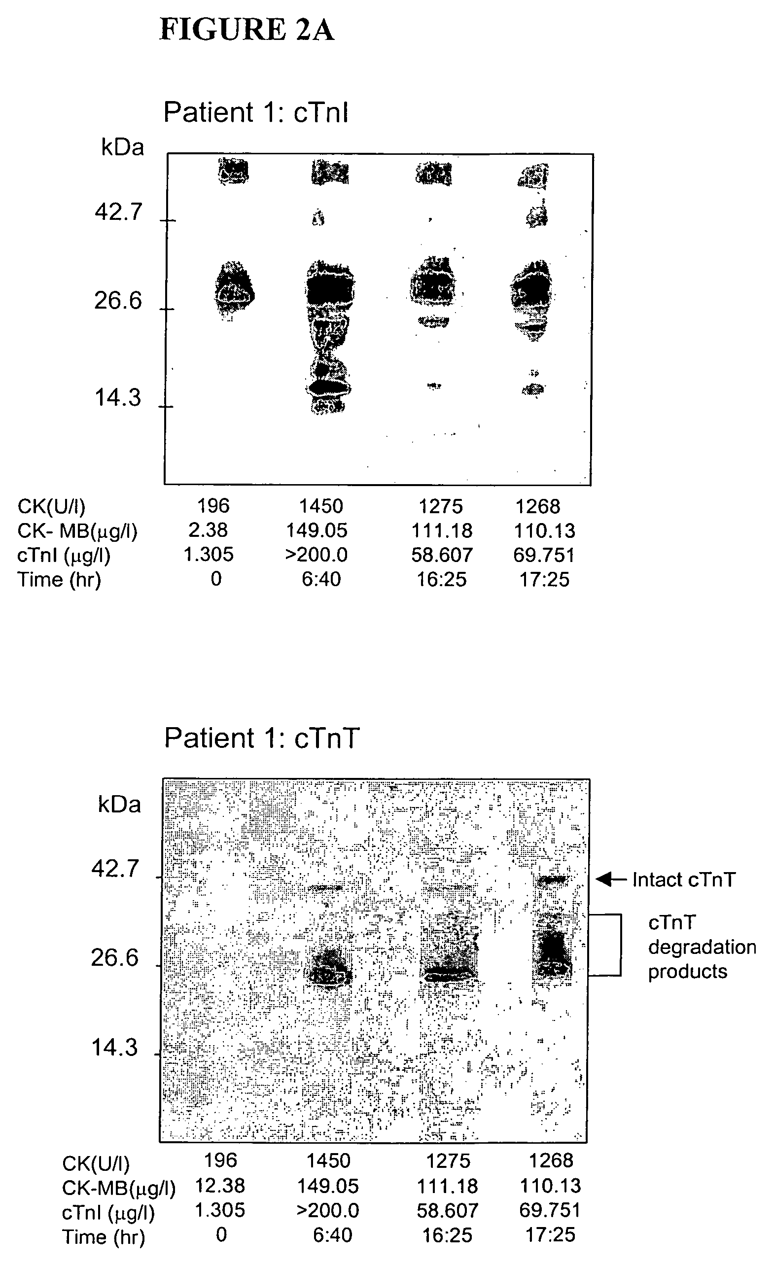
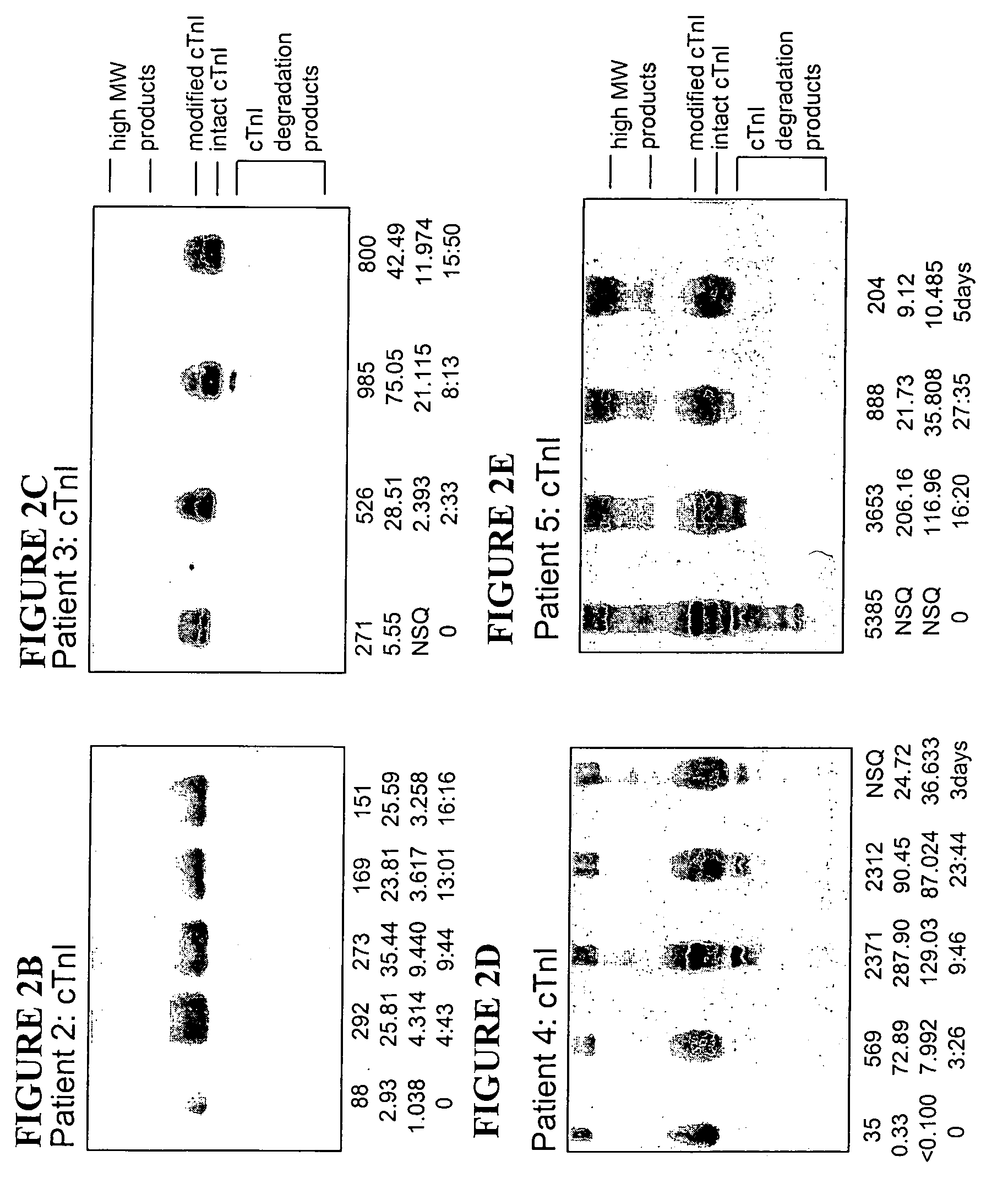
[0079]Blood samples from 12 patients admitted to a hospital emergency department, with chest pain and unequivocal signs of AMI (based upon electrocardiographic findings) were obtained. Serum sampling was performed according to routine care protocols and not by a defined study time course. This led to different intervals between successive blood samples, thus permitting study of the occurrence of biochemical markers in a “real-life” scenario.

Routine Biochemical Testing

[0080]Blood was collected in serum separator tubes, centrifuged and assayed immediately for routine biochemistry tests. Samples were then frozen until WB-DSA. Routine testing included total creatine kinase (CK, measured by CX7, Beckman Coulter, Inc., Fullerton, Calif.), its MB isoenzyme (CKMB) and cTnI (both measured by Technicon Immunol, Bayer Corporation, Tarrytown, N.Y.). A diagnosis of AMI was confirmed if there was a typical time profile observed for CK with at least a doubli...

example 2

Diagnosing Cardiac Muscle Damage

Patient Samples

[0084]Serum samples were obtained from a prospective case series of patients presenting, within four hours onset of symptoms of ACS, to a hospital emergency department. Serum samples of ten representative cases were selected from the first 45 cases of ACS enrolled, who also had non-diagnostic ECG and non-significant elevations in the biochemical cardiac markers CK, CK-MB, and cTnI using commercially available kits. Patients underwent a history and clinical examination, a 12 lead ECG was recorded and serial blood was drawn at presentation, and subsequently at 1, 2, 4, 6 and 16-24 hours for routine clinical testing of biochemical cardiac markers and for analysis by WB-DSA. Serum samples were stored at −80° C. until analyzed. Final discharge diagnosis from the emergency department was based on standard criteria of history, physical examination, ECG changes, and biochemical cardiac markers.

Analytical Biochemical Testing

[0085]Total CK levels...


PUM

PropertyMeasurementUnit
pHaaaaaaaaaa
molecular weightaaaaaaaaaa

Abstract

Methods and kits are provided for separating a mixture of proteins in a biological sample. Methods for detecting and profiling proteins in biological samples by the separation method and kits are also provided. These methods are particularly useful in assessing damage to cells such as cardiac and skeletal muscle cells and in the early clinical diagnosis of myocardial damage by detection of myofilament proteins in serum of a subject.

Description

[0001]This patent application is a continuation of U.S. patent application Ser. No. 09 / 934,297 filed Aug. 21, 2001 now abandoned, which claims the benefit of priority to U.S. Provisional Patent Application Ser. No. 60 / 226,588, filed Aug. 21, 2000 and U.S. Provisional Patent Application Ser. No. 60 / 308,948, filed Jul. 30, 2001, each of which are herein incorporated by reference in their entirety.FIELD OF THE INVENTION[0002]The present invention relates to methods and kits for separating a mixture of proteins in a biological sample by mixing the biological sample with a solution which substantially denatures proteins in the biological sample and subjecting the resulting mixture to a protein separation technique. Following separation, proteins of interest are preferably subjected to characterization. As demonstrated herein, the method of the present invention is useful in the development of highly sensitive analytical and diagnostic assays and kits for assessing cellular damage and dia...

Claims


Application Information

Patent Timeline
Patent Type & Authority Patents(United States)
IPC IPC(8): C12Q1/68G01N33/52G01N33/68
CPCG01N33/52G01N33/6803G01N33/6887G01N2550/00G01N2333/4712
Inventor VAN EYK, JENNIFER E.SIMPSON, JEREMY A. C.
Owner QUEENS UNIV OF KINGSTON
Why Eureka
Try Eureka