Lung device with sealing features

a technology of lung device and sealing feature, which is applied in the direction of prosthesis, catheter, application, etc., can solve the problems of not even recommending screening for persons of high risk, not knowing whether the use of spiral ct scan improves the prognosis for long-term survival, and many are acute and deadly, so as to mitigate pneumothorax and hemothorax, the effect of reducing the risk of lung diseas

Inactive Publication Date: 2010-08-03
EKOS CORP
View PDF296 Cites 54 Cited by
  • Summary
  • Abstract
  • Description
  • Claims
  • Application Information

AI Technical Summary

Benefits of technology

[0014]In another embodiment of the invention, a lung device system is provided comprising an elongated body adapted to make contact with an inner part of the lung or surrounding tissue of the lung through an access hole, a glutaraldehyde based sealant, and a hole closing element for closing the access hole adapted to deliver the glutaraldehyde based sealant through the access hole. The glutaraldehyde based sealant can be heat-treated. Further, the hole closing element can be adapted to mitigate air leakage into a space between the lung and pleural membrane. Mitigating leakage in the embodiments of the device mitigates pneumothorax and hemothorax. The device can, in some embodiments, be adapted for excising tissue, imaging tissue, and delivering pharmaceutical compositions. The sealants used in the embodiments of the invention can initiate or accelerate the clotting cascade. In some embodiments of the invention, the elongated body has a distal end and a proximal end, with the distal end comprising a cutting element. Further, a sleeve can be disposed about the elongated body. Sealants used in the embodiments of the invention are typically glutaraldehyde based sealants that are a tissue-bonding material. The glutaraldehyde based sealants suitable for the embodiments of the invention can comprise hydrogel, protein, polymer, and cross-linking agents. The adhesive force of the sealant is from 0.2 psi to 3.0 psi. It may be desirable to include a detectable label in some embodiments of the sealant. Typically, the sealant has a viscosity greater than 1.1 centipoise.

Problems solved by technology

While some pulmonary disorders are chronic (e.g., chronic obstructive pulmonary disease (COPD)), many are acute and deadly.
Taking chest x-rays and checking sputum under a microscope for the appearance of cancer cells had been performed for screening but was found to be unreliable, and thus is not even recommended screening for persons of high risk (e.g., those people who smoke).
However, at this juncture it is not known whether the use of spiral CT scans improve the prognosis for long-term survival by increasing the early detection of the disease.
Even with a scan indicating the possible presence of pre-cancerous tissue, the ability to take a biopsy for testing is difficult without causing the lungs to collapse, which can result in a required hospital stay.
Thus, with the current state of the art, any time a procedure requires an instrument to be inserted through an incision in the chest wall, the pleural layers surrounding the lung are pierced or compromised.
As a result of the propensity for transthoracic procedures to cause, for example, pneumothorax, there is a limitation on the outer diameter of the instruments that are used for these procedures.
This is a significant drawback for procedures such as percutnaeous transthoracic lung tissue biopsy, where the interventionalist introduces a biopsy needle through the chest wall.
Air leaks and bleeding frequently occur either during insertion or removal of the device through the opening in the pleural lining of the chest cavity.
For this reason, larger bore instruments (e.g., having a gauge of less than 19 and therefore a larger diameter) are not typically used to access the lung through the chest cavity, and practitioners are substantially limited in the amount of tissue accessible or treatable using a percutaneous procedure.
More importantly, practitioners do not fully benefit from minimally invasive techniques commonly used for diagnostic and therapeutic procedures that are easily performed in other areas of the body (e.g., the breast), when treating the lung.
However, each time the needle is reinserted, the chances for pneumothorax or bleeding increase.
Additionally, due to the small size of the multiple samples, the pathologist does not have the benefit of a larger sample size that would improve the accuracy of diagnosis.

Method used


Image

  • Lung device with sealing features
  • Lung device with sealing features
  • Lung device with sealing features

Examples


Embodiment Construction

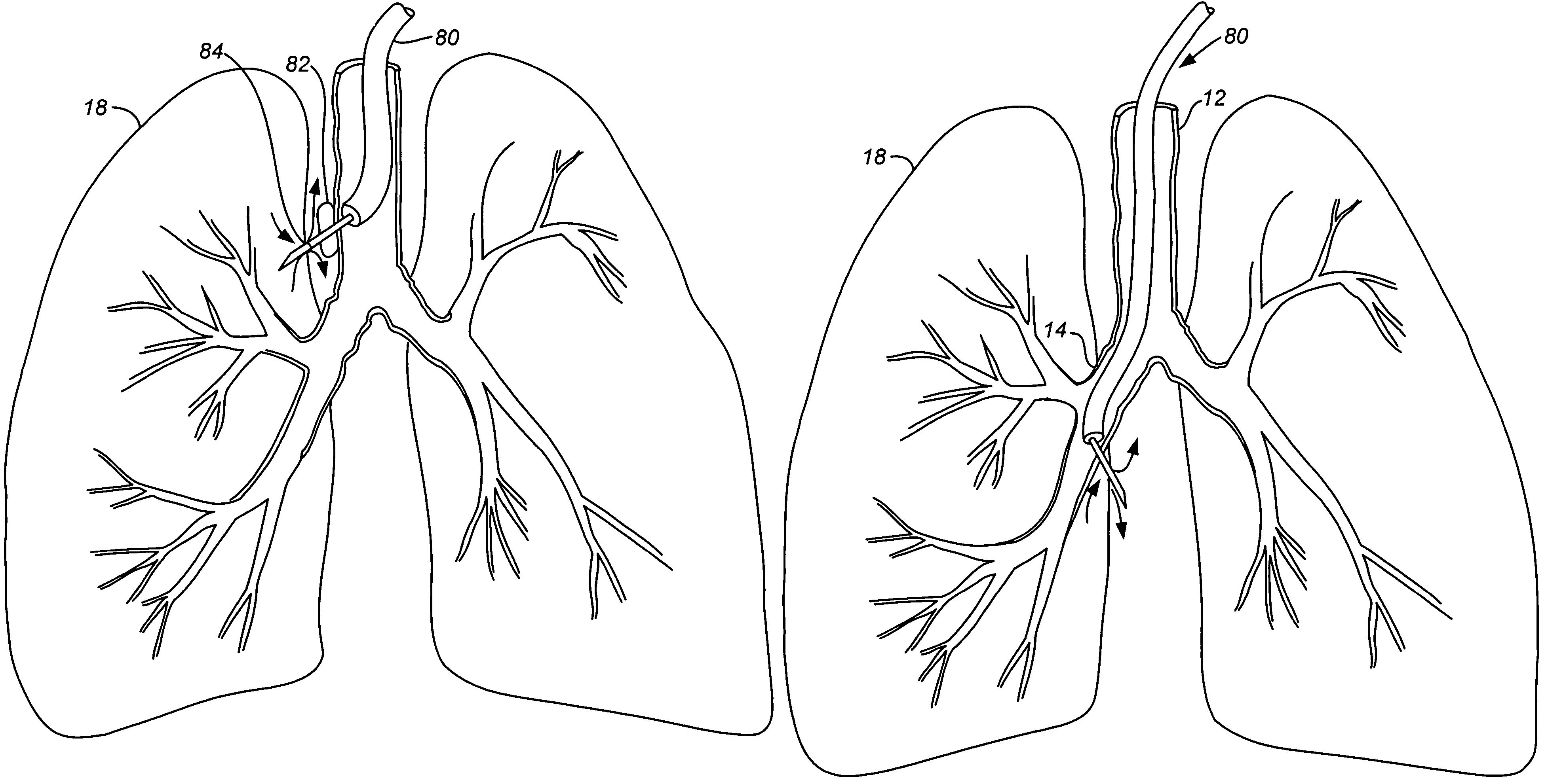
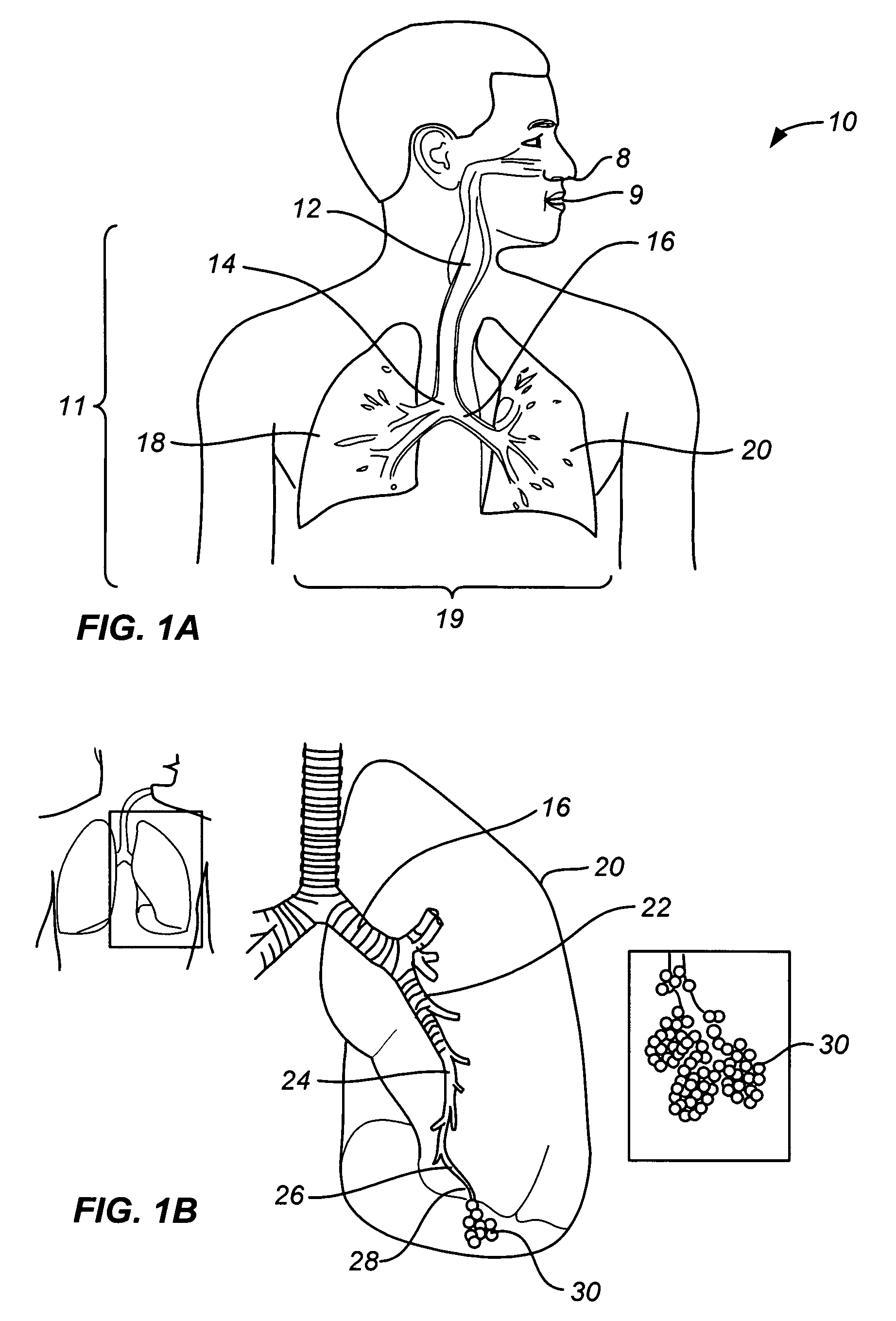
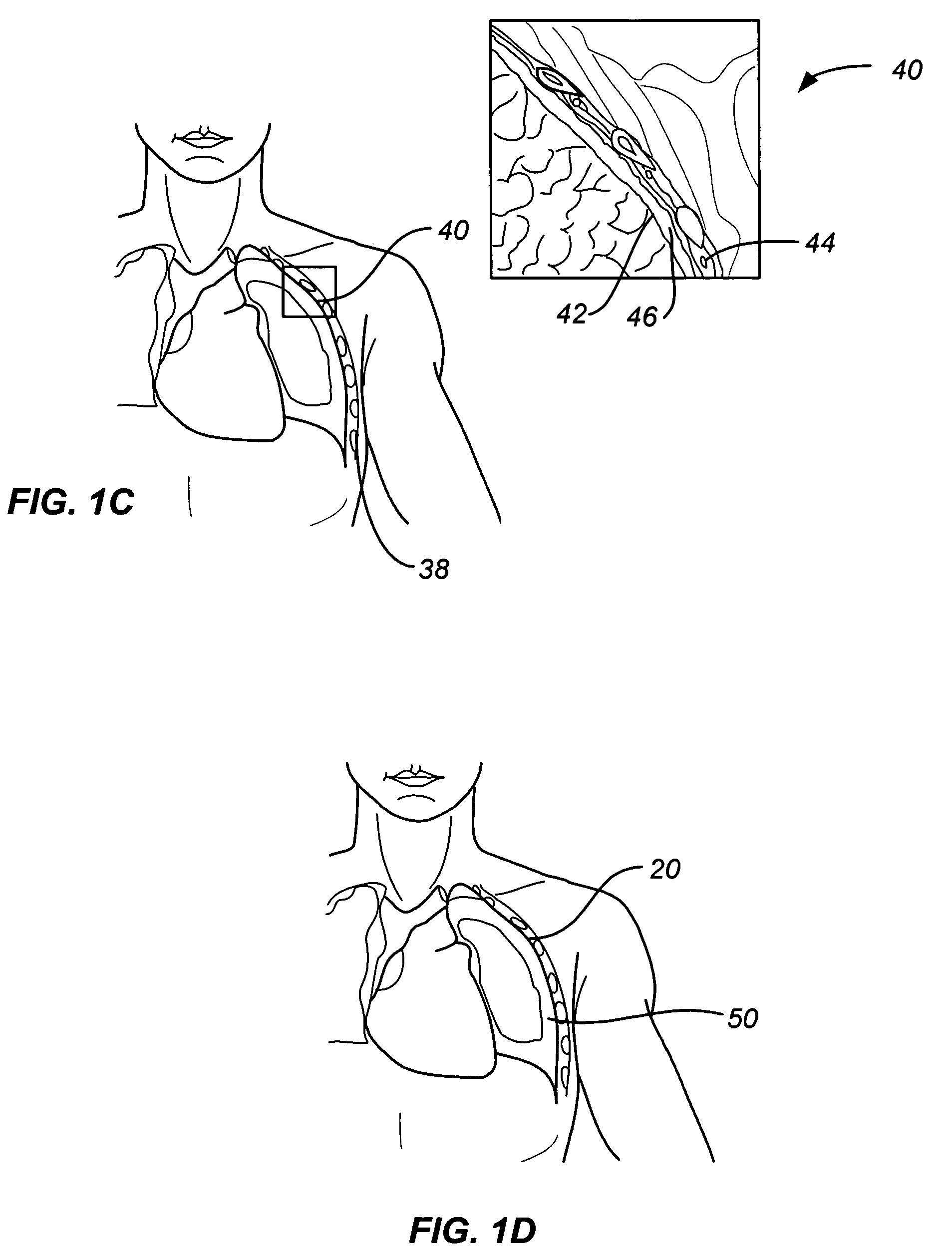
[0031]As noted above, a principal aspect of the present invention is the design of lung devices that can safely perform a transthoracic procedure without impacting the negative pressure required to maintain lung function. In particular, the present devices allow accessing the interior of the lung or the surrounding tissue to perform therapeutic or diagnostic functions while reducing the risk of complications associated with the accessing procedure. The devices and methods are used with adhesive compositions that have cross-linkable moiety and adhering moiety that enable the glue to adhere to lung tissue with low toxicity. Suitable adhesives, such as glue, act as a sealant to prevent the passage of liquid or gas.

[0032]The invention provides methods, materials and devices for providing diagnostic and therapeutic treatment to a target tissue, such as lung, using a suitable adhesive, such as glue, as a sealant to prevent the passage of liquid or gas. The materials used in the method inc...


PUM


Abstract

This invention relates generally to a design of lung devices for safely performing a transthoracic procedure. In particular, the invention provides devices and methods of using these devices to access the thoracic cavity with minimal risk of causing pneumothorax or hemothorax. More specifically, the invention enables diagnostic and therapeutic access to a thoracic cavity using large bore instruments. This invention also provides a method for diagnostic and therapeutic procedures using a device capable of sealing the wound upon withdrawal of the device. The invention includes a device comprising an elongated body adapted to make contact with a tissue of a subject through an access hole, and a sealant delivery element. The invention also includes a method of performing tissue treatment or diagnosis in a subject.

Description

CROSS-REFERENCE[0001]This application claims the benefit of U.S. Provisional Application No. 60 / 586,683, filed Jul. 8, 2004, which is incorporated herein by reference in its entirety.BACKGROUND OF THE INVENTION[0002]1. Field of the Invention[0003]This invention relates generally to a design of lung devices for safely performing a transthoracic procedure. In particular, the invention provides devices and methods of using these devices to access the thoracic cavity with minimal risk of causing pneumothorax or hemothorax. More specifically, the invention enables diagnostic and therapeutic access to a thoracic cavity using large bore instruments. This invention also provides a method for diagnostic and therapeutic procedures using a device capable of sealing the wound upon withdrawal of the device.[0004]2. Description of Related Art[0005]Pulmonary disorders affect millions of Americans, and many more individuals worldwide, each year. While some pulmonary disorders are chronic (e.g., chr...

Claims


Application Information

Patent Timeline
Patent Type & Authority Patents(United States)
IPC IPC(8): A61M31/00
CPCA61B17/00491A61B17/0057A61B17/32002A61B17/34A61B17/3415A61B2017/00278A61B2017/00495A61B2017/00637A61B2017/00641A61B2017/00809
Inventor MCGURK, ERINDIECK, RONALDMATHIS, MARKWARTCHOW, CHARLES
Owner EKOS CORP
Why Eureka
Try Eureka