
Reservoirs with subretinal cannula for subretinal drug delivery

a cannula and subretinal technology, applied in the field of eyes, can solve the problems of not being able to effectively treat the condition, not being able to provide specific therapy, and only being useful in about 20 percent of neovascular amd cases, so as to prevent the progression of the condition

Inactive Publication Date: 2010-09-14
DOHENY RETINA INST
View PDF6 Cites 32 Cited by
  • Summary
  • Abstract
  • Description
  • Claims
  • Application Information

AI Technical Summary

Benefits of technology

"The present invention features devices and methods for delivering therapeutic substances to the posterior segment of the eye through sub-retinal administration or sub-retinal prophylatic administration. This allows for more effective delivery of drugs and other therapeutic agents to the retina and choroid, while minimizing side effects and reducing the risk of complications. The therapeutic substances can include drugs, medications, antioxidants, and other molecules or agents that can be used to treat or prevent disorders or diseases of the eye. The methods can also involve instilling a therapeutic amount of a therapeutic medium into the sub-retinal space, which can disperse the therapeutic substance in the sub-retinal space and allow for sustained therapy. Overall, this invention provides a more effective and safe way to treat retinal and choroidal disorders."

Problems solved by technology

Roughly 80 percent of the AMD cases involve non-neovascular conditions, for which there are no effective treatments.
Perhaps the best known therapy is photodynamic therapy (PDT), however, while this therapy has received significant intention in both the ophthalmic and financial investment communities, it is useful in only about 20 percent of neovascular AMD cases.
In addition, this particular therapy is not a simple or inexpensive treatment.
It systemic side effects, however, include peripheral neuropathy, central nervous system depression, and embryptoxicity.
In addition, these systemic side effects have limited the dosage administered to patients for the treatment of sub-retinal neovascularization.
Systemic inhibition of angiogenesis in older patients can also interfere with the development of collateral circulation, which has a role in the prevention of central nervous system as well as cardiac ischemic events.
However, delivery of drugs, proteins and the like to the eye(s) of mammals so as to achieve the desired therapeutic or medical effect, especially to the retina and / or the choroids, has proven to be challenging, most of which is owed to the geometry, delicacy and / or behavior of the eye and its components.
Oral ingestion of a drug or injection of a drug at a site other than the eye can provide a drug systemically, however, such a systemic administration does not provide effective levels of the drug specifically to the eye.
In many ophthalmic disorders involving the retina, posterior tract, and optic nerve, adequate levels of the drug cannot be achieved or maintained by oral or parenteral routes of administration.
Such further and repeated administrations of such drugs, however, may produce undesired systemic toxicity.
This route of administration (i.e., topical administration), however, is only effective in treating problems involving the superficial surface of the eye and diseases that involve the cornea and anterior segment of the eye, such as for example, conjunctivitis.
Topical administration of drugs is ineffective in achieving adequate concentrations of a drug(s) in the sclera, vitreous, or posterior segment of the eye.
In addition, topical eye drops may drain from the eye through the nasolacrimal duct and into the systemic circulation, further diluting the medication and risking unwanted systemic side effects.
Furthermore, delivery of drugs in the form of topical eye drops is also of little utility because the drug cannot cross the cornea and be made available to the vitreous, retina, or other subretinal structures such as the retinal pigment epithelium (“RPE”) or choroidal vasculature and / or is highly unstable and therefore not easily formulated for topical delivery.
Direct delivery of drugs to the eye by a topical insert has also been attempted, however, this method is not desirable.
Consequently, this technique demands a certain degree of manual dexterity that can be problematic for geriatric patients who are particularly susceptible to certain eye disorders that appear age related (e.g., age related macular degeneration).
Also, in many instances such topical inserts may cause eye irritation and such inserts are prone to inadvertent loss due to eyelid laxity.
In addition, these devices provide a source of drug only to the cornea and anterior chamber, and thus do not provide any pharmacologic advantage over topical eye drops or ointments.
Thus, such devices have limited, if any at all, utility for providing an effective source of drugs to the vitreous or tissues located in the posterior segment of the eye.
This, in turn, presents problems because each additional intraocular injection carries with it a realistic risk of infection, hemorrhage and / or retinal detachment.
In addition, it also is well known that many therapeutic drugs cannot easily diffuse across the retina.
In addition to the relative effectiveness of drug delivery across the barrier, complications or side effects have been observed when using the direct injection into vitreous technique with some therapeutics.
When these compounds were used to treat neovscularization of the posterior segment by direct injection, these compounds were observed to cause undesirable side effects in many patients.
Moreover, a risk exists that the use of corticosteroids in patients with normal intraocular pressure will cause elevations in pressure that result in damage to ocular tissue.
Since therapy with corticosteroids is frequently long term, i.e., several days or more, a potential exists for significant damage to ocular tissue as a result of prolonged elevations in intraocular pressure attributable to that therapy.
Such a course of treatment also increases the duration and cost as well as the realistic risks of corneal ulceration, cataract formation, intraocular infection, and / or vitreous loss that accompany these procedures.
As noted in U.S. Pat. No. 5,273,530, the penetration of the anterior part or segment of the eye, using the transcorneal or the transscleral route creates the risks of corneal ulceration, cataract formation and other anterior penetration problems.

Method used


Image

  • Reservoirs with subretinal cannula for subretinal drug delivery
  • Reservoirs with subretinal cannula for subretinal drug delivery
  • Reservoirs with subretinal cannula for subretinal drug delivery

Examples


Embodiment Construction

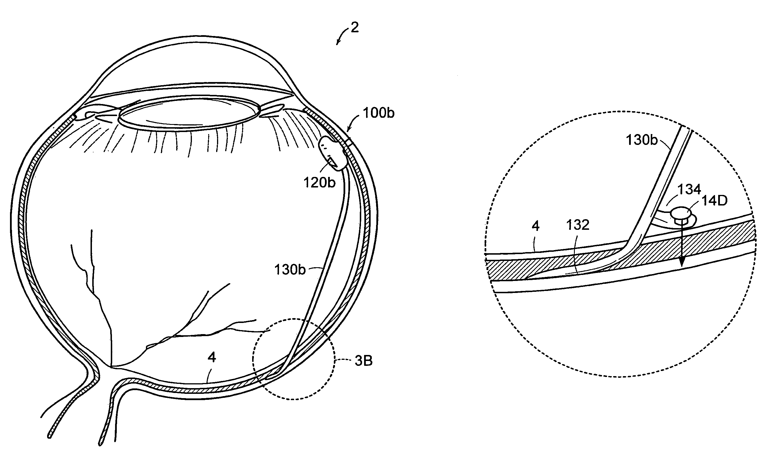
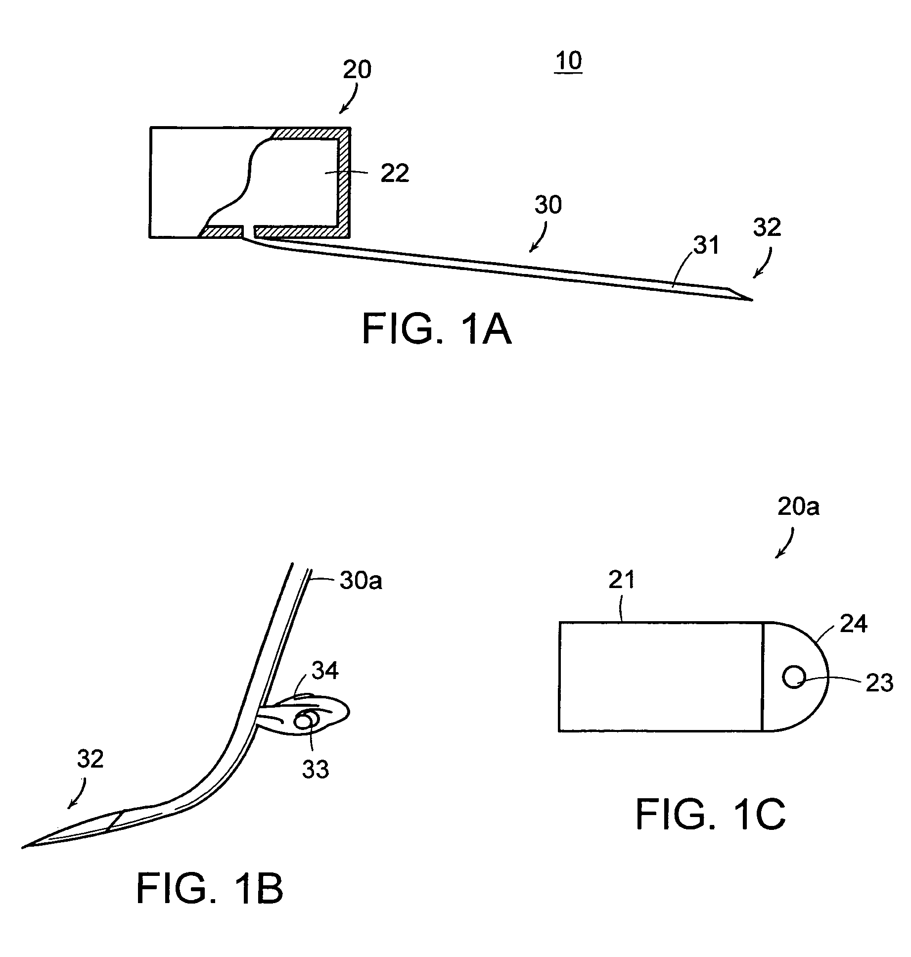
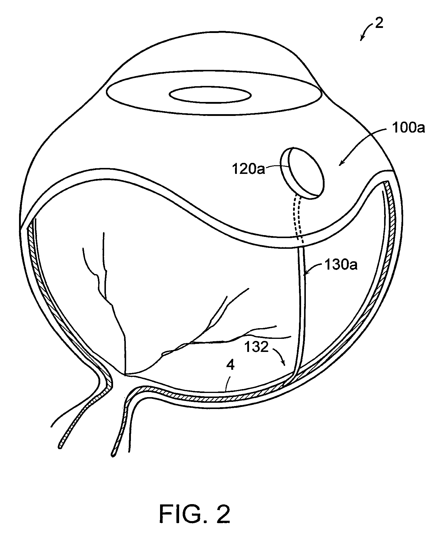
[0059]Referring now to the various figures of the drawing wherein like reference characters refer to like parts, there is shown in FIG. 1A a simplified block diagram or schematic view of a therapeutic medium delivery device 10 according to the present invention. As shown in FIG. 1A, the therapeutic medium delivery device 10 includes a main body 20 and a outlet member or cannula 30 that is fluidly coupled to the interior volume 22 or compartment of the main body. The cannula 30 also is arranged so as to extend outwardly from the main body and from any one of the bottom, side or top surfaces of the main body.

[0060]The main body 20, more specifically the main body interior volume, is sized so as to contain a desired amount of the therapeutic medium to be dispensed or instilled subretinally in the eye 2. In the case where the therapeutic delivery device 10 is disposed within the eye 2, the main body 20 also is preferably sized so as to not cause an appreciable impact on the vision of a ...


PUM


Abstract

Featured are devices and methods for administering a therapeutic medium to the posterior segment of an eye including instilling or disposing the therapeutic medium sub-retinally. Such instillation is accomplished by providing a reservoir containing a therapeutic medium to which is fluidly coupled a cannula and inserting a portion of the cannula sub-retinally, whereby the therapeutic medium within the reservoir is coupled to and dispersed subretinally via the cannula.

Description

[0001]This application claims the benefit of U.S. Provisional Application Ser. No. 60 / 442,323 filed Jan. 24, 2003, the teachings of which are incorporated herein by reference.FIELD OF INVENTION[0002]The present invention relates to devices and methods for treating eyes, such as eyes of mammals having eye disorders or diseases, more particularly to devices and methods for administering a therapeutic medium or agent sub-retinally, yet more particularly to devices and methods for administering such therapeutics or agents to the tissues of the eye so that the pharamacodynamic action of the such therapeutics / agents is localized at the choroid and the retina and more specifically to devices and methods for administering such therapeutics / agents using a device including a sub-retinal cannula through which such therapeutics / agents are administered subretinally.BACKGROUND OF THE INVENTION[0003]There are a number of vision-threatening disorders or diseases of the eye of a mammal including, bu...

Claims


Application Information

Patent Timeline
Patent Type & Authority Patents(United States)
IPC IPC(8): A61M35/00A61FA61F2/16A61F9/00A61M5/142A61M31/00
CPCA61F2/16A61F9/0017A61M5/1428A61M2210/0612A61F2250/0068
Inventor HUMAYUN, MARKDE JUAN, EUGENE
Owner DOHENY RETINA INST
Learn More