
Tumor lesion regression and conversion in situ into autologous tumor vaccines by compositions that result in anti-Gal antibody binding

a tumor lesion and anti-gal antibody technology, applied in the field of cancer treatment, can solve the problems of patient death, immune-mediated detection, regression, and/or destruction of micrometastases, and achieve the effect of reducing the growth of at least one of said untreated non-resectable solid tumors

Inactive Publication Date: 2010-10-26
UNIV OF MASSACHUSETTS MEDICAL SCHOOL
View PDF7 Cites 10 Cited by
  • Summary
  • Abstract
  • Description
  • Claims
  • Application Information

AI Technical Summary

Benefits of technology

"The present invention relates to a method for treating cancer by inducing the expression of α-gal epitopes on tumor cells. This can be achieved by introducing a glycolipid preparation containing α-gal epitopes into the tumor. The introduced glycolipid can induce local inflammation and lead to the regression or destruction of the tumor. The method can be performed using various procedures such as injection, imaging guided injection, endoscopy, bronchoscopy, cystoscopy, colonoscopy, laparoscopy, and catheterization. The treated tumor can also be converted into an autologous vaccine. The invention provides a promising method for treating cancer that targets the immune system and has the potential to induce tumor regression or destruction."

Problems solved by technology

The major cause of death in cancer patients with solid tumors is the recurrence of the cancer after surgery as multiple metastases that are nonresectable and / or refractory to any therapy.
As no treatment is available for them, many of these patients die within weeks or few months after detection of metastatic tumor lesions.
Such an immune response results in immune mediated detection, regression, and / or destruction of micrometastases which can not be detected visually and are not detectable by imaging.

Method used


Image

  • Tumor lesion regression and conversion in situ into autologous tumor vaccines by compositions that result in anti-Gal antibody binding
  • Tumor lesion regression and conversion in situ into autologous tumor vaccines by compositions that result in anti-Gal antibody binding
  • Tumor lesion regression and conversion in situ into autologous tumor vaccines by compositions that result in anti-Gal antibody binding

Examples


example 1

Extraction of α-Gal Glycolipids from Rabbit and Bovine Red Blood Cell Membranes

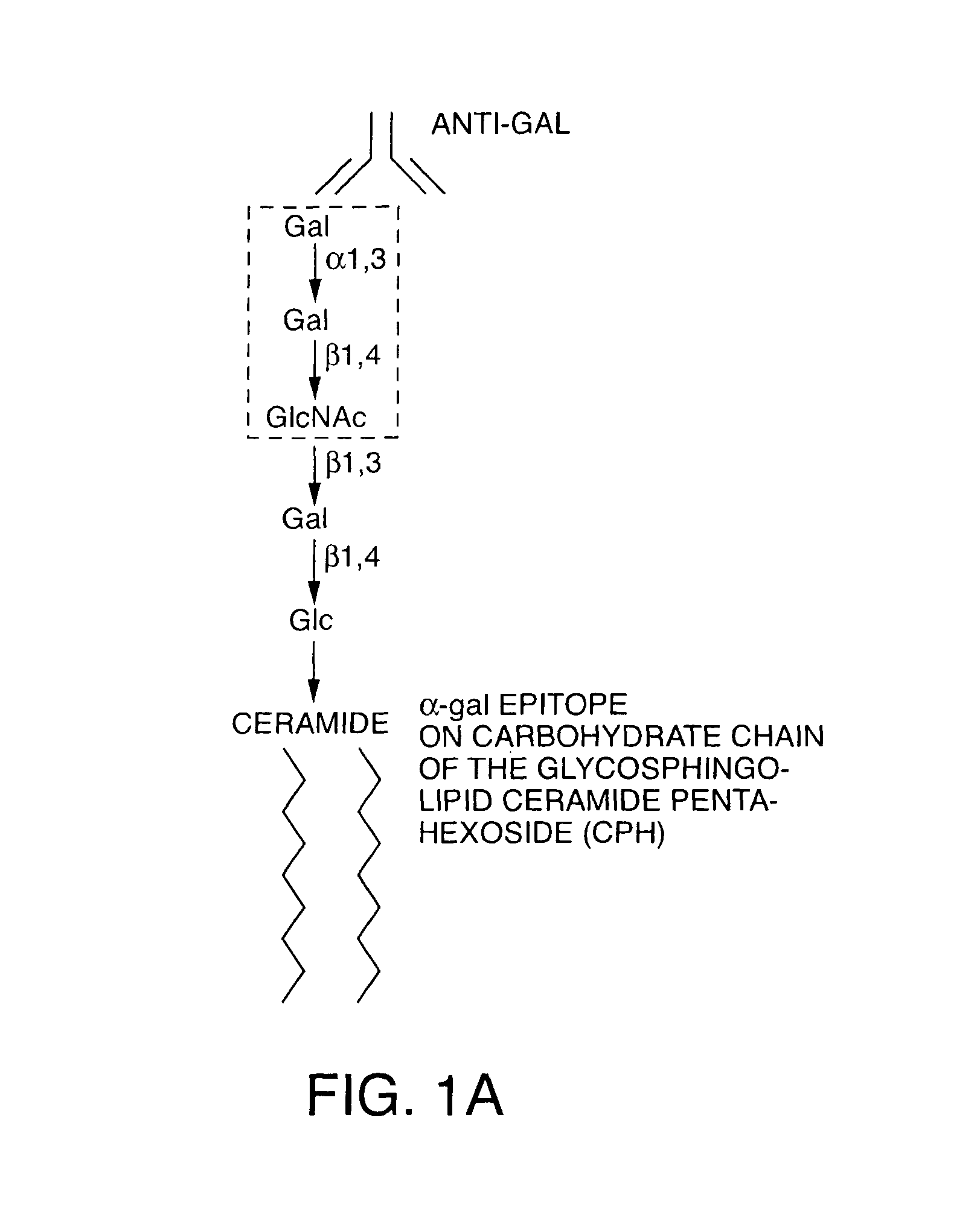
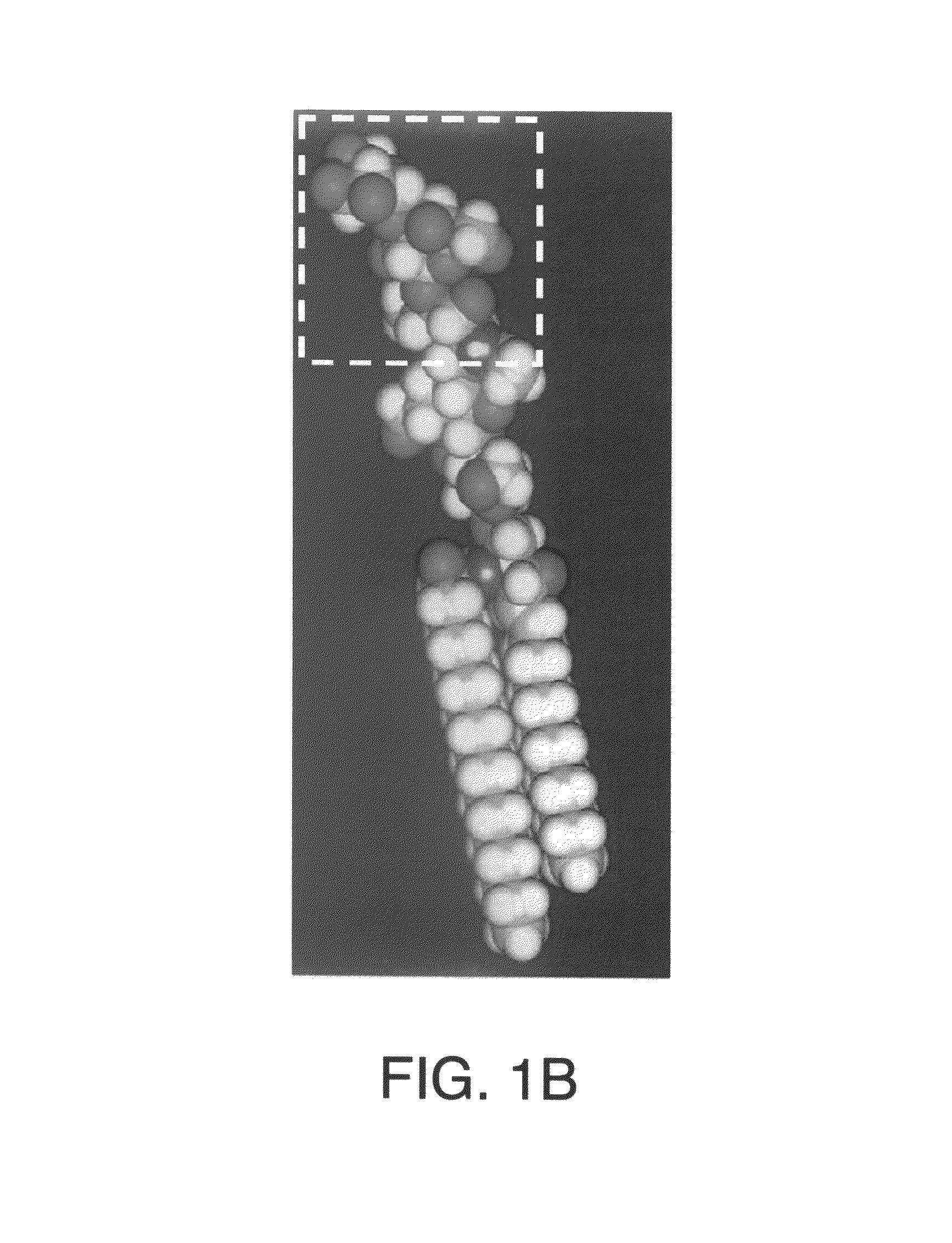
[0170]A rich source for α-gal glycolipids is rabbit red blood cells. The two major glycosphingolipids (GSL), i.e. glycolipids with ceramide tail, in rabbit red blood cell membranes are ceramide (Cer) trihexoside (CTH, with 3 sugars [hexoses], present also in human red blood cells) with the structure Galα1-4Galβ1-4Glc-Cer and ceramide pentahexoside (CPH, with 5 sugars [hexoses]) with α-gal epitopes as terminal part of the CPH structure of Galα1-3Galβ1-4GlcNAcβ1-3Galβ1-4Glc-Cer (Eto T, Iichikawa Y, Nishimura K, Ando S, Yamakawa T J. Biochem. (Tokyo) 1968; 64: 205-13, and Stellner K, Saito H, Hakomori S. Arch. Biochem. Biophys. 1973; 133: 464-72) (See FIG. 1 and FIG. 6A, lane 1). Immunostaining of rabbit α-gal glycolipids on thin layer chromatography (TLC) plates with human natural anti-Gal resulted in binding of the antibody to CPH but not to CTH (See FIG. 6A, lane 2). This immunostaining revealed also that...

example 2

Binding of Autologous Anti-Gal to Cells with Inserted α-gal Glycolipids and Subsequent Complement Mediated Lysis of Such Cells

[0175]Insertion of α-gal glycolipids into autologous cell membranes can be demonstrated by incubation of human red blood cells with 1 mg / ml of α-gal glycolipids for 2 h at 37° C. After extensive washing to remove unbound α-gal glycolipids, the expression of α-gal epitopes can be demonstrated by the incubation of the red blood cells with heat inactivated autologous serum that was diluted 1:2 in phosphate buffered saline (PBS). The binding of autologous anti-Gal to the inserted α-gal glycolipids was demonstrated by incubation of these red blood cells with phycoerythrin coupled anti-human IgG and analysis of staining by flow cytometry. The bound antibodies was indicated by the shift in binding of IgG antibodies in comparison with control untreated red blood cells (See FIG. 8A, thick line histogram and dotted line histogram, respectively). The antibodies binding ...

example 3

Insertion of α-Gal Glycolipids into Tumor Cell Membranes and Subsequent Complement Mediated Lysis of Cells with the Inserted α-Gal Glycolipids

[0177]This example demonstrates an in vitro mouse tumor model showing the insertion of α-gal glycolipids into tumor cell membranes and the subsequent binding of anti-Gal. This model utilizes B16 melanoma cells (mouse tumor cells lacking α-gal epitopes) that form tumor lesions in mice having an H-2b genetic background.

[0178]B16 cells in RPMI medium supplemented with 10% fetal calf serum were incubated with 1 mg / ml, 0.1 mg / ml, or 0.01 mg / ml of α-gal glycolipids for 2 h at 37° C. with constant rotation. After extensive washing to remove unbound α-gal glycolipids, the display of α-gal epitopes was demonstrated by binding mouse anti-Gal IgG purified on α-gal affinity column from serum of α1,3galactosyltransferase knockout mice that produce anti-Gal.

[0179]After 2 h incubation at 4° C. with the anti-Gal antibody the cells were washed and incubated wi...


PUM

PropertyMeasurementUnit
diameteraaaaaaaaaa
diametersaaaaaaaaaa
pHaaaaaaaaaa

Abstract

The present invention discloses that an intratumoral injection of: i) glycolipids with α-gal epitope; ii) gene vectors comprising an α1,3galactosyltransferase gene; or iii) a mixture of α1,3galactosyltransferase, neuraminidase, and uridine diphosphate galactose results in tumor regression and / or destruction. Binding of the natural anti-Gal antibody to de novo expressed tumoral α-gal epitopes induces inflammation resulting in an anti-Gal antibody mediated opsonization of tumor cells and their uptake by antigen presenting cells. These antigen presenting cells migrate to draining lymph nodes and activate tumor specific T cells thereby converting the treated tumor lesions into in situ autologous tumor vaccines. This therapy can be applied to patients with multiple lesions and in neo-adjuvant therapy to patients before tumor resection. In addition to the regression and / or destruction of the treated tumor, such a vaccine will help in the immune mediated destruction of micrometastases that are not detectable during the removal of the treated tumor.

Description

[0001]This application claims the benefit of U.S. Provisional Application No. 60 / 654,921, filed on Feb. 22, 2005.FIELD OF INVENTION[0002]The present invention is related to the field of cancer treatment. In one embodiment, the invention contemplates administering compounds to tumor lesions that induce local expression of α-gal epitopes within the tumor. In one embodiment, the administration induces regression and / or destruction of the treated tumor lesions. In another embodiment, the administration converts the treated tumor lesions into an autologous tumor vaccine. In one embodiment, the compounds comprise glycolipids having an α-gal epitope. In another embodiment, the compounds comprise a mixture of α1,3galactosyltransferase, neuraminidase, and uridine diphosphate galactose.BACKGROUND[0003]The major cause of death in cancer patients with solid tumors is the recurrence of the cancer after surgery as multiple metastases that are nonresectable and / or refractory to any therapy. The ma...

Claims


Application Information

Patent Timeline
Patent Type & Authority Patents(United States)
IPC IPC(8): A61K31/70A61K31/715C07H1/00C13K5/00C13K7/00
CPCA61K31/739
Inventor GALILI, URI
Owner UNIV OF MASSACHUSETTS MEDICAL SCHOOL
Learn More