Characterization of granulocytic Ehrlichia and methods of use
a technology of granulocytic ehrlichia and characterization method, which is applied in the field of granulocytic ehrlichia (ge) proteins, can solve the problems of impeded full classification of phagocytophila /i>species including antigenic relationships among individual isolates, and achieve the effects of reducing toxicity, reducing toxicity, and improving solubility of molecul
- Summary
- Abstract
- Description
- Claims
- Application Information
AI Technical Summary
Benefits of technology
Problems solved by technology
Method used
Image
Examples
example 1
PCR Amplification and Cloning of GE 16S rDNA
[0248]GE was cultivated in HL60 cells as described in Protocol A (supra). Cell extracts were prepared by lysis protocols as described supra. PCR primers (specific for the 16S ribosomal DNA of the genogroup comprising E. equi., E. phagocytophila, and the HGE agent used to amplify DNA from the cell extracts) were modified to include restriction enzyme recognition sites as follows:
forward primer, 5′-CTGCAGGTTTGATCCTGG-3′ (PstI site) (SEQ ID NO:40); reverse primer, 5′-GGATCCTACCTTGTTACGACTT-3′ (BamHI site) (SEQ ID NO:41).
[0249]These primers (0.5 μM) were added to a 104.1 reaction mixture containing 1×PCR buffer II (Perkin-Elmer Corp), 1.5 mM MgCl2 (Perkin-Elmer Corp.), 200 μM each dATP, dGTP, dCTP and dTTP, 2.5 U of Amplitaq DNA polymerase and 20 μl of USG3 DNA. Amplification was performed as described in Protocol F. The amplified 1500 bp fragment was digested with Pst I and Bam HI and ligated to pUC19 linearized with the same enzymes. The res...
example 2
Isolation of Clones Using Canine Sera
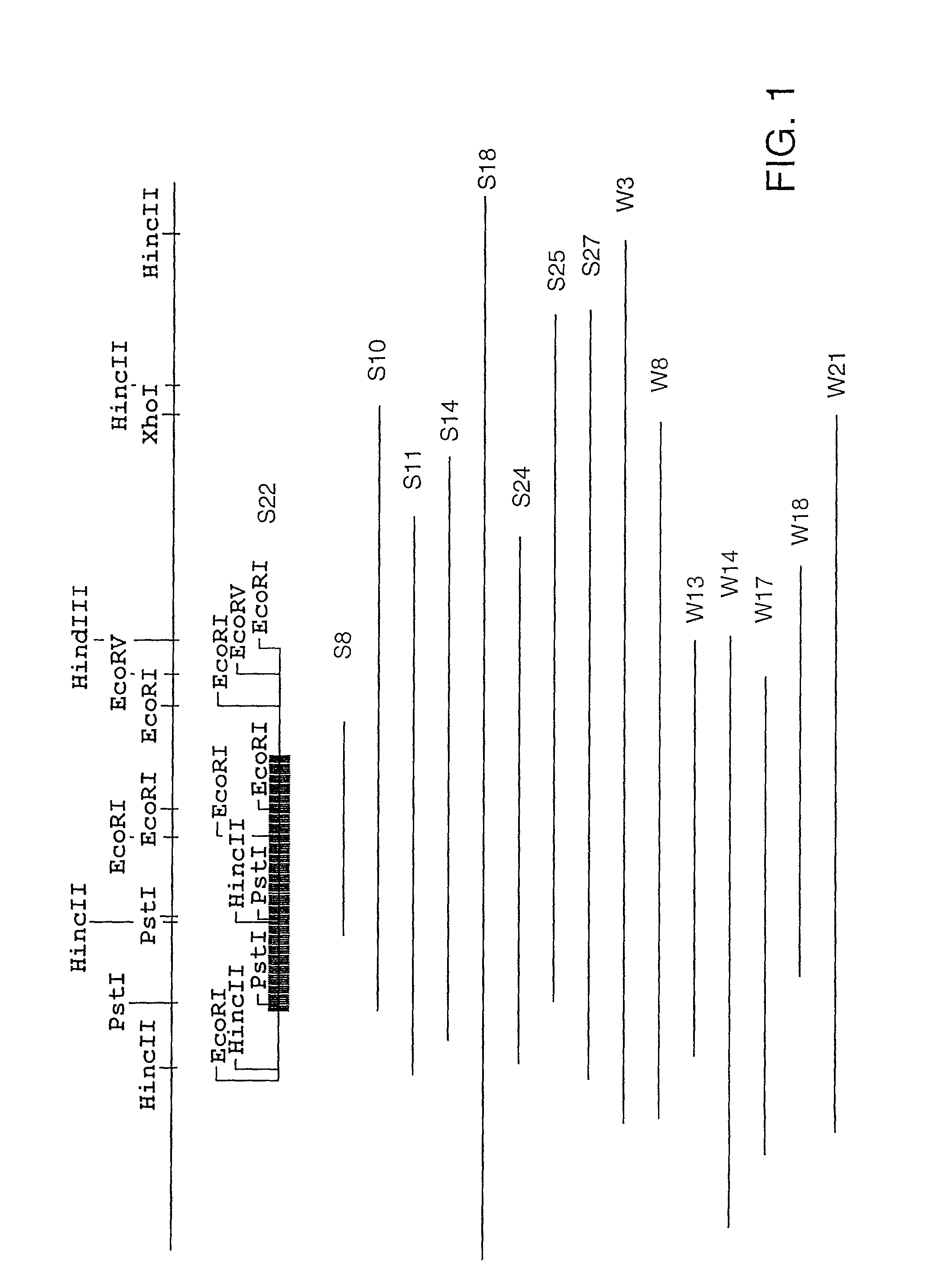
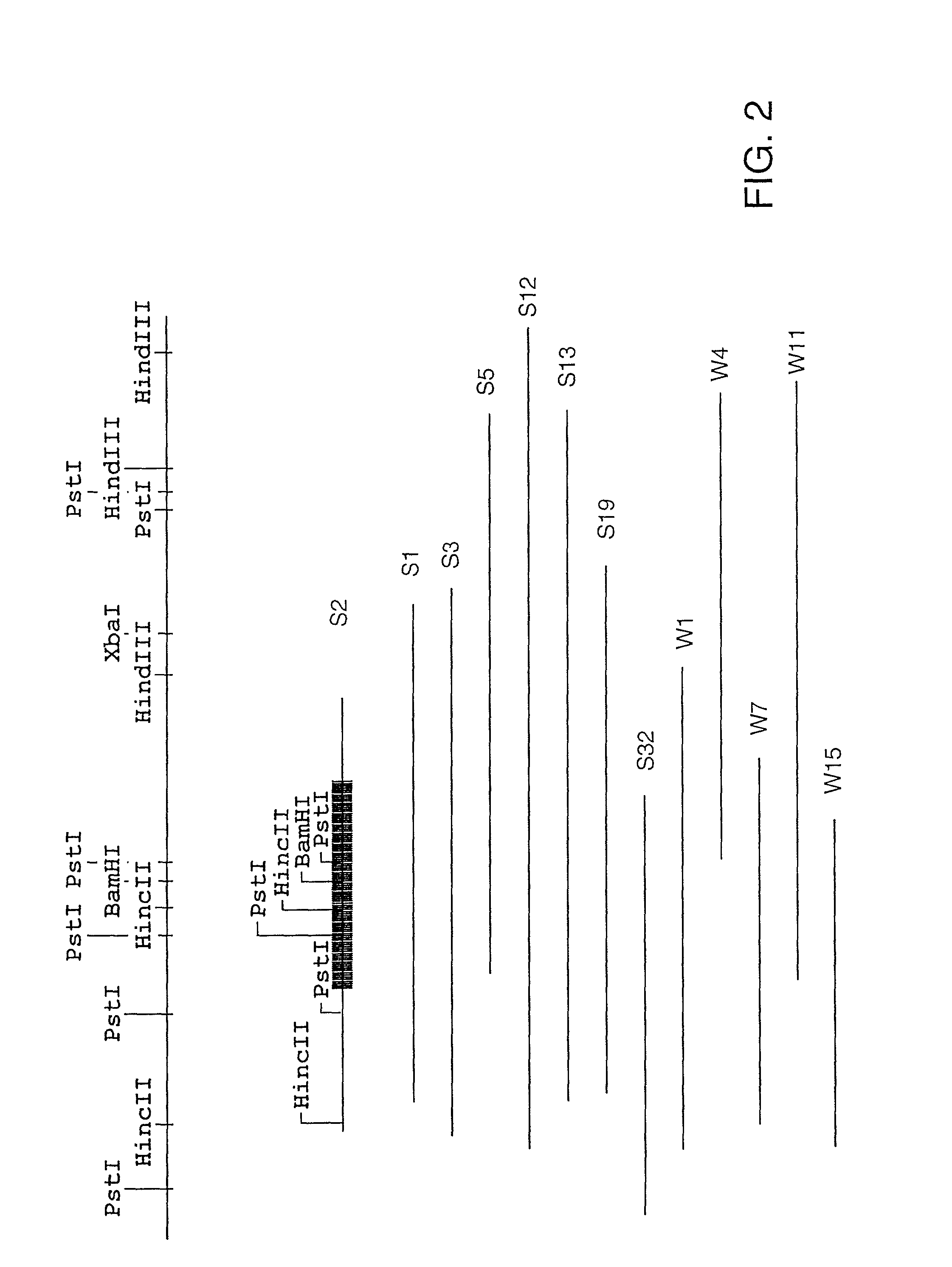
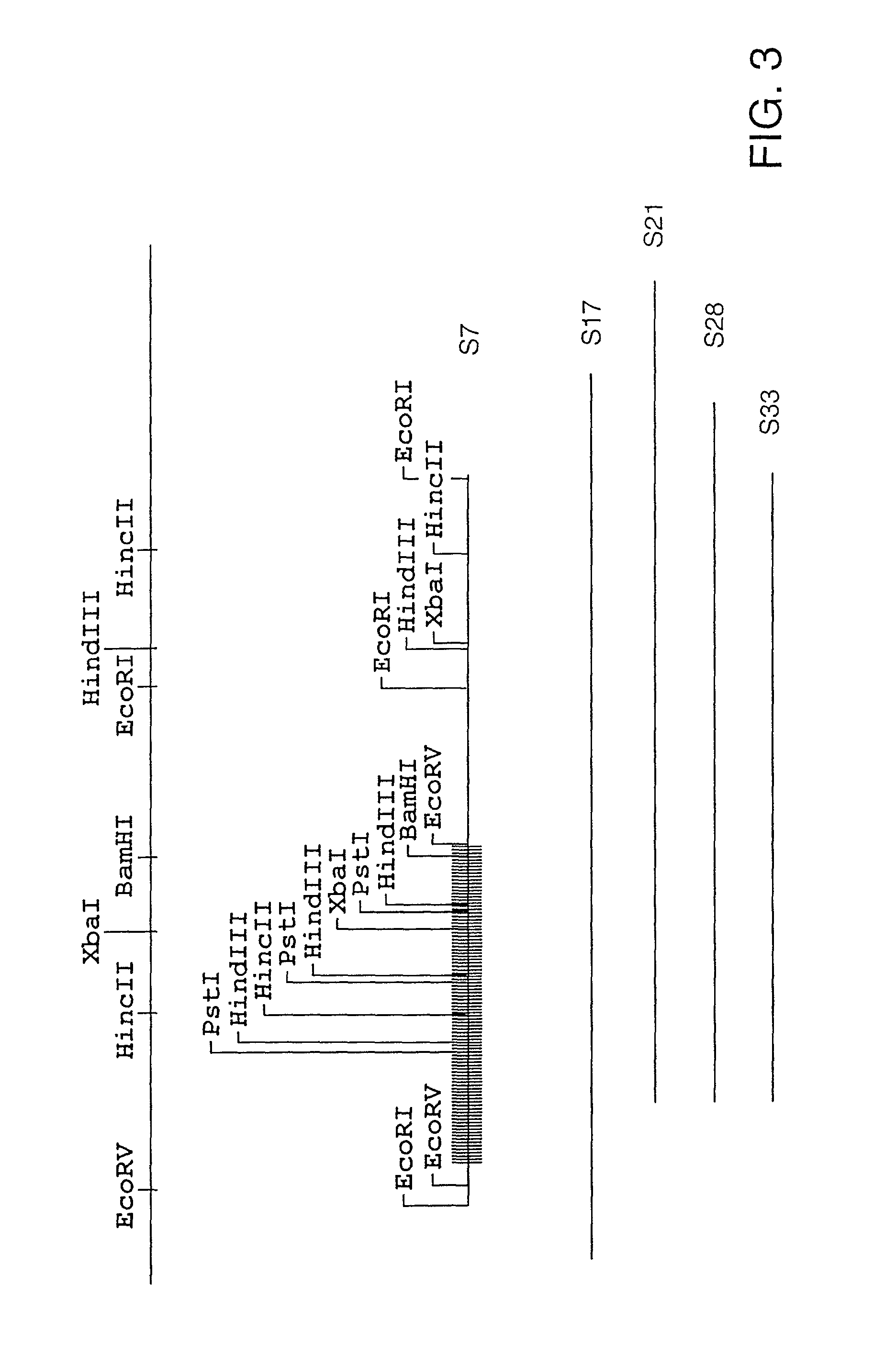
[0250]Western blot analysis of the individual recombinant plasmid was performed as described in Protocol G using canine sera prepared as described in Protocol D or a 1:1000 dilution of human sera prepared from two convalescent-phase sera from patients (No. 2 and 3, New York, kindly provided by Dr. Aguero-Rosenfeld) and from an individual in Wisconsin who was part of a seroprevalence study (No. 1, kindly provided by Dr. Bakken). Blots were washed and incubated with biotin-labeled goat anti-dog IgG (Kirkegaard & Perry Laboratories, Inc., Gaithersburg, Md.) followed by peroxidase labeled streptavidin (Kirkegaard & Perry Laboratories, Inc., Gaithersburg, Md.) or HRP conjugated anti-human IgG (Bio-Rad, Hercules, Calif.). After several additional washes, the dog sera blots were developed with 4-chloronapthol (Bio-Rad, Hercules, Calif.). Over 1000 positive clones were identified. Three hundred of these clones (both strong (S) and weak (W) immunoreactivi...
example 3
Characterization of Representative Clones S2, S7. S22, and S23
[0251]A representative clone was chosen for further characterization from each of the three groups (see Example 2, supra). These clones, S2, S7, and S22, were sequenced according to Protocol F. S23 was also sequenced since it did not appear to fall into one of these groups. The complete nucleic acid sequence of each of these clones is shown as follows: FIG. 4, group I (S22); FIG. 6, group II (S2); FIG. 8, group III (S7); FIG. 10, (S23). Sequence analysis (MacVector, Oxford Molecular Group) showed that each clone contained a single large open reading frame encoded by the plus strand of the insert and each one appeared to be a complete gene. The amino acid sequences encoded by each clone are shown in FIG. 5 (S22), FIG. 7, (S2), and FIG. 9 (S7), and FIG. 11 (S23). There are also two additional small open reading frames in the S23 DNA insert, one on the negative strand and the other on the positive strand. A comparison of the...
PUM
| Property | Measurement | Unit |
|---|---|---|
| temperature | aaaaa | aaaaa |
| weight | aaaaa | aaaaa |
| weight | aaaaa | aaaaa |
Abstract
Description
Claims
Application Information
 - R&D
- Intellectual Property
- Life Sciences
- Materials
- Tech Scout
- Unparalleled Data Quality
- Higher Quality Content
- 60% Fewer Hallucinations
Browse by: Latest US Patents, China's latest patents, Technical Efficacy Thesaurus, Application Domain, Technology Topic, Popular Technical Reports.
© 2025 PatSnap. All rights reserved.Legal|Privacy policy|Modern Slavery Act Transparency Statement|Sitemap|About US| Contact US: help@patsnap.com


