Reservoir device for intraocular drug delivery

a reserve device and intraocular drug technology, applied in the field of improved delivery devices, can solve the problems of limited ocular absorption, high risk of systemic toxicity for patients, and difficult to fill and refill, so as to achieve easy filling and refilling, reduce risk, and reduce the effect of invasiveness

Active Publication Date: 2011-02-08
THE JOHN HOPKINS UNIV SCHOOL OF MEDICINE
View PDF6 Cites 148 Cited by
  • Summary
  • Abstract
  • Description
  • Claims
  • Application Information

AI Technical Summary

Benefits of technology

[0016]The present delivery device and methods of use are minimally invasive. In particular, the delivery device has a small profile that allows it to be inserted though a small opening. Such an insertion procedure eliminates the risks associated with more invasive surgery, and, further, enables such procedures to take place in an office setting. Also, the device can be removed from a small insertion site, requiring few or no sutures for scleral closure. This is a dramatic improvement over state of the art technologies which require surgery for both implantation and removal.
[0...

Problems solved by technology

The delivery of drugs to the eye presents many challenges.
High systemic doses can penetrate this blood ocular barrier in relatively small amounts, but expose the patient to the risk of systemic toxicity.
Topical delivery of drugs often results in limited ocular absorption due to the complex hydrophobic/hydrophilic properties of the cornea and sclera.
However, these repeated intraocular injections carry the risk of infection, hemorrhage and retinal detachment.
Patients also find this procedure somewhat difficu...

Method used


Image

  • Reservoir device for intraocular drug delivery
  • Reservoir device for intraocular drug delivery
  • Reservoir device for intraocular drug delivery

Examples


Embodiment Construction

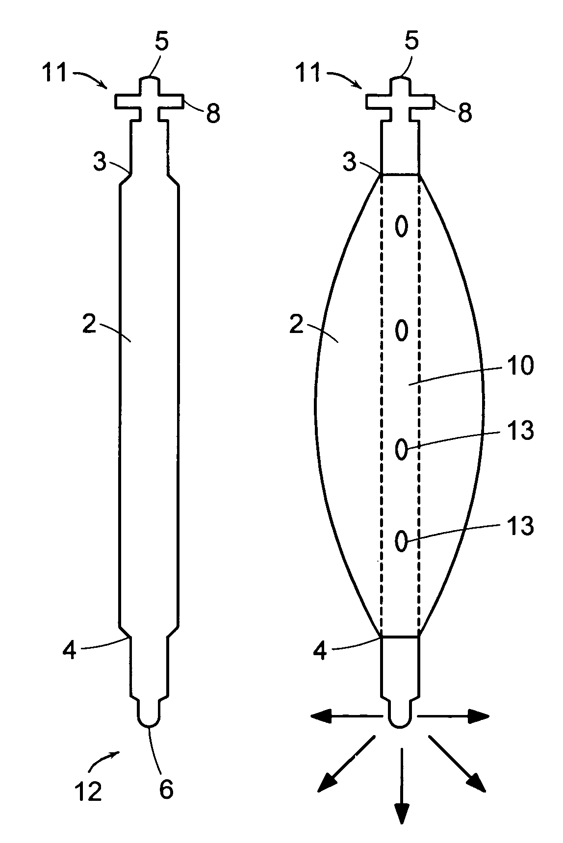
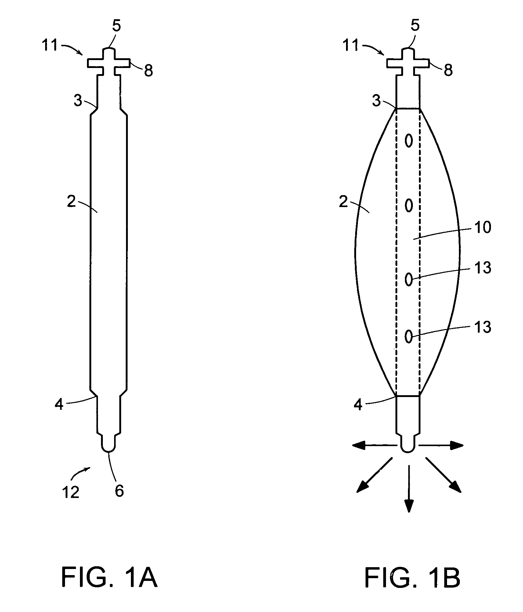
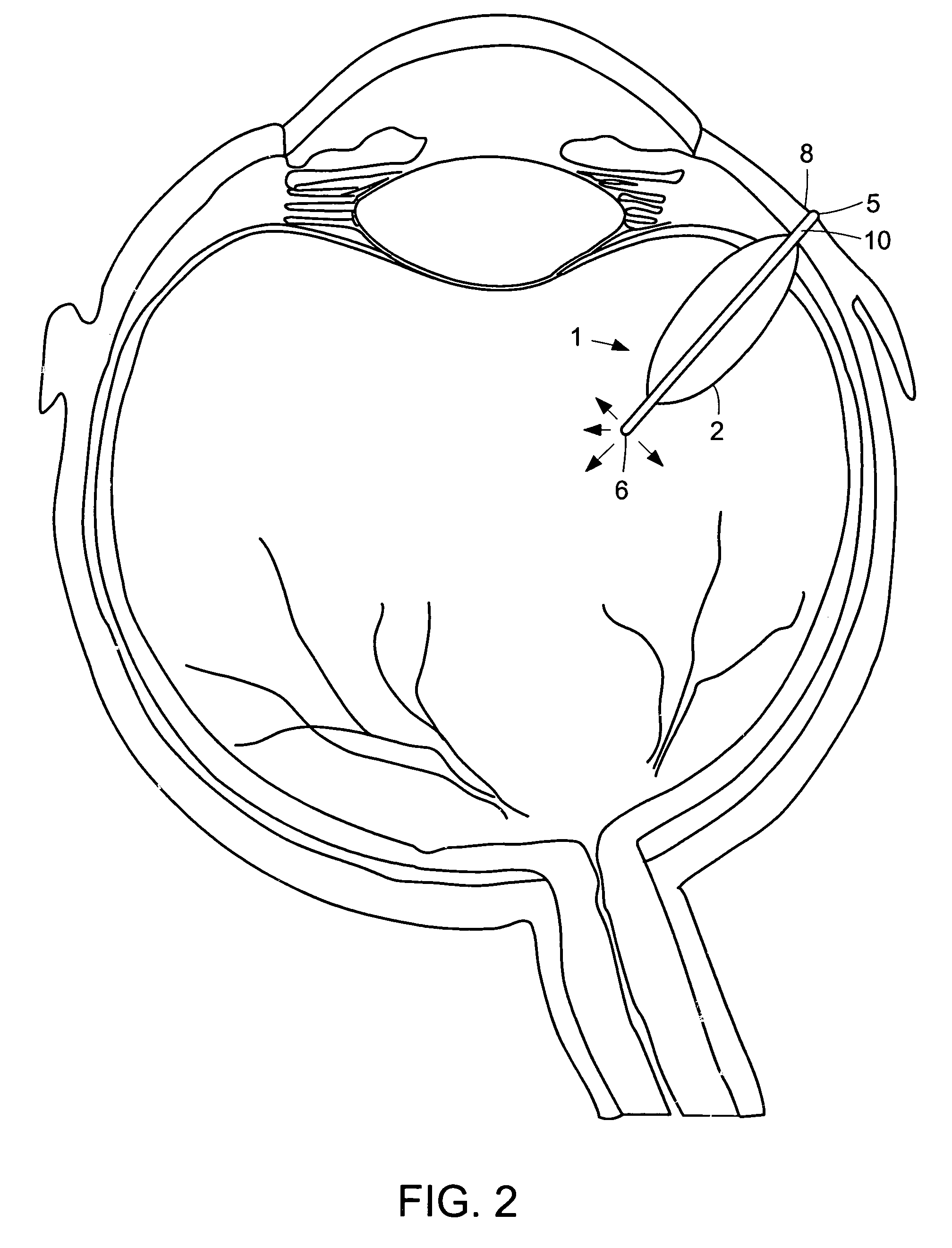
[0027]Referring now to the various figures of the drawing, wherein like reference characters refer to like parts, there is shown various views of a delivery device 1, in accordance with the invention.

[0028]As shown in FIGS. 1-3, the delivery device 1 includes a reservoir 2 having a proximal end 3 and a distal end 4. Located near the proximal end 3 of the reservoir 2 is an inlet port 5 for injection of a desired agent into the reservoir 1. Agent injected through the inlet port 5 is delivered to the treatment area through delivery mechanism 6.

[0029]The delivery device 1 may further include a hollow body or tube 10 housed at least partially within the reservoir 2. The hollow body or tube 10 has a proximal end 11 and a distal end 12. Preferably, the proximal end 11 of the hollow body or tube 10 extends outside the reservoir 2, as shown in FIGS. 1-3, and serves as the inlet port 5 through which the agent is injected into the device.

[0030]The materials used in fabricating the reservoir 2 ...


PUM


Abstract

A delivery device that allows for the sustained release of an agent, particularly useful for the sustained release of a therapeutic agent to limited access regions, such as the posterior chamber of the eye and inner ear. The delivery device is minimally invasive, refillable and may be easily fixed to the treatment area. The delivery device includes a hollow body with an inlet port at its proximal end for insertion of the agent, a reservoir for holding the agent and a delivery mechanism for the sustained delivery of the agent from the reservoir to the patient.

Description

[0001]The present application claims the benefit of U.S. provisional application No. 60 / 297,499 filed Jun. 12, 2001, which is incorporated by reference herein in its entirety.FIELD OF THE INVENTION[0002]The present invention relates to improved delivery devices and methods of use. More particularly, the present invention relates to minimally invasive, refillable, sustained release delivery devices particularly suitable for the delivery of therapeutic agents to limited access regions, such as the posterior chamber of the eye.BACKGROUND OF THE INVENTION[0003]The delivery of drugs to the eye presents many challenges. The ocular absorption of systemically administered pharmacologic agents is limited by the blood ocular barrier, namely the tight junctions of the retinal pigment epithelium and vascular endothelial cells. High systemic doses can penetrate this blood ocular barrier in relatively small amounts, but expose the patient to the risk of systemic toxicity. Topical delivery of drug...

Claims


Application Information

Patent Timeline
IPC IPC(8): A61F2/00A61F13/00A61K9/00A61F9/00A61F9/007
CPCA61F9/0017A61F2250/0067A61F2250/0068A61M31/002A61M39/0208A61M2039/0205A61M2205/04A61M2210/0612
Inventor VARNER, SIGNE ERICKSONDEJUAN, EUGENEBARNES, AARON CHRISTOPHERSHELLEY, TERRY HARRISONCOONEY, MICHAEL J.
Owner THE JOHN HOPKINS UNIV SCHOOL OF MEDICINE
Why Eureka
Try Eureka