Method for using photoplethysmography to optimize fluid removal during renal replacement therapy by hemodialysis or hemofiltration

a technology of hemodialysis and hemofiltration, applied in the field of non-invasive imaging, can solve the problems of abnormal decrease in patient's blood pressure, lack of fluid removal ability, hypovolemic shock, etc., and achieve the effect of avoiding patient hypotension, being more reliable, and being capabl

Active Publication Date: 2011-02-15
UNIV OF FLORIDA RES FOUNDATION INC
View PDF15 Cites 44 Cited by
  • Summary
  • Abstract
  • Description
  • Claims
  • Application Information

AI Technical Summary

Benefits of technology

[0016]In one regard, superior RRT monitoring methods would be able to provide real-time continuous measurements of signals that would be analyzed to provide relative blood flow and / or volume, blood pressure, and pulse rate. Such superior RRT monitor systems would utilize pulse oximetry / photoplethysmography at a highly perfused central tissue site, such as the lip, tongue, nares, cheek. Optionally, a second additional may be located at a typically less perfused area such as a finger or toe. It is desired to have such superior monitoring methods, which are more reliable, are capable of averting patient hypotension, allow cross-site comparisons of blood flow and / or volume and provide non-invasive feedback based control methods that continuously and automatically manipulate the ultrafiltration rate to achieve an optimal ultrafiltration rate.
[0017]Methods, systems and devices have been developed for optimizing fluid removal during renal replacement therapy (RRT) in which the ultrafiltration rate is adjusted in real-time during a patient's treatment and further not requiring human monitoring and / or interaction. The present invention senses a fall in carotid blood flow or failure of compensatory mechanisms resulting in cerebral hypoperfusion. By monitoring blood flow and / or volume status, via photoplethysmography, the system detects a decrease of central blood volume that precedes the onset of hypotension and maintains a safe filtration rate by reducing the ultrafiltration rate when the blood flow and / or volume status feedback signal decreases, indicating that hypotension may occur. Adjustment of the ultrafiltration rate may be manually performed by the healthcare practitioner or adjustment may be automated using a real-time blood flow and / or volume status feedback signal, thereby averting hypotension before it occurs.
[0018]The present invention provides a method of optimizing fluid removal during renal replacement therapy, said method comprising securing a pulse oximeter / photoplethysmography probe to a central source site of a patient wherein said probe is configured to generate a signal stream indicative of blood flow and / or volume status at said central source site; processing said signal stream received from said probe to obtain a separate pulsatile cardiac component (PCC) signal and venous capacitance component (VCC) signal; evaluating said PCC signal, or VCC signal, or both, to determine the occurrence of a change in blood flow and / or volume status; and adjusting the ultrafiltration rate of said patient's renal replacement therapy responsive to said change in blood flow status. The method allows for the comfortable and non-invasive monitoring of a patient's blood flow and / or volume status during renal replacement therapy and further provides optimization of the ultrafiltration rate capable of averting hypotension. This invention also measures compensatory mechanisms such as vascular tone, which in turn is used as a diagnostic tool to detect deficiencies in such mechanisms.

Problems solved by technology

Dialysis patients require removal of excess water from their blood because they lack the ability to rid their bodies of fluid through normal kidney function.
One of the potential risks to health associated with RRT is hypotension, which is an abnormal decrease in the patient's blood pressure.
An abnormally high or uncontrolled ultrafiltration rate may result in hypovolemic shock, hypotension, or both.
If too much water is removed from the patient's vascular compartment, such as might occur if the ultrafiltration rate is too high or uncontrolled, the patient could suffer hypotension and / or go into hypovolemic shock.
However, refilling of the plasma is often not completed during a RRT session.
The delay in refilling the plasma can lead to insufficient blood volume in a patient.
When patients are treated at a faster fluid removal rate, they begin to experience symptomatic hypotension.
A mismatch occurs when the ultrafiltration rate exceeds the plasma refilling rate.
However, these controllers do not always protect the patient from hypotension.
For example, the operator may set the fluid removal rate too high.
If the excessive rate is not reduced, the patient may still suffer from hypotension, even while the controller operates properly.
None of these proposed designs led to significant commercialization owing to the high cost of sensors, high signal to noise ratio or lack of economic incentive for manufacturers.
However, as realized by the inventors, this and similar technology have failed to show clinical relevance due to failure to measure compensatory mechanisms and importance of regional (e.g. cerebral) blood flow.

Method used


Image

  • Method for using photoplethysmography to optimize fluid removal during renal replacement therapy by hemodialysis or hemofiltration
  • Method for using photoplethysmography to optimize fluid removal during renal replacement therapy by hemodialysis or hemofiltration
  • Method for using photoplethysmography to optimize fluid removal during renal replacement therapy by hemodialysis or hemofiltration

Examples


example 1

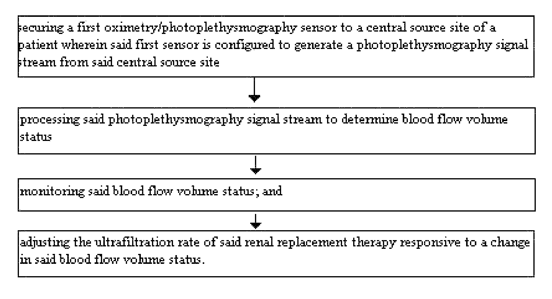
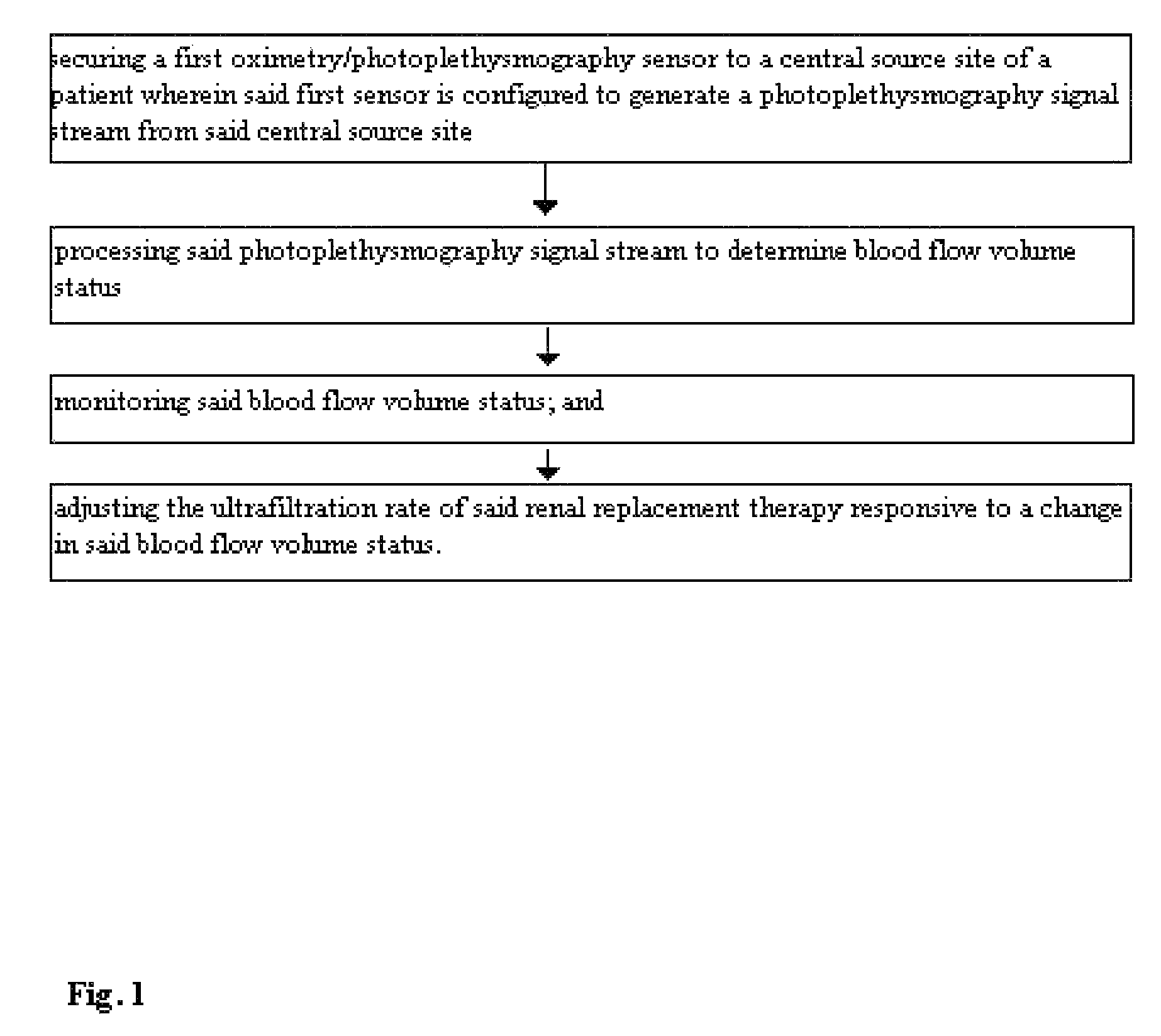
[0065]FIG. 1 demonstrates a method for optimizing fluid removal in a patient undergoing RRT. A central pulse oximetry / photoplethysmography sensor is placed on the cheek, nasal septum, alar nares, or tongue. An optional peripheral pulse oximetry / photoplethysmography sensor may be placed on the patient's finger or toe. Before and during dialysis, a number of derived parameters are monitored. At the start of dialysis, the amplitude and area under the curve (AUC) of the fast component of the photoplethysmograph are calculated from one or more monitoring sites and a ratio of the amplitudes and / or AUC is calculated. This represents the relative blood flow to the head (brain) and the optional peripheral site. As fluid is removed during RRT, this ratio may change (i.e. greater flow to the head than finger) or since many dialysis patients lose the normal reflexive responses to fluid depletion, there may be an equal or greater loss in amplitude and the AUC from the probes monitoring the head....

example 2

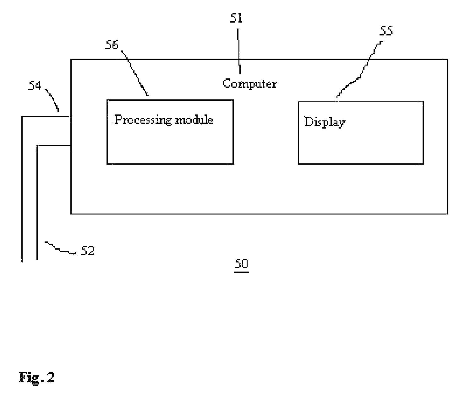
[0067]In FIG. 2, there is shown a system 50 for obtaining and processing photoplethysmography data from a patient for purposes optimizing fluid removal during RRT to avert hypotension. The system comprises a computer 51 that is configured to receive and process signals from lines 52 and 54, which are distally connected to one or more pulse oximeter probes (not shown) located on the patient. Those skilled in art will appreciate that the signals may be preprocessed to some degree by a separate signal processor and subsequently sent as one signal stream to the computer 51. Thus, the computer 51 is configured to receive signals from either lines 52 or 54 or a combination of both. Typically, one of the lines will carry power from the computer 51 to the pulse oximeter probe, while the other line carries signals back to the computer 51. The computer 51 comprises a processing module 56 with program code module(s) and / or electrical / circuitry components associated therewith to direct the proc...

example 3

[0070]FIG. 4 demonstrates a method for optimizing fluid removal during RRT by use of photoplethysmography to monitor a patient's blood flow and / or volume status. The method comprises determining the arterial component signal and / or the venous capacitance component signal in said patient utilizing photoplethysmography; and adjusting the ultrafiltration rate so as to modulate the arterial component signal and / or the venous capacitance component signal to maintain a patient's blood flow and / or volume status at a target level.


PUM

PropertyMeasurementUnit
wavelength rangeaaaaaaaaaa
frequencyaaaaaaaaaa

Abstract

Disclosed herein are methods, systems and devices to monitor vascular volume status during renal replacement therapy utilizing at least one oximetry / photoplethysmography sensor. The methods, systems and devices provide an alternative to conventional vascular volume monitoring methods during renal replacement therapy while enabling reliable, non-invasive, and automatic monitoring of vascular volume to avert patient hypotension. The methods, systems and devices may be employed in the context of both inpatient and outpatient dialysis facilities and may also be incorporated into conventional hemodialysis and hemofiltration techniques and equipment.

Description

BACKGROUND[0001]1. Field of the Invention[0002]This invention relates to non-invasive methods for monitoring a patient's vascular volume during renal replacement therapy by means of at least one pulse oximetry / photoplethysmography sensors. Further, this invention relates to monitoring a patient's vascular volume, vascular tone, and / or regional blood flow (e.g. carotid blood flow as a surrogate for cerebral blood flow) during renal replacement therapy using photoplethysmography methods and adjusting the rate of ultrafiltration real-time during the patient's treatment.[0003]2. Discussion of the Art[0004]Renal Replacement Therapy (RRT) has evolved from the long, slow hemodialysis treatment regime of the 1960's to a diverse set of therapy options, the vast majority of which employ high permeability membrane devices and ultrafiltration control systems.[0005]Biologic kidneys remove metabolic waste products, other toxins, and excess water. They also maintain electrolyte balance and produce...

Claims


Application Information

Patent Timeline
Patent Type & Authority Patents(United States)
IPC IPC(8): A61M37/00
CPCA61B5/02007A61B5/0261A61B5/0295A61B5/6819A61M1/34A61M1/3609A61M2205/3306A61M2205/50A61M1/3403A61M1/341
Inventor ROSS, EDWARD ALLANMELKER, RICHARD J.
Owner UNIV OF FLORIDA RES FOUNDATION INC
Why Eureka
Try Eureka