
Patterned gradient wound dressing and methods of using same to promote wound healing

a gradient wound dressing and gradient technology, applied in the field of wound healing, can solve the problems of increasing the cost of reapplication of growth factors, reducing the reducing the available amount of topically applied basic fibroblast growth factors in solution, etc., to achieve the effect of improving the promotion of wound healing

Active Publication Date: 2011-05-03
WISCONSIN ALUMNI RES FOUND
View PDF4 Cites 2 Cited by
  • Summary
  • Abstract
  • Description
  • Claims
  • Application Information

AI Technical Summary

Benefits of technology

The present invention is about a new type of wound dressing that promotes directed cell migration during dermal wound healing. This dressing has a support substrate with a growth factor gradient immobilized on it. The growth factor gradient is made up of a series of increasing concentrations of growth factor that promotes cell migration. The dressing can be made with a variety of growth factors, including but not limited to epidermal growth factor, insulin-like growth factor, basic fibroblast growth factor, platelet-derived growth factor, vascular endothelial growth factor, keratinocyte growth factor, transforming growth factor beta, and mixtures thereof. The support substrate can be made of various polymeric materials or natural products. The growth factor concentration across the gradient can be controlled using a power equation. The patterned gradient wound dressing can be used to treat acute and chronic dermal wounds. The invention also provides a method for screening for agents that promote directed cell migration.

Problems solved by technology

In fact, the available amount of topically applied basic fibroblast growth factor in solution decreases by 50% within 4 hours of contact with sterile gauze,21 and reapplication of growth factors is often cost prohibitive.
Furthermore, growth factors in the physiological environment can be rapidly degraded or otherwise rendered inactive before reaching their target.
Covalent tethering of growth factors to biomaterials has the potential to ameliorate many of these problems, and possibly result in increased availability of active growth migration signals, coupled with precise control over cell migration direction.
Acceleration of wound closure not only results in decreased patient suffering and cost of wound treatment but may also minimize scarring and lead to formation of a more stable closed wound.9,34 A recent review of the use of growth factors in wound dressings concluded that the clinical outcomes of these materials have been “generally disappointing.”17 It is widely accepted that these lackluster results have been directly attributable to the manner in which growth factors were incorporated into the dressings, namely that the delivery methods did not allow for sustained growth factor availability or bioactivity.
Another major obstacle in implementing growth factor-based therapies has been the expense of the biomolecules, meaning that a system that improves healing responses using a reduced amount of growth factor could make growth factor based wound healing therapies more widely available.

Method used


Image

  • Patterned gradient wound dressing and methods of using same to promote wound healing
  • Patterned gradient wound dressing and methods of using same to promote wound healing
  • Patterned gradient wound dressing and methods of using same to promote wound healing

Examples


example 1

Materials and Methods

[0081]All chemicals were obtained from Sigma-Aldrich (St. Louis, Mo.) unless otherwise noted.

[0082]Cell culture. Immortalized human keratinocytes (HaCaTs, courtesy of N. Fusenig, DKFZ, Heidelberg, Germany) were cultured and maintained in Dulbecco's modification of eagle's medium (DMEM), 10% fetal bovine serum (FBS), 1% glutamine, and 1% penicillin / streptomycin at 37° C., 5% CO2.

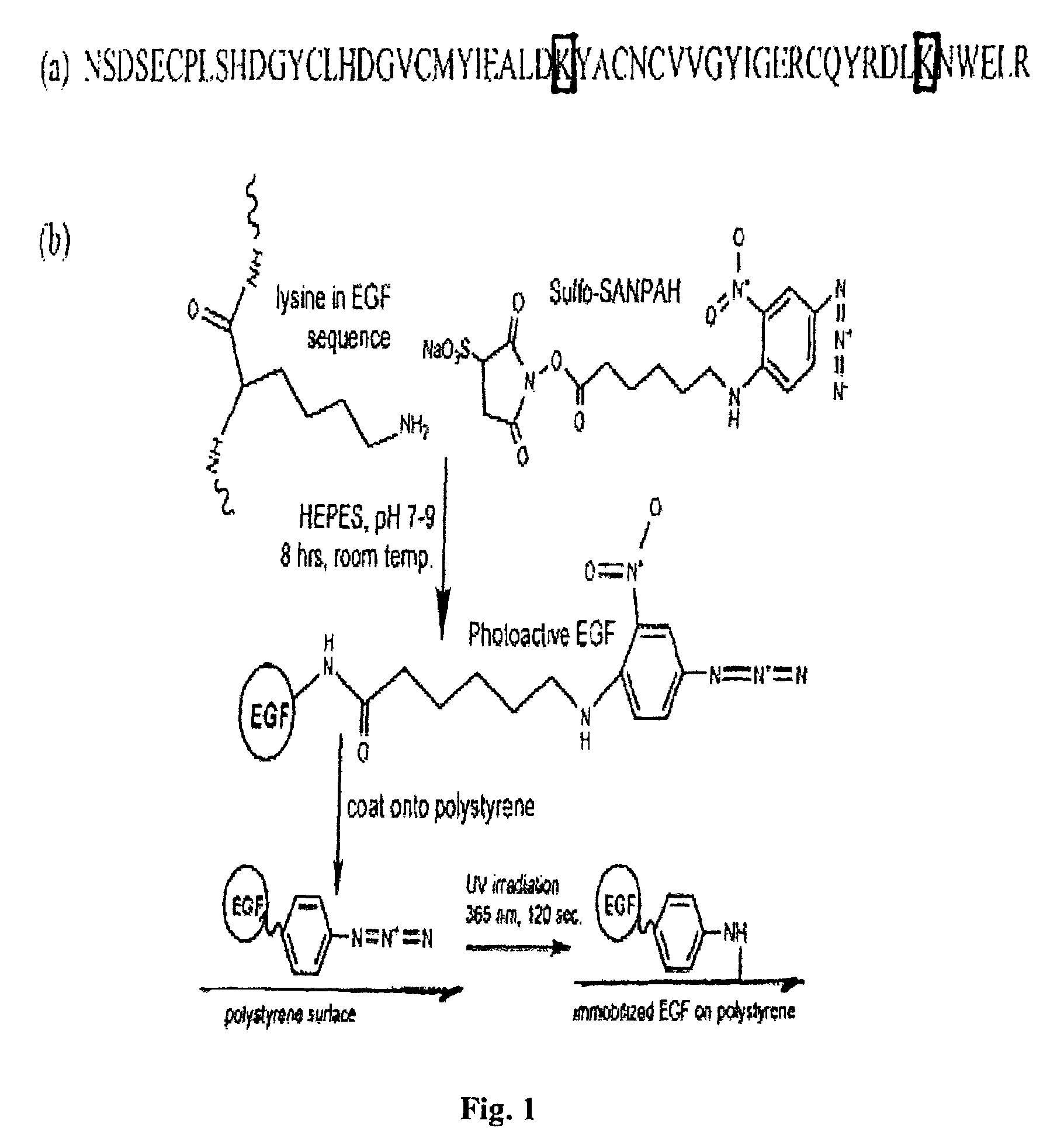
[0083]EGF modification. Recombinant human EGF (Peprotech Inc., Rocky Hill, N.J.) having the amino acid sequence of SEQ ID NO:1 (see FIG. 1) was rendered photoactive via conjugation to Sulfo-SANPAH (sulfosuccinimidyl-6-[40-azido-20-nitrophenylamino]hexanoate; Pierce Biotechnology Inc., Rockford, Ill.). Sulfo-SANPAH (SS) is a heterobifunctional crosslinker containing a photosensitive phenyl azide group on one end and an amine-reactive N-hydroxysuccinimide on the other (FIG. 1). Photoreactive EGF was synthesized via the reaction of primary amine groups of EGF with the N-hydroxysuccinimide fu...

example 2

EGF Modification

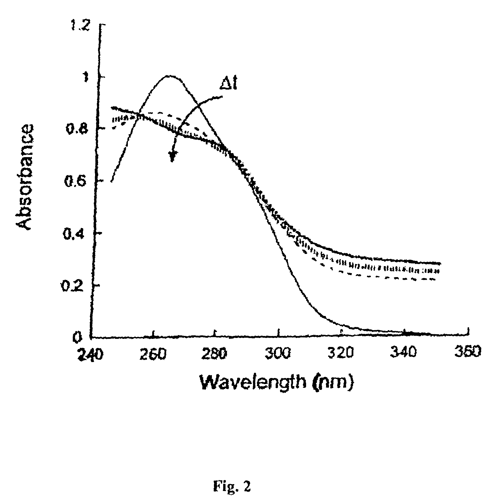
[0092]Spectroscopy was used to confirm conjugation of SS to EGF, as well as determine the optimal UV exposure time for complete photolysis (i.e., tethering) of the SS. Conjugation of SS to EGF was confirmed by an absorption spectrum displaying peaks at 262 nm (corresponding to the photoactive phenyl azide of SS) and 280 nm (corresponding to EGF). Owing to the breadth of the 262 nm peak and the coupling of multiple SS groups to a single EGF molecule, the EGF peak appears as a shoulder, rather than as an isolated peak (FIG. 2). To optimize the UV exposure time needed for photolysis of the phenyl azide (and hence covalent tethering to our surface), the disappearance of the 262 nm peak was measured as a solution of SS-EGF was exposed to UV light for varying amounts of time. The loss of the phenyl azide peak of SS upon UV exposure is a standard measure of phenyl azide photolysis.30,31 Within 1 minute of UV exposure, the peak dropped dramatically, indicating partial photol...

example 3

Gradient Characterization

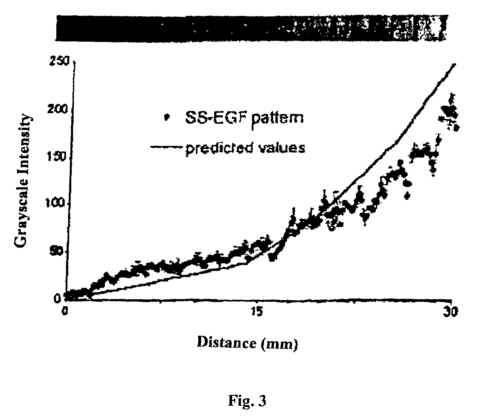
[0093]Photo-immobilization of fluorescently labeled SS-EGF to polystyrene plates via the phenylazide functionality of SS was successfully completed in the desired gradient pattern (FIG. 3). The actual slope of the EGF gradient (as measured by image quantification) correlated well with the predicted gradient slope (i.e., the gradient slope of the photomask). Immobilized EGF increased steadily along the length of the gradient path (i.e., along the x-axis), while remaining homogeneous across the width of the path (i.e., along the y-axis), as desired (FIG. 3).

[0094]An ELISA for hEGF yielded a quantitative description of the total amount of EGF tethered across each gradient pattern. Combining this information with gradient intensity curves, the concentration of EGF at any point along the gradient can be calculated. FIG. 4 illustrates the experimentally calculated EGF concentration at a few example points along the gradient paths used in the cell migration experim...


PUM

PropertyMeasurementUnit
pHaaaaaaaaaa
wavelengthaaaaaaaaaa
concentrationaaaaaaaaaa

Abstract

The present invention provides a wound dressing that includes a patterned gradient of immobilized growth factor molecules that promote directed migration of cells during dermal wound healing. Growth factor is immobilized on a support substrate to present a gradient pattern of increasing growth factor concentration to migrating cells. Methods of promoting wound healing using the patterned gradient wound dressing and fabrication methods of same are also provided.

Description

STATEMENT REGARDING FEDERALLY SPONSORED RESEARCH[0001]This invention was made with government support under Grant no. EB005440 awarded by the National Institutes of Health. The government has certain rights in the invention.CROSS-REFERENCE TO RELATED APPLICATIONS[0002]Not applicable.FIELD OF THE INVENTION[0003]The present invention relates to the field of wound healing. More particularly, the present invention relates to patterned gradient wound dressings and their use in the promotion of wound healing.BACKGROUND OF THE INVENTION[0004]Approximately 5-7 million Americans are afflicted with chronic skin wounds that account for billions of dollars in medical expenses each year.1,2 The incidence of chronic wounds is expected to increase dramatically3 due to an increased elderly population and incidence of diabetes, a disease that is accompanied by wound-healing deficiencies.4 In the United States, at least 82,000 lower-limb amputations are performed annually due to diabetic ulcers.5 [00...

Claims


Application Information

Patent Timeline
Patent Type & Authority Patents(United States)
IPC IPC(8): A61F13/00A61F15/00A61L15/16
CPCA61F13/00012G01N33/5029A61F13/00063A61L27/54A61L27/60G01N33/5008A61F13/00017A61F13/069A61F2013/00157A61F2013/00229A61F2013/00519A61F2013/00523A61F2013/0054A61F2013/00927A61L2300/414A61L2400/18A61P17/02A61F13/01017A61F13/01012
Inventor MASTERS, KRISTYN SIMCHASTEFONEK, TRACY JANE
Owner WISCONSIN ALUMNI RES FOUND
Learn More