Methods and devices for active bioassay

a bioassay and active technology, applied in the field of active bioassay methods, can solve the problems of limited assay time and inability to solve the problem completely, and achieve the effect of reducing background and increasing bioassay sensitivity

Inactive Publication Date: 2011-06-14
GEORGE MASON INTPROP INC
View PDF23 Cites 15 Cited by
  • Summary
  • Abstract
  • Description
  • Claims
  • Application Information

AI Technical Summary

Benefits of technology

[0019]The methods of the present invention have several advantages over standard assays controlled by diffusion: (i) the methods can be performed in seconds and minutes, rather than in hours, (ii) the methods provide reduction in background using active washing as a means to discriminate between specifically and non-specifically bound ligands, and (iii) the methods increase bioassay sensitivity by concentrating analyte molecules in the vicinity of probe molecules. It is also illustrated herein, that the active assay methods and procedures of the present invention are especially advantageous when combined with microarrays of probe molecules. This combination allows multiple assays to be performed in a short period of time.

Problems solved by technology

In most practical cases of heterogeneous analysis assay time is limited by the diffusion of analyte molecules to the surface with immobilized probes.
However, these approaches cannot solve the problem completely: as seen from the Eqn.
Though blocking the surface with proteins (BSA, dry milk and casein in immunoassay) and grafting inert polymers to surface (Chapman et al., 2000) considerably reduce the background, they never remove it completely.

Method used


Image

  • Methods and devices for active bioassay
  • Methods and devices for active bioassay
  • Methods and devices for active bioassay

Examples


example 1

Electrophoretic Acceleration of the Reaction in the Standard Elisa and Suppression of Convection

[0217]This example illustrates that electrophoretically enhanced ELISA increases the rate of signal formation and provides increased signal levels for very low concentrations of probe.

[0218]Electrophoretic Acceleration of the Reaction in the Standard ELISA.

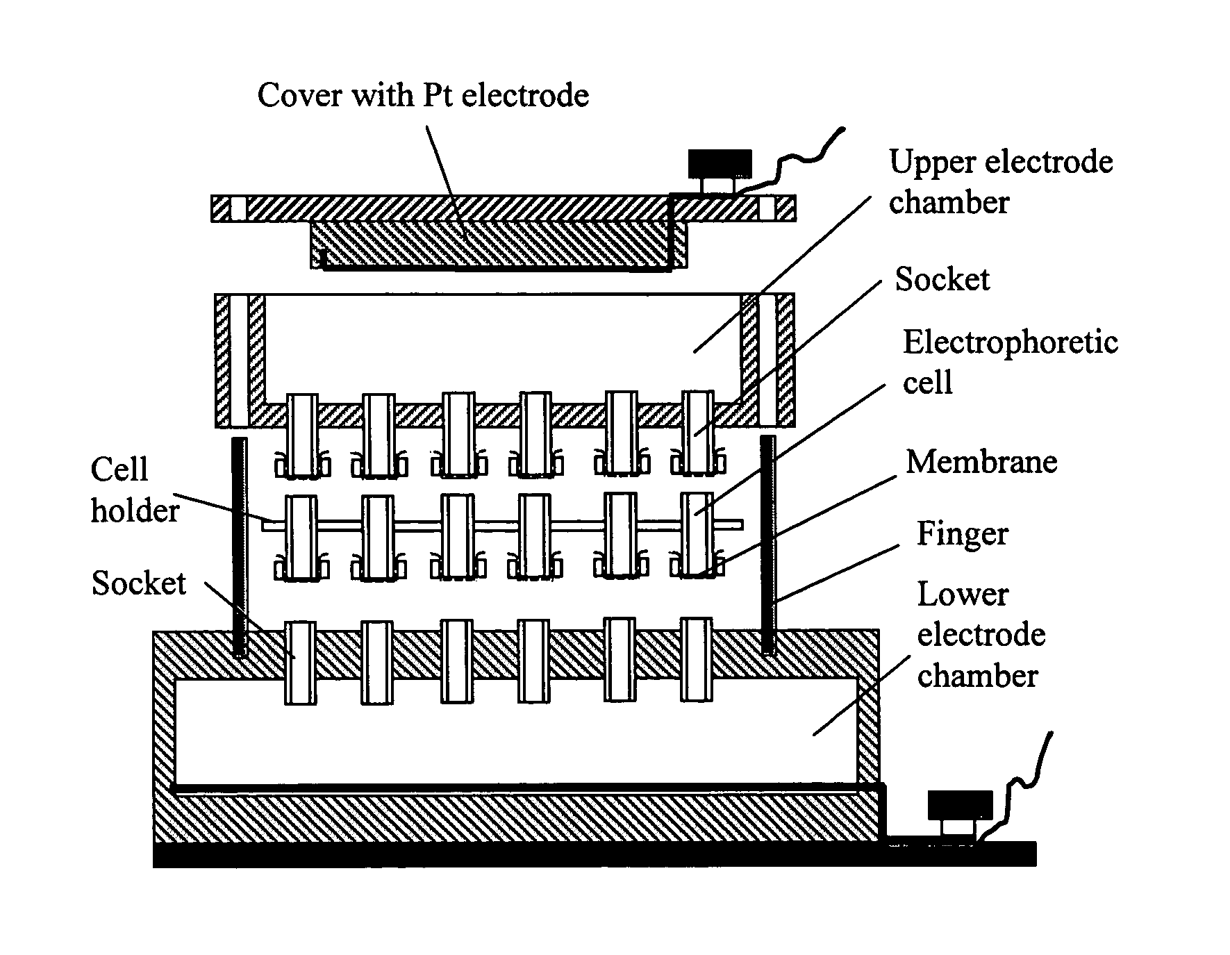
[0219]A simple device was designed as illustrated schematically in FIG. 2. It includes upper and lower electrode chambers and electrophoretic cells. The lower chamber has an array of open outlets electrically connecting electrophoretic cells with the lower chamber. The upper electrode chamber has also array of outlets. These outlets are covered with a dialysis membrane to prevent leakage of buffer and to allow electric current to pass. Different types of the electrophoretic cells were tested with this basic device. Some of them were made of standard 0.7 mL Eppendorf tubes with their bottom parts and the cover caps cut off. Dialysis film...

example 2

Device for Performing Electrophoretically Assisted ELISA

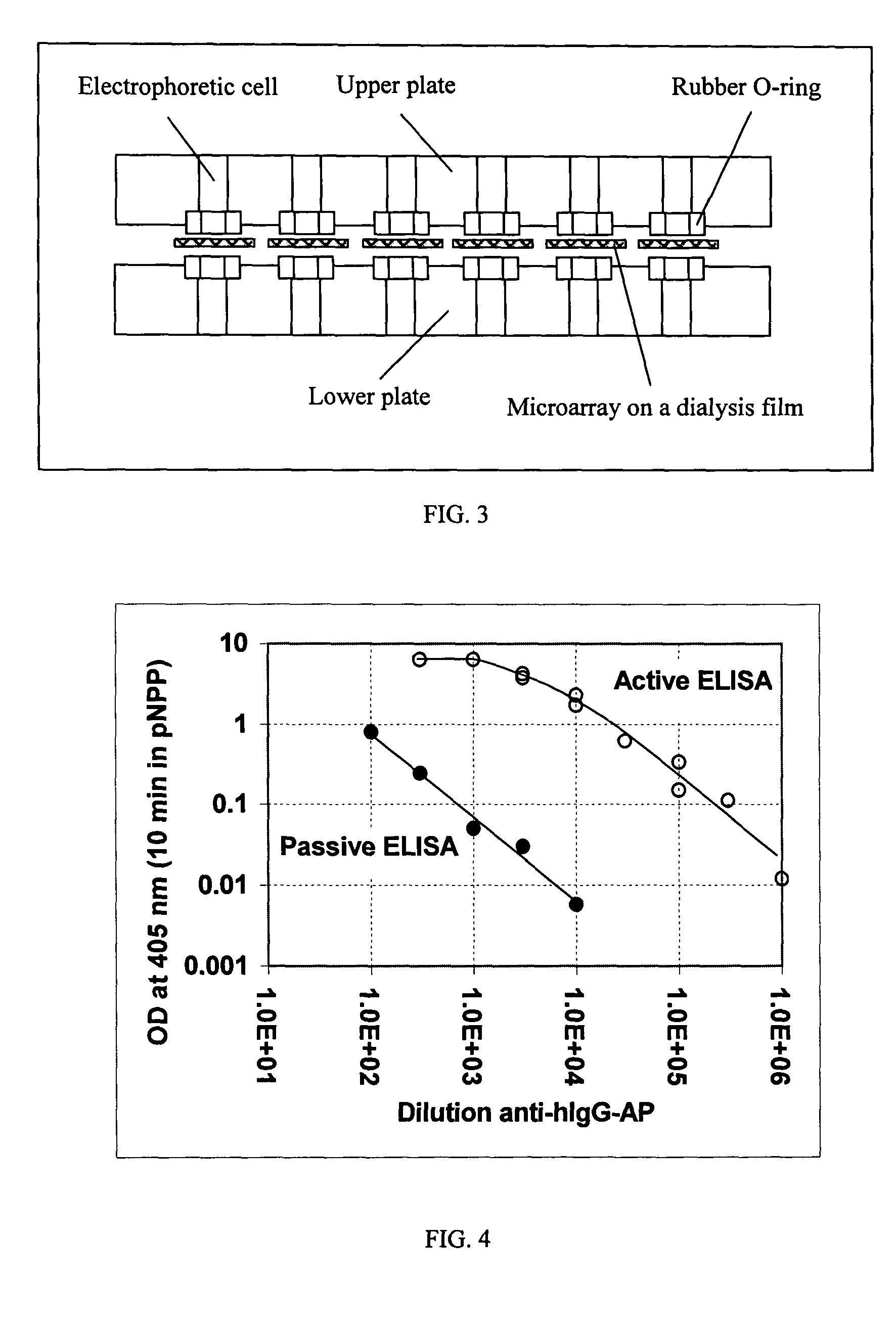
[0238]This example illustrates a device that can be used to perform an electrophoretically-assisted ELISA assay. An apparatus schematically presented in FIG. 8 allows to simultaneously run 96 independent active assays. Electrophoretic micro-plate was manufactured from a commercial CellScreen plate (Millipore) in which microfilter membranes was removed with a sand paper and replaced with dialysis membranes from regenerated cellulose (Sigma product). The plate was treated in plasma discharge for 50 sec. Dialysis membrane was also treated in plasma discharge for 30-50 sec. Cyanoacrylate glue was evenly distributed over the edges of wells, the membrane was firmly pressed to the wells and allowed to settle for 3-5 min. To prevent deposition of cyanoacrylate vapor onto the membrane, a flow of air was directed into each well via array of sockets prepared from standard 0.2 mL tips. The glued membrane was cut between the wells to reduce...

example 3

Enhanced Sensitivity in the Electrophoretically Assisted ELISA as Compared to ELISA in Commercial Microplates

[0240]This example illustrates that electrophoretically-assisted ELISA provides increased sensitivity. In this example electrophoretically-assisted ELISA was performed using the apparatus described in Example 2 and protocol described in the Example 1. FIG. 9 presents a comparison of the active assay with the standard assay on a commercial polystyrene microplate. The data presented in FIG. 9 clearly demonstrate that active ELISA is substantially more sensitive as compared with the standard ELISA performed under the most optimal conditions (long time of coating and reaction, optimal pH and salt composition in the reaction buffer).


PUM

PropertyMeasurementUnit
angleaaaaaaaaaa
Dissociation constantsaaaaaaaaaa

Abstract

The present invention provides an active assay method for detecting a biological analyte. According to the method, a probe molecule is immobilized on a surface. An analyte is then placed in fluidic connection with the probe molecule on the surface. A force is then applied to the analyte to move it toward the surface to facilitate contact and possibly binding of the analyte to the probe. Optionally, another force can be applied or the force can be reversed, to remove unbound or weakly bound analyte from the surface. Analyte that remains bound to the surface is then detected. The detection can include rolling or sliding beads over an analyte and/or probe on a substrate, and detecting bound beads. The present invention furthermore, provides devices, such as electrophoresis apparatuses and biochip assemblies, for carrying out the methods of the invention.

Description

RELATED APPLICATION DATA[0001]This application claims the benefit of priority under 35 U.S.C. §119(e) of U.S. Ser. No. 60 / 412,664, filed Sep. 20, 2002, the entire content of which is incorporated herein by reference.BACKGROUND OF THE INVENTION[0002]1. Field of the Invention[0003]The invention relates generally to assay methods for detection of biological molecules, viruses, and cells, and more specifically to solid-phase assay methods for detection of biological molecules, viruses, and cells in solution, based on active motion of analytes to and from probe molecules under action of electrostatic, magnetic, centrifugal (gravitation) or hydrodynamic forces.[0004]2. Background Information[0005]There are several principal limits in conventional bioassay methods: (i) diffusion limit for transport of analyte to immobilized probes, (ii) limits for the assay sensitivity due to limited affinity of probe molecules and, (iii) limits for the assay sensitivity due to unspecific binding of analyt...

Claims


Application Information

Patent Timeline
Patent Type & Authority Patents(United States)
IPC IPC(8): G01N27/00G01N33/00G01N33/48G01N33/50G01N33/543G01N33/553C12Q1/68G01NG01N33/53G01N33/561
CPCG01N33/561G01N33/54333
Inventor MOROZOV, VICTORMOROZOVA, TAMARA
Owner GEORGE MASON INTPROP INC
Why Eureka
Try Eureka