Device for the picture-providing diagnosis of tissue using one of at least two diagnosis modes

a tissue diagnosis and tissue technology, applied in the field of tissue diagnosis devices, can solve the problems of insufficient exploitation of the given long time frame, small sensitivity of light endoscopy, and insufficient time frame for therapeutic purposes, so as to improve differentiation, improve sensitivity, and improve colour contrast

Inactive Publication Date: 2011-09-13
RICHARD WOLF GMBH
View PDF23 Cites 11 Cited by
  • Summary
  • Abstract
  • Description
  • Claims
  • Application Information

AI Technical Summary

Benefits of technology

[0028]It is therefore an object of the invention to provide a diagnosis device for the picture-providing diagnosis of tissue—in particular for the examination of the bronchial region of a patient—with the selective application of the picture-providing white light diagnosis and the picture-providing autofluorescence diagnosis. The mode of the picture-providing autofluorescence diagnosis has an increased sensitivity, i.e., an improved colour contrasting and thus an improved differentiation between healthy and pre-malignant or early-malignant tissue. In the mode of the autofluorescence diagnosis only, the assessment of the autofluorescence light itself is used which permits an increased specifity, i.e., an improved differentiation possibility of pre-malignant and early-malignant tissue above all also with respect to healthy tissue having an uneven structure and therefore light-swallowing surface which leads relatively little fluorescence light to the receiver. At the same time additionally the colour impression of the tissue arising at the observer in the autofluorescence mode is to be largely independent of the observation angle, i.e. of the angle of the endoscope with respect to the surface of the tissue. Furthermore it is to be possible to be able to fall back on existing diagnosis devices without having to make significant modifications. This means above all things that for both working modes as a detection unit there may be applied a 3-chip camera which is conventional with regard to the optical specifications of the camera head. The retention of the optical specifications means that in the working mode of the white light diagnosis the picture does not undergo any compromise and retains its usual good colour quality. Furthermore the system is to be constructed such that a simple change between the picture-providing white light diagnosis and the picture-providing autofluorescence diagnosis may be realized.
[0033]Simultaneously however in a second step in the diagnosis mode of the picture-providing autofluorescence, the red amplification of the camera must be reduced or the red signal damped—according to the intensity of the additional red light or according to the ratio of the fluorescence excitation light (in the blue / violet) to the additional red light. The high intensity of the additional red light, which is selected sufficiently high so that the red fluorescence with respect to the red remission at the tissue becomes negligible or almost negligible, and the accordingly strong red remission at the tissue would otherwise—without reduction of the red amplification or without damping of the red signal in the camera—dominate the green fluorescence of diseased tissue and the healthy tissue such that the tissue would in each case appear indeed red or at least reddish. This would worsen the differentiation on account of the transmitted colour impression.
[0035]This inventive procedure significantly increases the sensitivity for detection of pre-malignant or early-malignant tissue compared to when only fluorescence light and no red light additionally emitted from the illuminating and fluorescence exciting light source and remitted at the tissue is detected.
[0036]The red part share detected by the camera is now no longer or at least minimally dependent on the malignancy degree of the tissue and therefore no longer or is only minimally reduced by an increasing degree of malignancy, diseased tissue. This produces a more clearer red shifting and, with a suitable setting of the camera damping, irradiates more strongly (since the camera damping ideally is set such that the constant red part share remitted by the tissue and constant with regard to tissue atypia produces a clearly stronger signal than the reduced red fluorescence with diseased tissue). Accordingly, the diseased tissue appears in a stronger red and the colour of the diseased tissue may be perceived brighter and thus better than when exclusively fluorescence light is observed.

Problems solved by technology

Unfortunately this time frame until now for therapeutic purposes could only be insufficiently exploited which is reflected is the small five-year survival rate with patients with a bronchial carcinoma which has remained unchanged for decades.
The reason why these exploitation possibilities in the given long time frame have not been sufficient up to now lies in the fact that the sensitivity of the conventional diagnosis forms—including white light endoscopy—is very small for pre-malignant and early-malignant lesions.
With the conventional white light diagnosis, it is difficult to differentiate premalignant and early-malignant tissue from benign tissue which is why it may remain undetected.
The white light diagnosis for this has an insufficient sensitivity.
From this there results problems which may be comparable with those which have already been described above.
The difficulties in assessing such tissue renders obligatory the taking of a sample, in order to detect where possible all potential early-malignant or malignant sources.
However, the sample taking incurs additional time and costs.
Moreover, additional biopsies necessitated by the samples likewise create additional costs.
Furthermore each taking of a sample may lead to bleeding which inhibits further examination.
For including an evaluation of colour shifting into the tissue differentiation under the given circumstances it is a disadvantage that with an increasing degree of malignancy or with an increasing atypia of the tissue not only does the fluorescence intensity in the green spectral region decline but also the fluorescence intensity in the red spectral region—even if less marked (see FIG. 1).
However, a detection unit having two cameras must be applied, in front of whose sensors there are mounted optical band-width filters which represent specially manufactured products in that they do not correspond to the filter specifications of conventional 3-chip cameras suitable for white light endoscopy.
A disadvantage of U.S. Pat. No. 5,590,660 is that the cameras to be used or their sensors may not be part of a conventional 3-chip camera since the first detection region comprises the wavelength region of the whole autofluorescence light which consists of green light but also of red light, whilst the second detection region does not intersect with this first region and accordingly may only lie in the long-wave red spectral region.
The device of U.S. Pat. No. 5,590,660 is necessarily complicated, awkward to use, and expensive.
The fact that the wavelength region led to the first camera is so wide or reaches so far into the red spectral region is disadvantageous in that tissue differentiation between the fluorescence curves for the different tissue conditions towards the longwave region, in particular where the yellow and red spectral region approach one another, becomes more and more washed.
Although an increased sensitivity for pre-malignant or early-malignant lesions is achieved, there arise the above mentioned disadvantages entailed when one makes do without the red reference such as the reduced specificity by way of an increased false-positive rate.
Furthermore making do without the detection of red light (fluorescence as well as red light remitted at the tissue) means a loss of brightness.
Thus the system therefore also uses large, heavy and awkward image intensifiers in order to achieve a sufficiently bright picture.
This method is disadvantageous because, in contrast to red light, blue light has a lower tissue penetration depth.
The tissue assessment is thus dependent on the incidence angle of the illumination and excitation light which of course is not at all desirable.

Method used


Image

  • Device for the picture-providing diagnosis of tissue using one of at least two diagnosis modes
  • Device for the picture-providing diagnosis of tissue using one of at least two diagnosis modes
  • Device for the picture-providing diagnosis of tissue using one of at least two diagnosis modes

Examples


Embodiment Construction

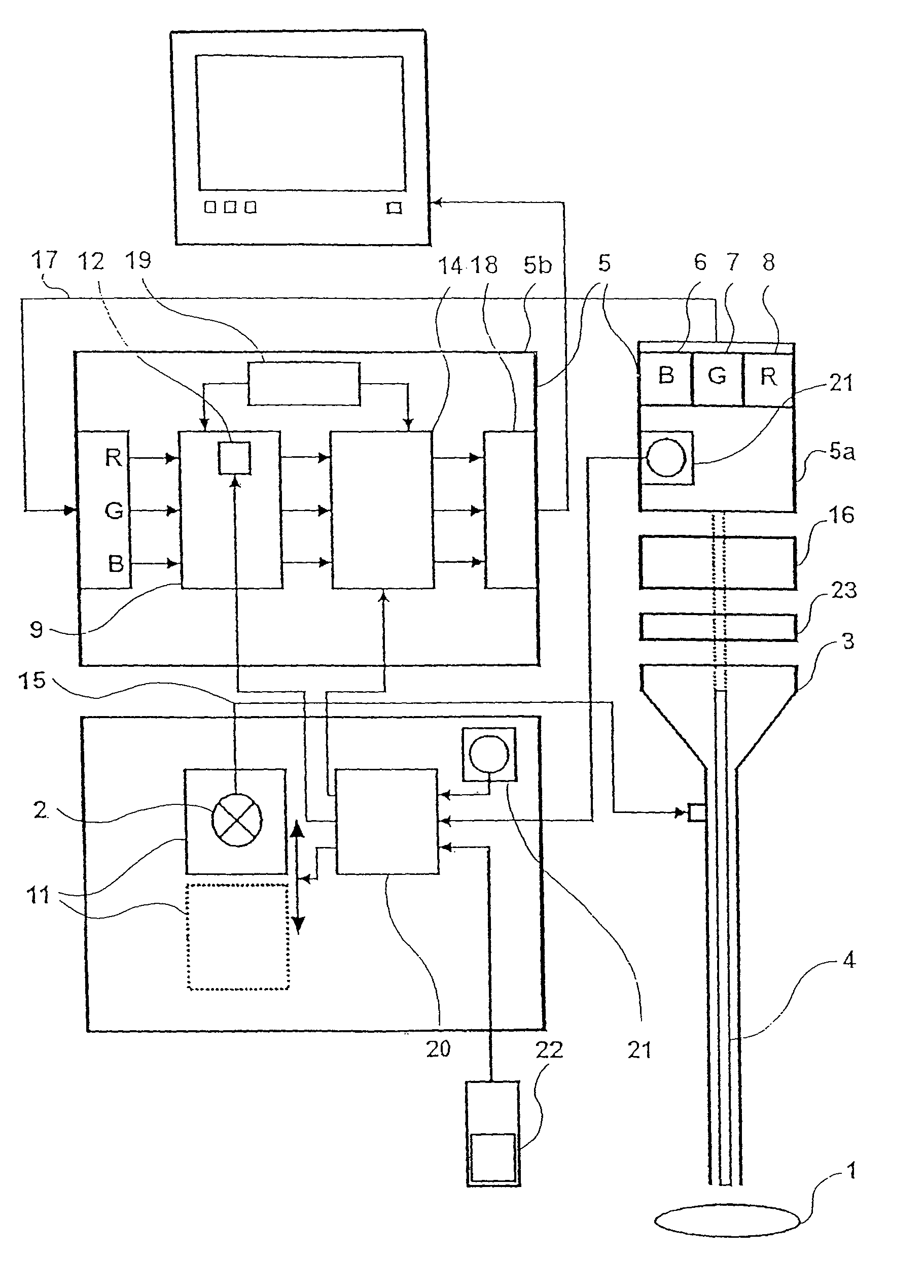
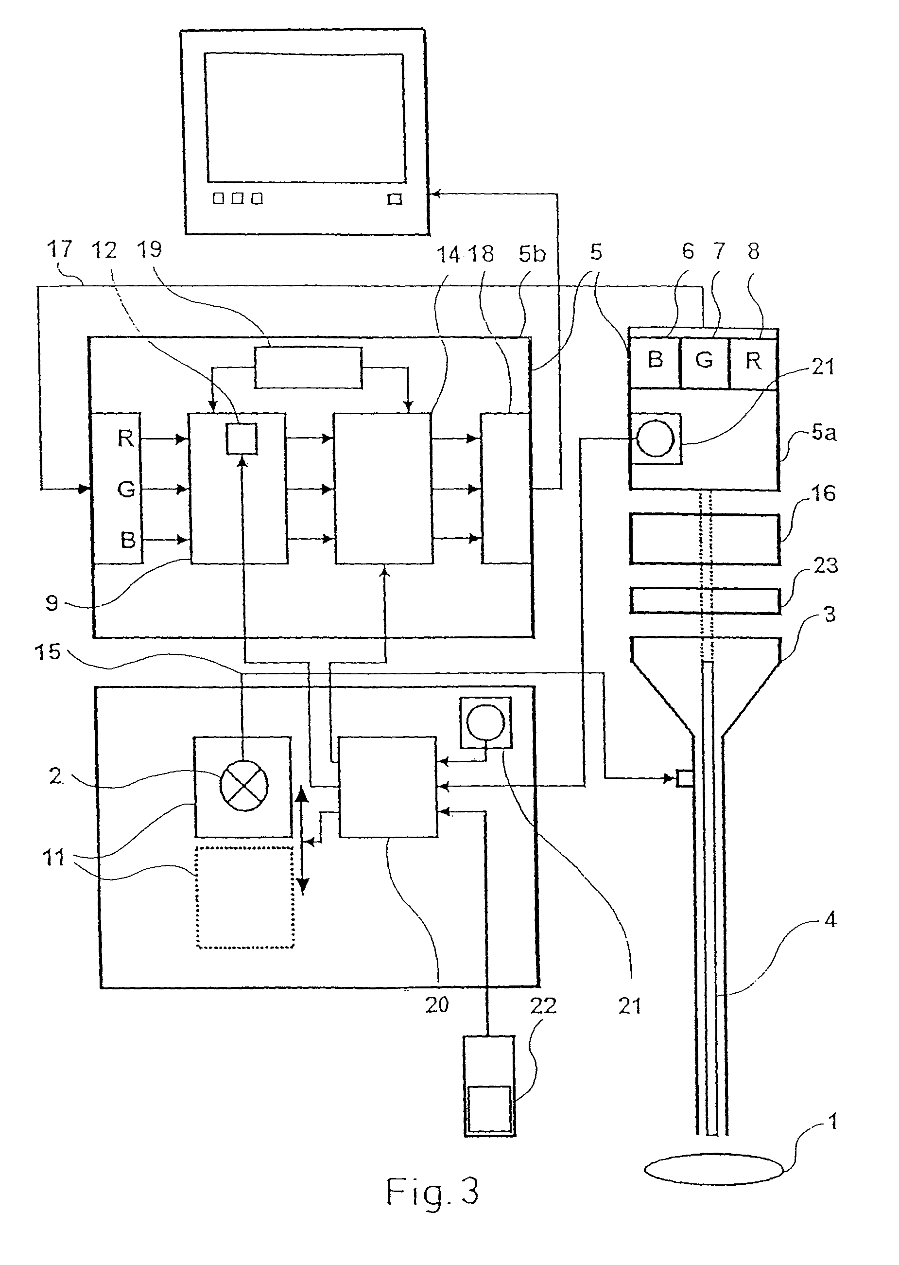
[0047]FIG. 3 shows a diagnosis device for the combined picture-providing white light diagnosis and picture-providing autofluorescence diagnosis of tissue 1. The device for examining the tissue 1 comprises a light source 2, wherein an incoherent, broad-band light source emitting in the visible spectral region is particularly suitable. The light source 2 emitting preferably white light may be a short arc lamp such as, for example, a xenon lamp a gas discharge lamp with mercury part shares or a mercury high pressure or even mercury highest pressure lamp. The latter have a broad irradiation base continuum in the visible and the line typical for mercury in the blue and violet. These partly ideally lie in the absorption spectrum for fluorescence excitation of the body-intrinsic fluorochrome relevant to the autofluorescence diagnosis. Alternatively a halogen lamp may be used.

[0048]All mentioned lamps have the advantage that on the one hand they have an irradiation part share in the violet / ...


PUM

PropertyMeasurementUnit
excitation wavelengthaaaaaaaaaa
wavelengthsaaaaaaaaaa

Abstract

A device for picture-providing diagnosis of tissue is selectively operated in a picture-providing white light diagnosis mode and a picture providing auto-fluorescence mode. A color camera having red, green, and blue sensors provides a monitor which picture signals. A light source emits fluorescence excitation light and additionally emits so much red light that the red light remitted by the tissue dominates the red fluorescence light in the picture providing auto-fluorescence mode. The signal from the red sensor of the camera is damped so that normal time appears green and pre-malignant and early malignant tissue appears red.

Description

BACKGROUND OF THE INVENTION[0001]1. Field of the Invention[0002]The invention relates to a device for the picture-providing diagnosis of tissue with the selective application of a picture-providing white light diagnosis mode and a picture-providing auto-fluorescence mode, the device having a light source whose light is led to the tissue by an endoscope, and a picture transmission unit arranged in the endoscope which transmits the picture of the tissue to a colour camera, wherein the colour camera comprises three sensors for the three colour regions red, green and blue which in each case are in connection with a picture processor unit which provides a monitor with the picture signals.[0003]2. Description of the Prior Art[0004]The best chance of healing cancerous lesions is to treat the lesions in the pre-malignant and early-malignant stages of the diseased lesions. The prospects for success with a curative treatment decreases when the lesion reaches the invasive carcinoma stage. Ther...

Claims


Application Information

Patent Timeline
Patent Type & Authority Patents(United States)
IPC IPC(8): A61B5/05A61B1/00A61B1/04G01N21/64A61B1/267A61B1/273A61B5/00
CPCA61B1/043A61B1/0638A61B5/0071A61B5/0075A61B5/0084
Inventor WEBER, BERND CLAUSGOLL, THOMASSCHMIDT, OLAFEIDNER, PHILIPPMULLER, STEFANVAN DEN BERGH, HUBERTGOUJON, DIDIERWAGNIERES, GEORGES
Owner RICHARD WOLF GMBH
Why Eureka
Try Eureka