
Method for the ablation of cartilage tissue in a knee joint using indocyanine

a knee joint and cartilage tissue technology, applied in the field of knee joint cartilage ablation using indocyanine, can solve the problems of high cost of excimer laser, large amount of damaged cartilage, and inability of ordinary skill in the art to perform cartilage ablation in vivo

Active Publication Date: 2011-10-11
RAVUSSIN PIERRE
View PDF18 Cites 1 Cited by
  • Summary
  • Abstract
  • Description
  • Claims
  • Application Information

AI Technical Summary

Benefits of technology

"The present invention provides a method for ablating human or animal cartilage by applying an exogenous absorber to a portion of the surface of the cartilage and then using a laser diode to direct electromagnetic energy at a narrow spectrum to the absorber. This results in the ablation of at least a portion of the cartilage. The invention also includes a system for ablating cartilage using a laser diode and an optical conduit to transmit the laser beam to the cartilage surface. The exogenous absorber can be applied as a dye or a solution containing a compound that selectively absorbs the electromagnetic energy. The invention has the advantage of minimizing damage to the surrounding tissue and can be used in the treatment of joint disorders such as arthritis."

Problems solved by technology

However, these mechanical cutting tools may damage a significant amount of healthy cartilage while removing damaged or diseased tissue.
However, Prodoehl's article does not enable one of ordinary skill in the art to perform ablation of cartilage in vivo.
Furthermore, the Excimer laser is expensive and relatively bulky so it is not suitable for use in a hospital or outpatient surgery clinical environment.
While cheaper lasers that are relatively compact and light are known, such as laser diodes for example, these lasers are not suitable for arthroscopic surgical ablation of cartilage because their wavelength energies are poorly absorbed by cartilage.
However, there are several drawbacks to the application of infrared laser technology to arthroscopic surgery.
Unfortunately, the aqueous liquid must be pierced by the infrared laser beam in order to reach the cartilage.
This means that the infrared laser beam must have considerable energy, which then generates undesirable thermal effects.
These undesirable thermal effects increase as the power of the laser increases, which makes quick ablation impossible without damaging a significant amount of healthy joint tissue.
A second drawback of infrared lasers relates to the difficulties encountered when selecting a laser that operates at a wavelength not absorbed well by the aqueous environment.
It turns out that a laser that is not absorbed well by the aqueous environment utilized during arthroscopic surgery is also not absorbed well by articular cartilage because of the water content of articular cartilage.
However, previous applications of energy absorbing dyes to therapeutic methods have been limited.
However, U.S. Pat. No. 6,221,068 does not teach, or even suggest, applying the ICG / diode laser combination to effect tissue ablation.
Furthermore, U.S. Pat. No. 6,221,068 does not teach or suggest that the tissue “solder” could be applied to a wound in an aqueous environment.
Furthermore, photodynamic therapy can only be applied to vascularized or neovascularized tissues, which most articular cartilage tissue (e.g., a hyaline cartilage of the knee) is not.

Method used


Image

  • Method for the ablation of cartilage tissue in a knee joint using indocyanine
  • Method for the ablation of cartilage tissue in a knee joint using indocyanine
  • Method for the ablation of cartilage tissue in a knee joint using indocyanine

Examples


Embodiment Construction

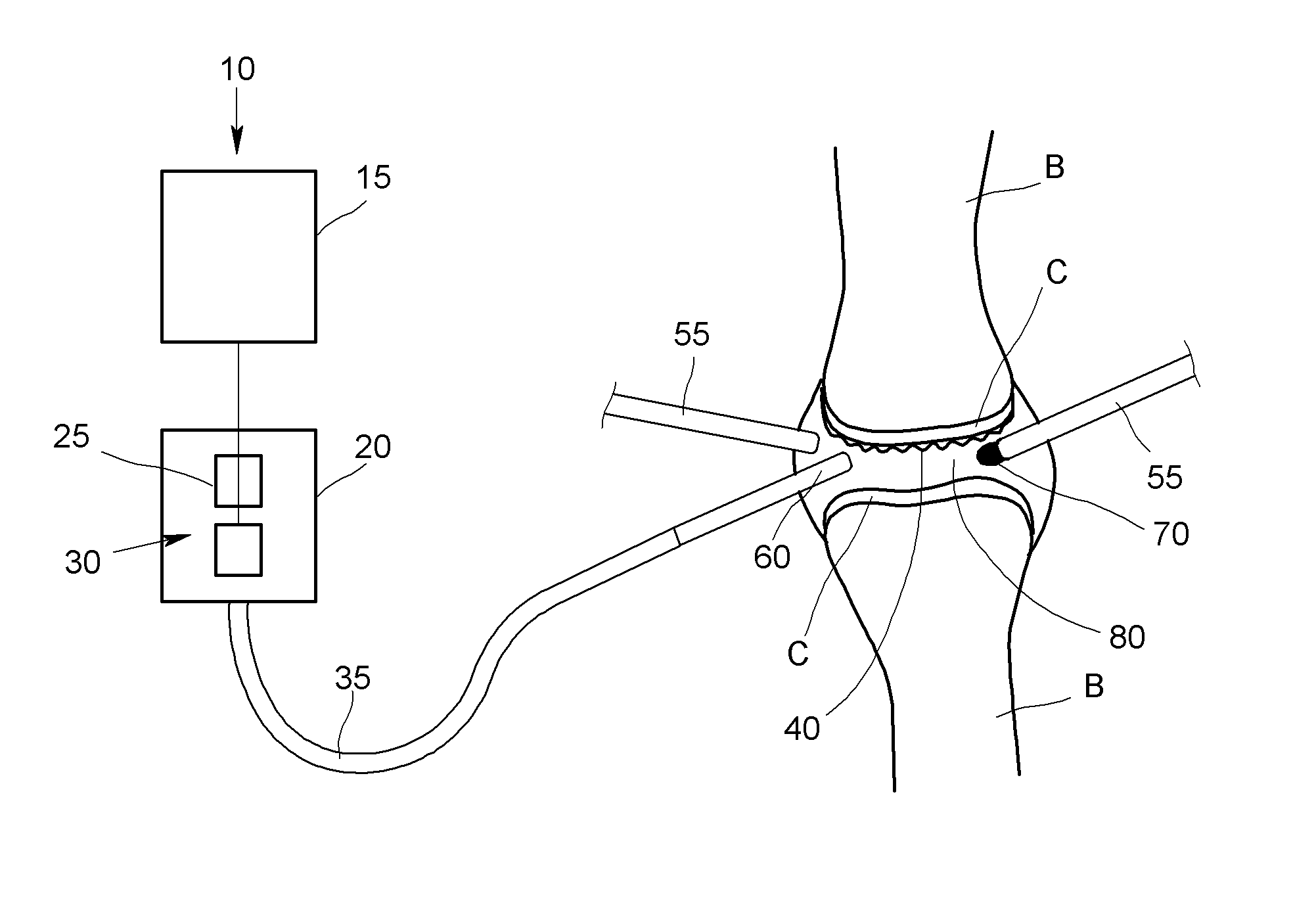
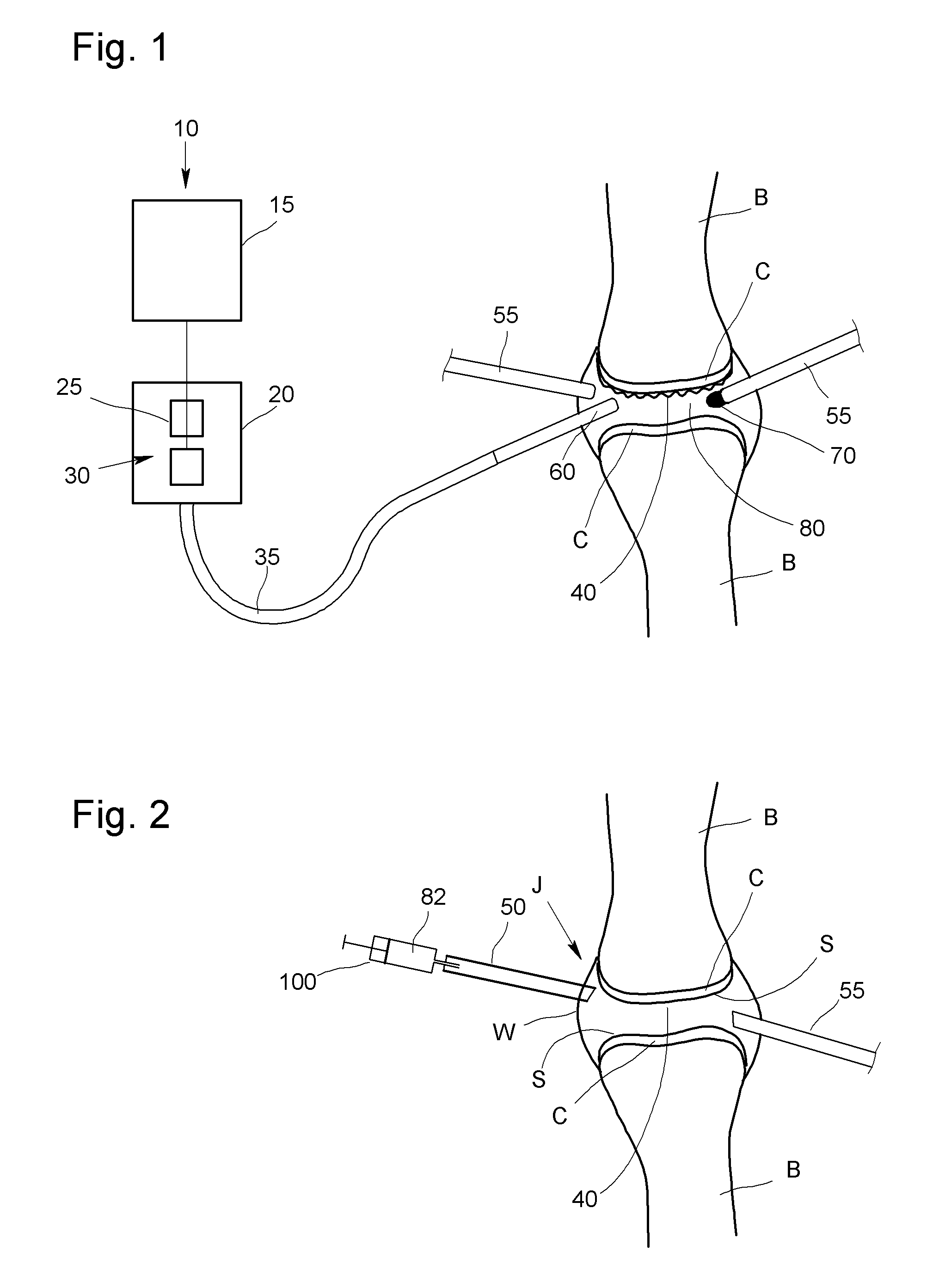
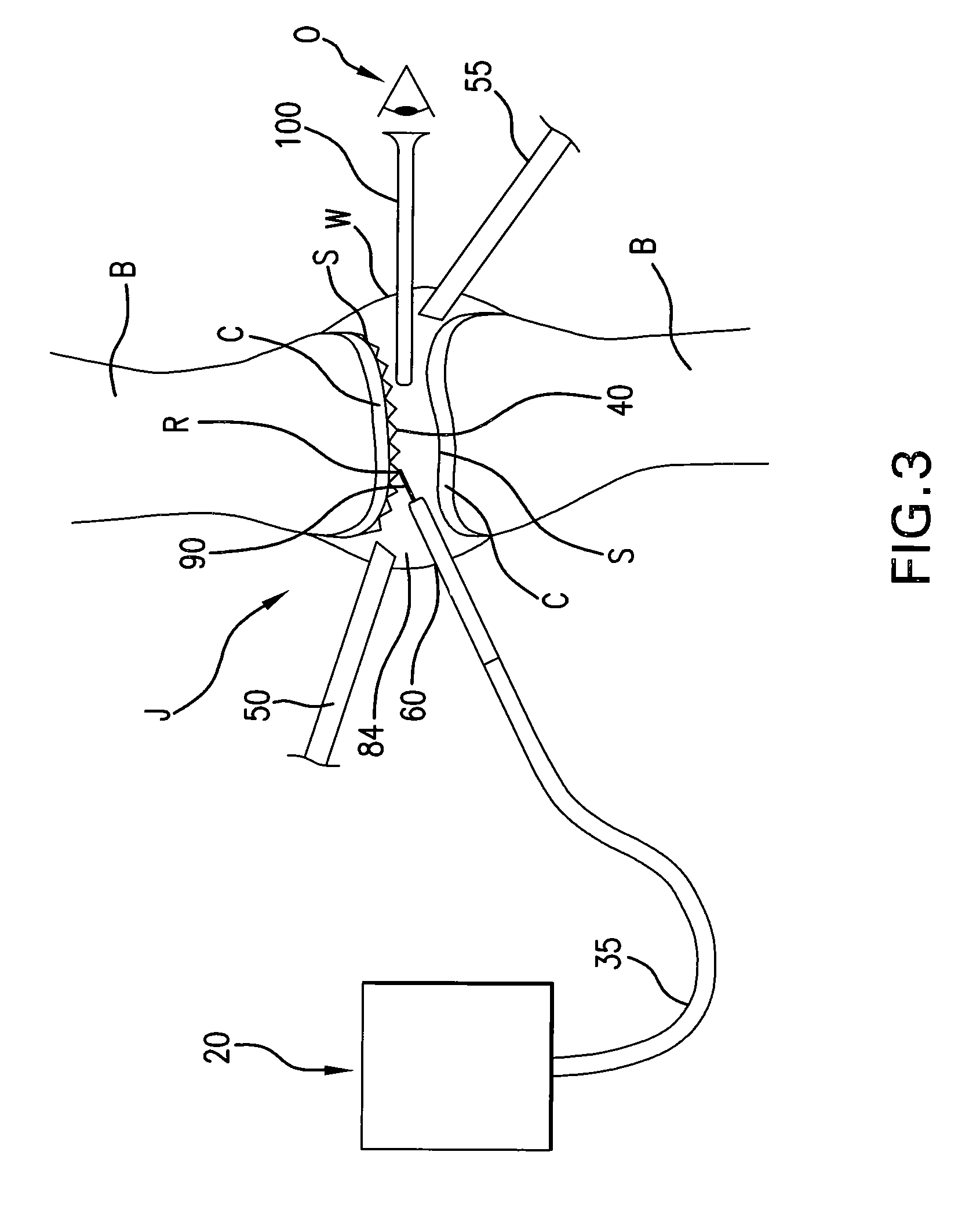
[0026]The apparatus of the present invention is a system for the ablation of cartilage tissue in vivo in a human or animal treatment subject. The present invention also includes a method for the ablation of cartage tissue in vivo in a human or animal treatment subject. While the present invention is described with respect to application to ablation of cartilage of the knee joint, the present invention is not limited in application to ablation of knee joint cartilage. The present invention may be applied to the ablation of human and / or animal cartilage in other joints as well, and even to nonarticular cartilage or to any tissue that fixes the dye.

[0027]The present invention is described with reference to FIGS. 1 to 4 where like parts are referred to using like character references. In addition, a non-limiting illustrative apparatus embodiment, in accordance with the present invention, will be described first followed by a description of a non-limiting method embodiment of the inventi...


PUM


Abstract

A method for the ablation of human or animal articular cartilage is disclosed that employs an exogenous absorber, such as a compound having the molecular formula C43H47N2NaO6S2, to coat or color the surface of the articular cartilage. The coated or colored cartilage surface then has an energy absorption peak at a specific wavelength due to the exogenous absorber, which makes it possible to employ a simple and cheap laser diode to generate a pulsed laser beam at the specific wavelength so as to superheat the coated or colored cartilage thereby causing at least partial ablation of cartilage.

Description

FIELD OF THE INVENTION[0001]The invention pertains to a method, and system for performing the method, for the ablation of cartilage tissue in a joint, such as, for example, a knee joint, of a human or animal. More particularly, the invention pertains to a method for the ablation of cartilage tissue that utilizes an exogenous absorber applied to the surface of the cartilage tissue so as to selectively absorb electromagnetic energy from a diode laser and thereby effect ablation of cartilage tissue coated with the exogenous absorber. The present invention includes a system, including a diode laser and an exogenous absorber, for performing the method of the present invention.BACKGROUND OF THE INVENTION[0002]In the orthopedic arts, it is known that cartilage tissue in a joint, such as a knee joint, has to be reshaped in order to treat certain joint disorders such as a hyaline cartilage surface irregularities. Generally, arthroscopic surgery is preferred due to the lower rates of morbidit...

Claims


Application Information

Patent Timeline
Patent Type & Authority Patents(United States)
IPC IPC(8): A61N5/067A61B18/28
CPCA61B18/24A61N5/0601A61N5/062
Inventor RAVUSSIN, PIERRE
Owner RAVUSSIN PIERRE
Learn More