Detection of early stages and late stages HPV infection

a technology of hpv infection and early stage, which is applied in the field of early stage and late stage hpv infection detection, can solve the problems of poor inter- and intra-observer agreement, high risk of progression toward invasive cervical cancer, and difficulty in obtaining samples

Active Publication Date: 2012-10-02
HEER MEDICAL TECH DEV CO LTD
View PDF113 Cites 5 Cited by
  • Summary
  • Abstract
  • Description
  • Claims
  • Application Information

AI Technical Summary

Problems solved by technology

These lesions, preferentially observed in women aged 35-40 yrs, have a high risk of progression toward invasive cervical cancer.
However, due to subjective test criteria, there are various drawbacks for pap smear tests: difficulty in obtaining samples, poor inter- and intra-observer agreement, high rates of false negatives and false positives, the requirement of specialized labs staffed with highly trained personnel, and inability to identify the majority of HPV-infected human subjects.
Nucleic acid tests, such as “DNA Hybrid Capture”, have been developed with high assay sensitivity, but these tests are still not ideal, due to not only their high cost, assay operation procedures, the requirements for facility, equipment, and highly trained personnel, but also their very low positive predictive value (PPV) in cervical intraepithelial neoplasia (CIN) testing samples.
In addition, DNA testing can not differentiate disease stages after HPV infection nor can it diagnose different cell lesions (e.g., cannot diffrentiate LSIL from HSIL, nor CIN lesions from non-transforming latent or remissive viral infection).
Known protocols for producing monoclonal antibodies are generally unsuitable for the production of anti-HPV monoclonal antibodies and cannot be used in immunocytochemical diagnostic tests performed on human subjects of the general population.
In addition, three problems exist in clinical HPV detection.
Secondly, there are too many HPV types and most HPV types present in clinical samples are not known or systemically identified due to the lack of available antibodies.
Third, HPV virus can not be cultured in labs by standard tissue culture techniques.
Thus, there are no available HPV proteins purified to large quantities as immunogens for generating anti-HPV antibodies, and there are no available HPV proteins or purified anti-HPV antibodies to recognize anti-viral antibodies or viral proteins present in clinical samples for clinical HPV detection.
However, some progressive cervical cancer cases are reported to be infected by low risk HPV types, while infection of some high risk HPV types will never progress into cervical cancer.

Method used


Image

  • Detection of early stages and late stages HPV infection
  • Detection of early stages and late stages HPV infection
  • Detection of early stages and late stages HPV infection

Examples


examples

A. Detecting HPV Proteins from Biological Samples Using One Anti-HPV Antibody

1. Direct EIA: One or More HPV Proteins Coated on Microtiterplate to be Detected by One or More Anti-HPV Antibodies

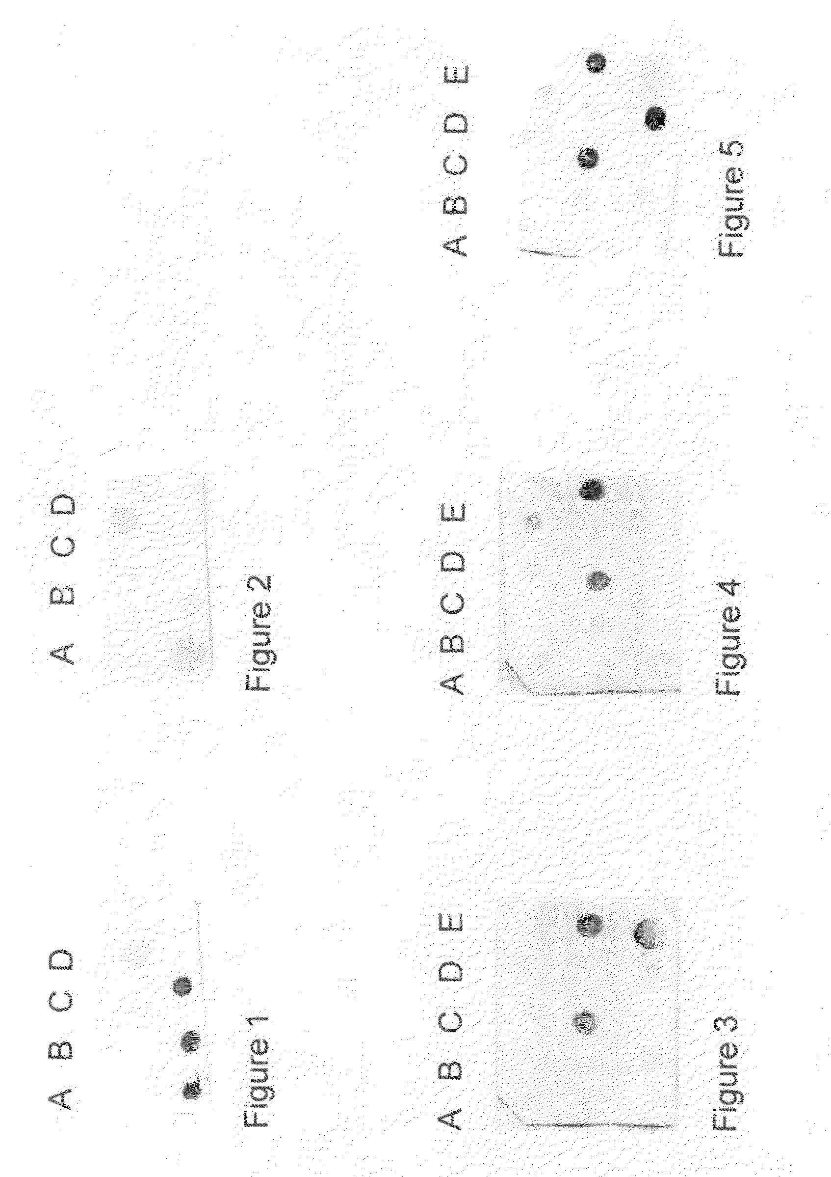
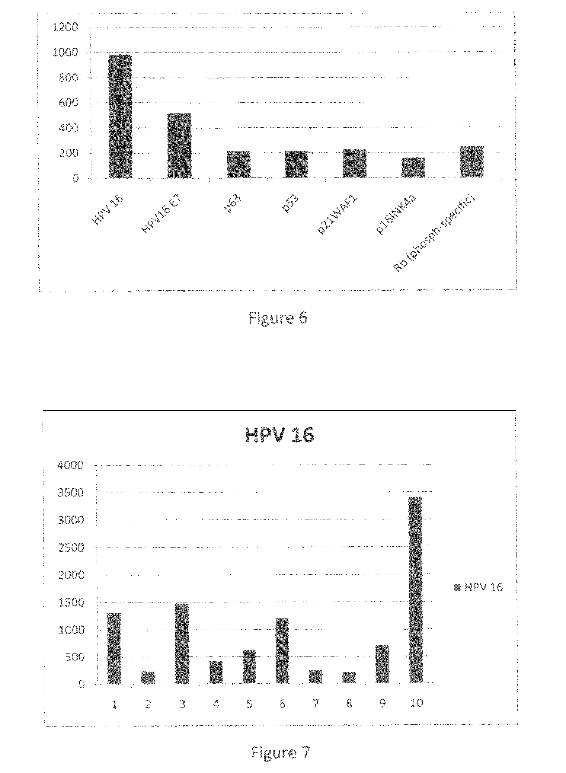
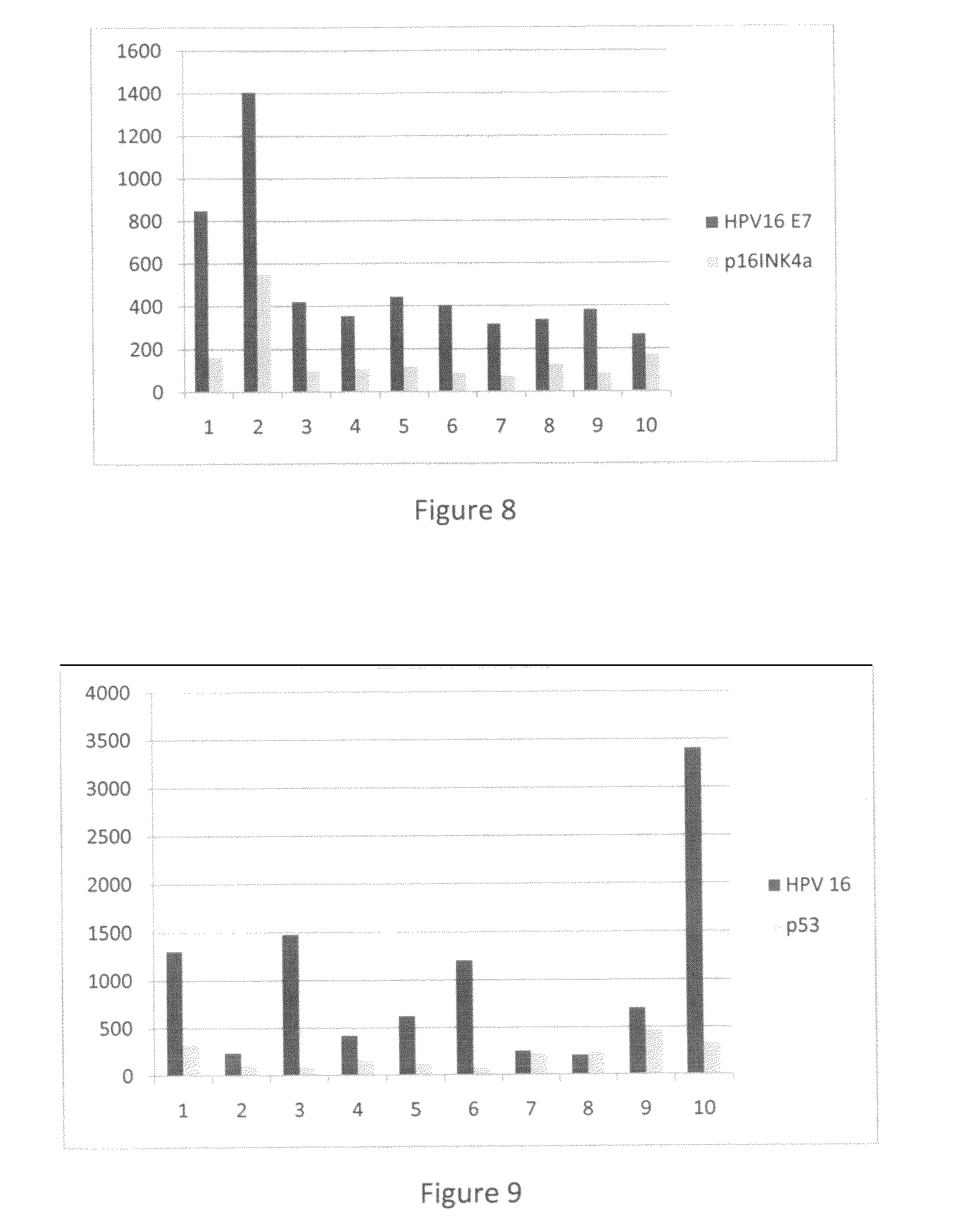
[0120]Clinical samples from cervical scrapes were obtained for detection of HPV E6, E7 or L1 proteins on direct EIA. Cervical cells from various sample sources included cervical scrape cells in liquid based cytology solution, cervical scrape cells in transport medium (used for HPV DNA test sample), or cervical scrape cells in lysis buffer. To perform the direct EIA described herein, specimens were processed, centrifuged, washed, and lysed to generate cell lysate as analyte. The proteins in the cell lysate were quantitated and coated to a microtiterplate with the same amount of protein in each well. The plate was blocked, and detected by each HPV monoclonal antibody followed by HRP-conjugated secondary antibody (anti-mouse IgG or anti-rabbit IgG for example). TMB substrate was added followed by ...


PUM


Abstract

Embodiments of the invention provide methods, monoclonal antibodies, polyclonal antibodies, assays, and kits for detecting HPV infection and HPV related cancer diagnosis, including infection by various HPV genotypes, early and / or late stage HPV-associated or HPV-specific cancers. Various specific or pan monoclonal antibodies recognizing specific epitope for specific HPV protein or HPV type, or common epitope for various HPV proteins or HPV types are obtained. The invention also provides one or more solid surface to coat the testing cell lysate. Also, the anti-HPV antibody can be coated on the solid surface of the invention to capture HPV proteins and detect HPV infection.

Description

CROSS-REFERENCE TO RELATED APPLICATIONS[0001]This application claims benefit of U.S. provisional patent application Ser. No. 61 / 131,991, filed Jun. 13, 2008, and U.S. provisional patent application Ser. No. 61 / 192,912, filed Sep. 22, 2008. Each of the aforementioned related patent applications is herein incorporated by reference.BACKGROUND OF THE INVENTION[0002]Infection by human papillomaviruses (HPV) at specific epithelium cells to induce epithelial proliferations plays an important role for cervical carcinogenesis. About 99 percent of confirmed cervical cancer cases are found to be associated with HPV infection with biopsy-confirmed squamous intraepithelial lesions (SIL) or cervical intraepithelial neoplasia (CIN). The incidence of HPV infection, primarily transmitted through sexual contact, is highest among young women and about 20 million sexually active men and women worldwide are currently infected. Approximately 1% of the population has genital warts and 4% of women have cer...

Claims


Application Information

Patent Timeline
Patent Type & Authority Patents(United States)
IPC IPC(8): C12Q1/00A61K39/42G01N33/53G01N33/542G01N33/00A61K39/395C12Q1/70
CPCC07K16/084C12Q1/708G01N33/571G01N33/56983G01N2469/10G01N2333/025
Inventor CHENG, SHULING
Owner HEER MEDICAL TECH DEV CO LTD
Why Eureka
Try Eureka