Glycoproteomic probes for fluorescent imaging of fucosylated glycans in vivo
a fluorescent imaging and glycan technology, applied in the field of glycoprotein probes for fluorescent imaging of fucosylated glycans in vivo, can solve the problems of complex glycan structure, inability to genetically manipulate glycans in a similar fashion to dna and proteins, and complex glycan structure identification, etc., to achieve specific covalent labeling, rapid identification, and versatile
- Summary
- Abstract
- Description
- Claims
- Application Information
AI Technical Summary
Benefits of technology
Problems solved by technology
Method used
Image
Examples
example 1
Analysis of Fluorescent Labeling at the Cell Surface by Flow Cytometry
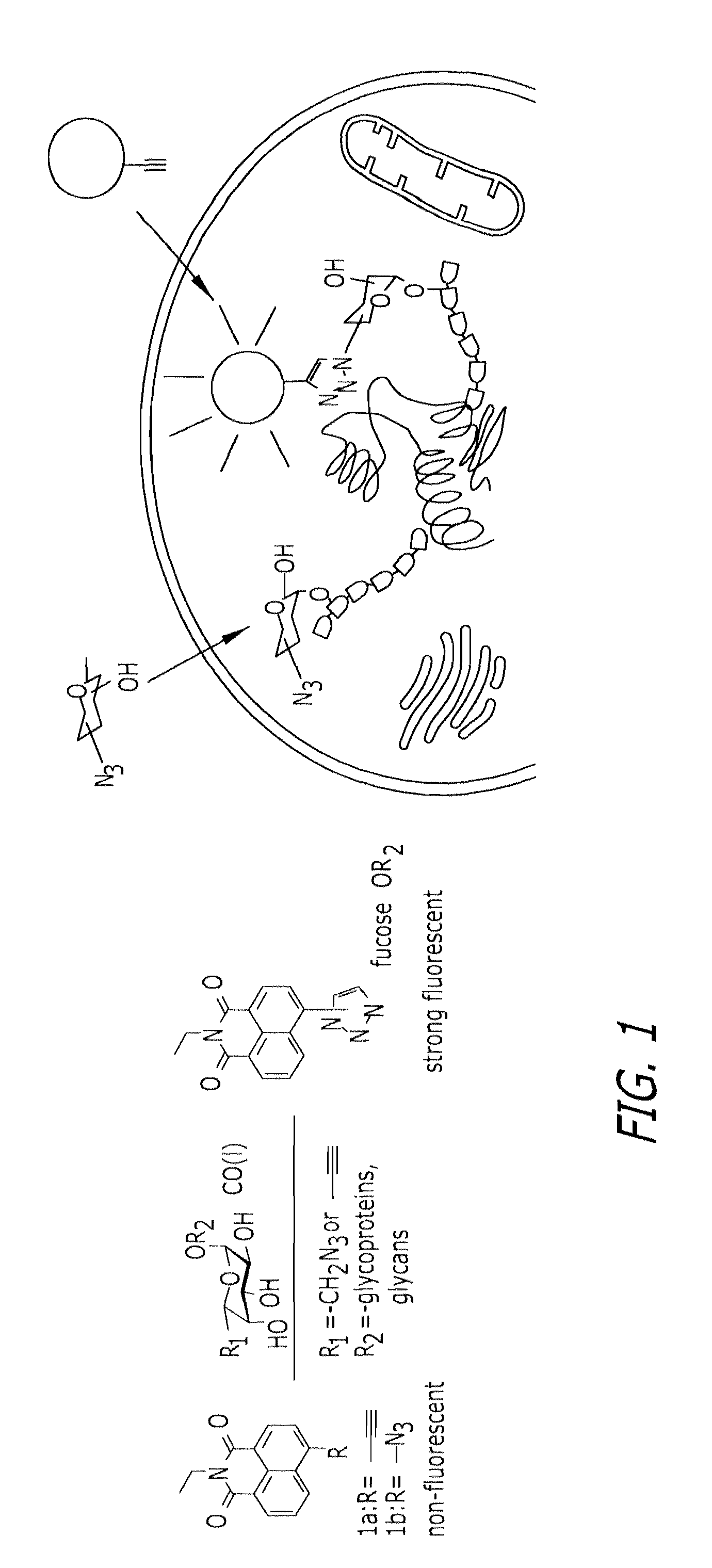
[0096]Jurkat cells were cultured in RPMI medium 1640 (Invitrogen, Carlsbad, Calif.), supplemented with 10% FCS and peracetylated fucose 11 or azidofucose 12 (200 μM) at a density of 2×10⁵ cells per ml for 3 days, in the presence or absence of tunicamycin (5 μg / ml). After washing with 0.1% FCS / PBS, 106 cells were resuspended in 100 μA of a reaction solution (0.1 mM probe 1a or biotinylated alkyne 13 / 0.2 mM Tris-triazoleamine catalyst / 0.1 mM CuBr in PBS) at room temperature for 30 min, followed by washing with 0.1% FCS / PBS. For the biotinylation experiment, cells were stained with 0.25 μg of UltraAvidin-Fluorescein (Leinco Technologies, St. Louis, Mo.) in 50 ml of staining buffer (1% FCS / 0.1% NaN3 in PBS) for 30 min at 4° C., followed by three washes with staining buffer. The fluorescence intensity was detected and acquired by BD LSR II (BD Biosciences, San Jose, Calif.) and FACSDiva software (BD Biosciences). Twent...
example 2
Fluorescence Microscopy and Imaging
[0097]The human hepatocellular carcinoma cell line, Hep3B (American Type Culture Collection, Manassas, Va.), was cultured in Opti-MEM (Invitrogen) supplemented with 0.1% FCS and treated with natural fucose or peracetylated azidofucose 12 (200 μM) for 3 days. The cells then were transferred to a coverslip glass slide and cultured overnight in the same medium. The cells were fixed by acetone and labeled as follows. Fixed cells were incubated with 0.2 mM probe 1a / 2.0 mM Tris-triazoleamine catalyst / 1.0 mM CuSO4 / 2.0 mM sodium ascorbate in PBS at room temperature overnight. After labeling, cells were washed with PBS, and fluorescence images were obtained by using Axiovert 200M (Carl Zeiss, Inc., Thornwood, N.Y.). For counter staining of Golgi compartments, the fixed cells were stained by using Alexa Fluor 594 conjugated WGA lectin (Invitrogen), and each fluorescent dye was imaged by using Bio-Rad (Carl Zeiss) Radiance 2100 Rainbow laser scanning confocal...
example 3
4-Ethynyl-N-ethyl-1,8-naphthalimide (1a)
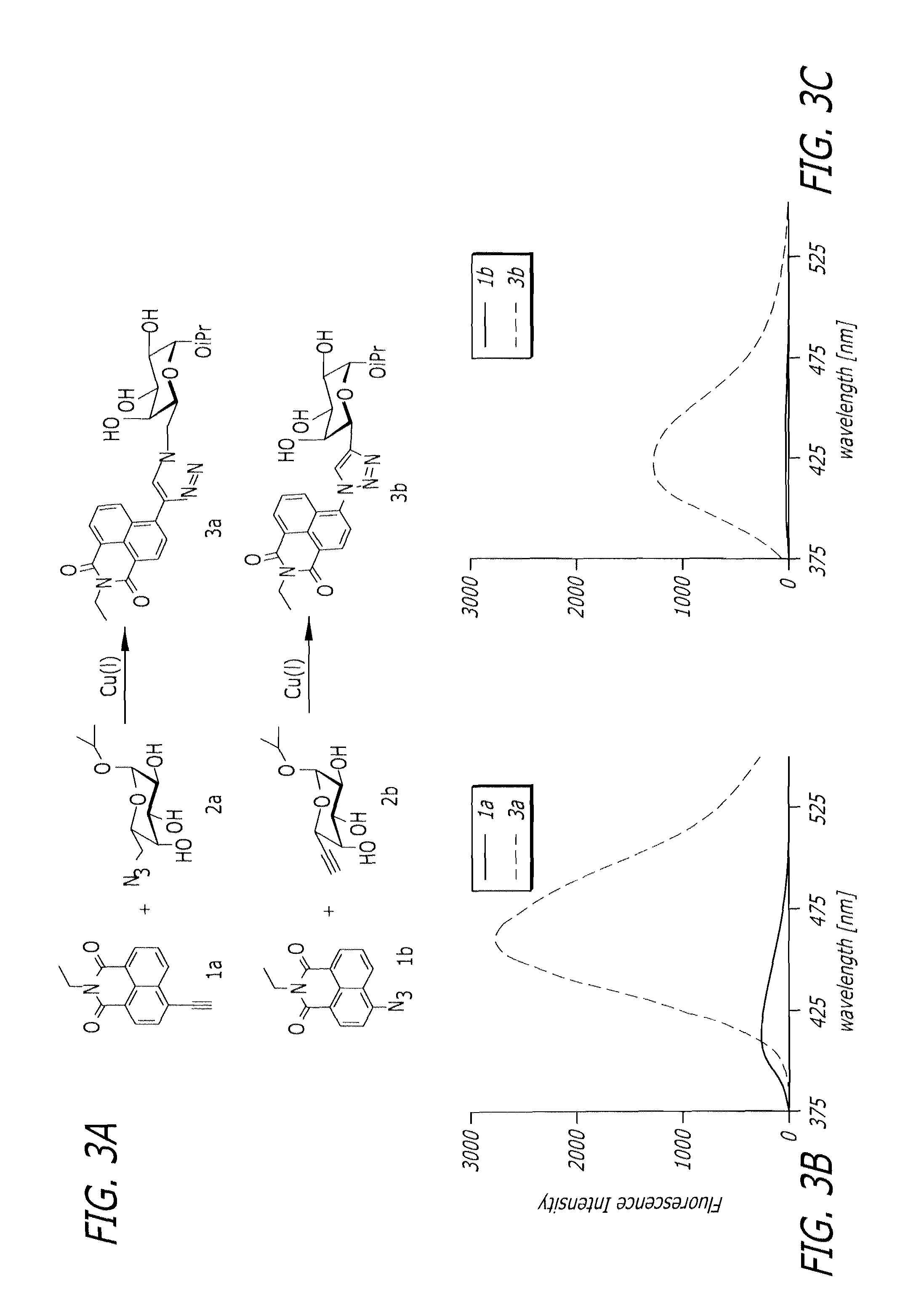
[0099]As shown in Scheme 2, to a solution of 4-bromo-N-ethyl-1,8-naphthalimide 15 (234 mg, 0.77 mmol) in 10 mL of THF was added tetrakis(triphenylphosphine)palladium (90 mg, 0.078 mmol), CuI (30 mg, 0.16 mmol), trimethylsilylacetylene (0.54 mL, 3.82 mmol) and N,N-diisopropylethylamine (0.5 mL, 2.87 mol) under argon gas. The mixture was stirred at room temperature overnight. The reaction mixture was diluted with AcOEt, and washed with saturated NH4Cl solution, dried over Na2SO4, and evaporated. The residue was purified partially by flash column chromatography on silica gel (AcOEt / hexane 1:10) to give the corresponding trimethylsilyl compound (180 mg). To a solution of this compound (180 mg) in 25 mL of MeOH was added 1M tetrabutylammonium fluoride solution in THF (2 mL, 2 mmol), and the mixture was stirred at 60° C. for 15 min. The reaction mixture was diluted with water, and the precipitates were collected by filtration. The solids were purifi...
PUM
| Property | Measurement | Unit |
|---|---|---|
| wavelengths | aaaaa | aaaaa |
| density | aaaaa | aaaaa |
| pH | aaaaa | aaaaa |
Abstract
Description
Claims
Application Information
 - R&D
- Intellectual Property
- Life Sciences
- Materials
- Tech Scout
- Unparalleled Data Quality
- Higher Quality Content
- 60% Fewer Hallucinations
Browse by: Latest US Patents, China's latest patents, Technical Efficacy Thesaurus, Application Domain, Technology Topic, Popular Technical Reports.
© 2025 PatSnap. All rights reserved.Legal|Privacy policy|Modern Slavery Act Transparency Statement|Sitemap|About US| Contact US: help@patsnap.com


