
Method of diagnosing and treating interstitial cystitis

a technology of interstitial cystitis and diagnosis method, which is applied in the direction of antibody medical ingredients, instruments, drug compositions, etc., can solve the problems of c-fiber activation, irritating the bladder wall, and leaky epithelium lining the bladder wall, so as to reduce the amount or production of et-1 and inhibit the effect of action

Active Publication Date: 2013-10-22
THE TRUSTEES OF THE UNIV OF PENNSYLVANIA
View PDF3 Cites 2 Cited by
  • Summary
  • Abstract
  • Description
  • Claims
  • Application Information

AI Technical Summary

Benefits of technology

[0011]In another aspect, a method is provided for monitoring progression of IC in a mammalian subject suffering from that disorder. In this method the level of expression of ET-1 in a biological sample from a mammalian subject having IC is measured and compared to the level of expression of ET-1 of a temporally earlier biological sample of the same subject. In this method, a decreased expression level of the ET-1, particularly in the urine or urethelium of the subject, compared to that in an earlier biological sample of the same subject is indicative of regression or improvement in the disorder. Conversely, an increase in ET-1 in the later sample is indicative of progression or increase in severity of the disorder. As one embodiment, this method can be applied to a subject being treated for IC. In this circumstance, the method enables a determination of the efficacy of the treatment.
[0016]In yet another aspect, there is provided a use of a composition that inhibits the action of ET-1 or the binding of ET-1 with its receptors, or reduces the amount or production of ET-1, in a method for treating interstitial cystitis.

Problems solved by technology

The loss of the integrity of the urothelium is suspected to lead to leaky epithelium lining the bladder wall and allow toxic substances in urine to irritate the bladder wall.
This can lead to activation of C-fibers, release of substance P and mast cell degranulation and subsequent injury to the submucosal tissue.
These tissue changes lead to neuronal activation, leading to urinary urgency / frequency and pelvic pain associated with IC10.
It is difficult to make a clear diagnosis of IC because clinicians are hampered by the lack of biomarkers to either evaluate the symptoms, pathological changes, and pathogenesis of the condition and / or to provide appropriate treatments.
Even when a mode of treatment is selected and tried, it is difficult to evaluate the effect of treatment for inducing beneficial changes in the bladder wall, the urothelium and the suburothelial regions, which are severely affected in IC.

Method used


Image

  • Method of diagnosing and treating interstitial cystitis
  • Method of diagnosing and treating interstitial cystitis
  • Method of diagnosing and treating interstitial cystitis

Examples


example 1

General Experimental Methods

[0079]A. Statistics

[0080]Data are described appropriately before proceeding to analysis. Biomarker expression and all other continuous data are presented as sample means (±SEM). Graphical methods are used to examine the distribution and quality of the data. Group comparisons are performed using ANOVA. Categorical variables are summarized by proportions and compared across groups using standard chi-square tests of association. Exact p-values from Fisher's exact test are calculated for these tests. Logistic regression models are used to identify which patient demographics, study sites, and other clinical factors are predictive of particular phenotypes. In addition, statistical tests for univariate associations between clinical phenotypes and biomarkers are computed using Statistical analysis software (SAS; SAS, Cary, N.C.). Adjustments for clinical sites are made for most analyses. Separate multivariable predictive models are developed for each clinical phe...

example 2

The Expression of ET-1 in the Urinary Bladder Epithelium

[0093]The expression of ET-1 in the urothelium in the bladder biopsies from IC patients is analyzed at the mRNA and protein level by RT-PCR (and Real-time PCR) and Western blot analysis, respectively. The relative increase in the expression of the endothelin at the mRNA level is assessed by quantitative real-time PCR using the published procedure37. Immunofluorescence microscopy is used for in situ localization of the endothelin expression to the urothelial cell layer. For immunofluorescence microscopy, formalin-fixed tissue sections of bladder biopsies previously obtained for the 1CDB (Interstitial Cystitis Database) were prepared as described above. The detailed procedure for performing the immunofluorescence microscopy is provided in Example 1. Briefly, Anti-ET-1 antibody (Calbiochem, San Diego, Calif.; cat. #CP44) is used at a dilution of 1:400 and anti-mouse IgG-Cy3 secondary antibody (Sigma, St. Louis, Mo.; C-2181) at a d...

example 3

Expression of ET-1 in the Urine of Patients with IC

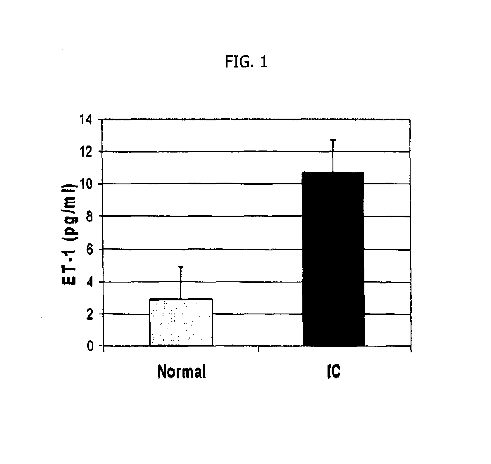
[0098]Endothelin expression is quantitated at the protein level by enzyme linked immunosorbent assay (ELISA). ELISA is performed using the human big Endothelin-1 (ET-1) Enzyme Immunometric Assay kit (Assay Designs, Inc., Ann Arbor, Mich.) to determine human big ET-1 peptide levels in urine samples from five female IC patients. The normal control group was urine samples from four normal healthy females. The ET-1 primary antibody shows little to no cross-reactivity with related endothelin peptides including ET-2 and ET-3. The immunoassay kit has a sensitivity as low as 0.14 pg / ml. Because the two-site “Sandwich” ELISA assays require two antibodies, they can be more specific than RIA assays40.

[0099]Briefly the appropriate number of wells in a 96 well microtiter plate coated with rabbit antibody specific to human ET-1 are washed twice with wash buffer containing phosphate buffered saline with detergent. Next 100 μL of either standard, c...


PUM

PropertyMeasurementUnit
volumesaaaaaaaaaa
volumesaaaaaaaaaa
volumesaaaaaaaaaa

Abstract

A method for diagnosing, or differentially diagnosing, interstitial cystitis (IC) involves detecting or measuring increased expression of a biomarker Endothelin 1 (ET-1) in a biological sample from a mammalian subject, particularly in the urine or urothelial tissue. An increased level of expression of ET-1 above the level of expression in the same sample of a healthy mammalian subject is an indication of a diagnosis of IC. Such diagnosis may further involve identify other clinical symptoms of IC. Additionally the method may use additional biomarkers, such as Hb-EFG, EGF, APF, IL-8, IL-6, and cGMP. Assay methods and diagnostic reagents and kits for such diagnosis are provided. Methods and compositions for treating IC by reducing the action, production or synthesis of ET-1 in the urine or urothelium and / or inhibiting its binding to its ETA and / or ETB receptors are also provided.

Description

CROSS-REFERENCE TO RELATED APPLICATIONS[0001]This application is a national stage of International Patent Application No. PCT / US2010 / 049914, filed Sep. 23, 2010, which claims the benefit of the priority of U.S. Provisional Patent Application No. 61 / 246,273, filed Sep. 28, 2009 (expired), which applications are incorporated herein by reference.BACKGROUND OF THE INVENTION[0002]Interstitial cystitis, also known as painful bladder syndrome, is a pathological condition that affects as many as 1 million patients in the U.S. with a high incidence in females. A heterogeneous array of symptoms varies from case to case and even in the same individual. Such symptoms can include mild discomfort, pressure, tenderness, intense pain in the bladder and pelvic area, increased urinary frequency / urgency, nocturia or a combination of these symptoms12. Pain may change in intensity as the bladder fills with urine or as it empties. People with interstitial cystitis also have a defect in the protective lin...

Claims


Application Information

Patent Timeline
Patent Type & Authority Patents(United States)
IPC IPC(8): G01N33/48G01N33/53
CPCC12Q1/6883G01N33/6893C12Q2600/158G01N2800/34C12Q2600/136A61P13/10A61P29/00
Inventor CHACKO, SAMUEL K.
Owner THE TRUSTEES OF THE UNIV OF PENNSYLVANIA
Learn More