Method of producing virus-like particles of picornavirus using a small-ubiquitin-related fusion protein expression system
a technology of ubiquitin-related fusion and picornavirus, which is applied in the direction of fusion with degradation motif, polypeptide with his-tag, and anti-microbial ingredients
- Summary
- Abstract
- Description
- Claims
- Application Information
AI Technical Summary
Benefits of technology
Problems solved by technology
Method used
Image
Examples
example 1
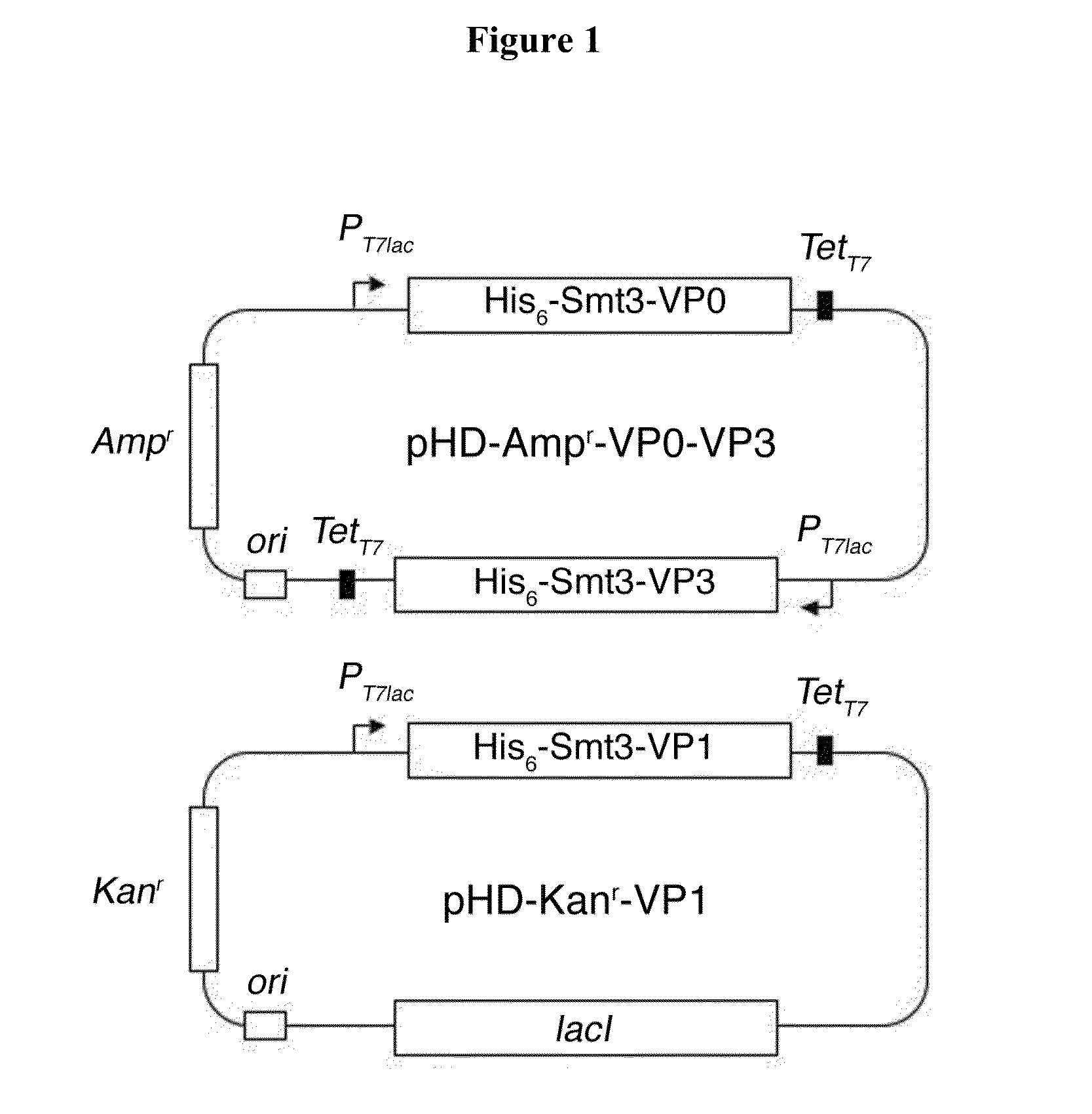
Preparation of Protein Complex Containing His6-Smt3-VP0, His6-Smt3-VP1, and His6-Smt3-VP3 Fusion Proteins
[0030]cDNAs encoding FMDV VP0, VP1, and VP3 were amplified by the sticky-end polymerase chain reaction cloning method described in Shih et al. Protein Sci. 11:1714-1719 (2002). The amino acid sequences of the three FMDV capsid proteins are shown below:
[0031]
Amino acid sequence of FMDV capsid protein VP0(SEQ ID NO: 2)N T G S I I N N Y Y M Q Q Y Q N S M D T Q L G D NA I S G G S N E G S T D T T S T H T N N T Q N N DW F S K L A N T A F S G L F G A L L A D K K T E ET T L L E D R I L T T R N G H T T S T T Q S S V GV T Y G Y A T A E D F V S G P N T S G L E T R V VQ A E R F F K T H L F D W V T S D P F G R C H L LE L P T D H K G V Y G S L T D S Y A Y M R N G W DV E V T A V G N Q F N G G C L L V A M V P E L R SI S K R E L Y Q L T L F P H Q F I N P R T N M T AH I T V P Y L G V N R Y D Q Y K V H K P W T L V VM V A A P L T V N N E G A P Q I K V Y A N I A P TN V H V A G E L P S K EAmino acid s...
example 2
Preparation of FMDV-Like Particles
[0038]The His6-Smt3-VP0 / His6-Smt3-VP1 / His6-Smt3-VP3 ternary protein complex described in Example 1 above was treated with U1P1 protease to remove the His6-Smt3 moiety, following the procedures described in Lee et al., Protein Sci. 17:1241-1248 (2008). SDS-PAGE analysis showed that the protease-digested product contains three proteins having molecular weights ˜32 kDa, 29 kDa and 26 kDa, which correspond to the theoretical molecular weights of VP0, VP3, and VP1, respectively. An Edman degradation assay was then performed to determine the N-termini amino acid residues of these three proteins. Results thus obtained showed that they were identical to the N-termini amino acid residues of VP0, VP1, and VP3. It confirms that the proteins contained in the protease-digested products were indeed FMDV capsid proteins VP0, VP1, and VP3.
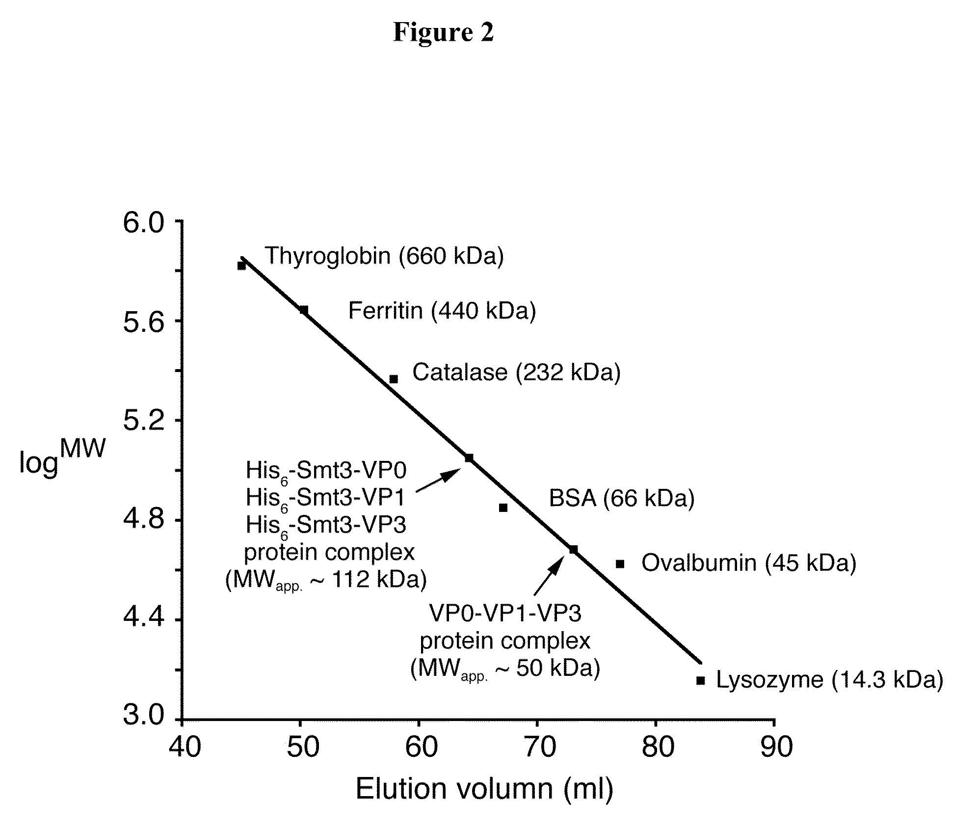
[0039]The protease-treated product was then subjected to the HPLC assay described above. The results showed that VP0, VP1, and V...
example 3
Immune Responses Elicited by FMDV-Like Particles
[0042]The VLPs of FMDV prepared by the method described in Example 2 above was examined as follows to determine their activity in inducing immune responses.
[0043]First, HEK293 cells, transfected with p5×NFκB-Luc, EGFP-N1 (as a control plasmid), pcDNA3.1-TLR2, or pcDNA3.1 (as a blank control), were treated with (a) 1 μm Pam3csk4 (a TLR2 ligand; a positive control), (b) Tris buffer (20 mM Tris-Cl, pH 8.0, and 100 mM NaCl; a blank control), (c) 90 μg / ml BEI-inactivated FMDV (a positive control), and (d) VLPs at various doses (i.e., 0.6, 1, and 3 μg / ml). Twenty-four hours after treatment, the luciferase activities, indicating NFκB activation, were determined in the cells and normalized against the EGFP levels in the same cells. The results thus obtained indicate that, similar to the treatment of Pam3csk4 or inactivated FMDV, treatment of the VLPs induced NFκB activation.
[0044]Next, activation of bone marrow derived dendritic cells (BMDCs) ...
PUM
| Property | Measurement | Unit |
|---|---|---|
| pH | aaaaa | aaaaa |
| pH | aaaaa | aaaaa |
| molecular weights | aaaaa | aaaaa |
Abstract
Description
Claims
Application Information
 - R&D
- Intellectual Property
- Life Sciences
- Materials
- Tech Scout
- Unparalleled Data Quality
- Higher Quality Content
- 60% Fewer Hallucinations
Browse by: Latest US Patents, China's latest patents, Technical Efficacy Thesaurus, Application Domain, Technology Topic, Popular Technical Reports.
© 2025 PatSnap. All rights reserved.Legal|Privacy policy|Modern Slavery Act Transparency Statement|Sitemap|About US| Contact US: help@patsnap.com


