
Human monoclonal antibodies and methods for producing the same

a monoclonal antibody and human antibody technology, applied in the field of cell biology and immunology, can solve the problems of “serum sickness, large time and expense for humanizing antibodies, and untapped potential for therapeutic and diagnostic agents

Active Publication Date: 2014-05-06
MUSC FOUND FOR RES DEV
View PDF46 Cites 6 Cited by
  • Summary
  • Abstract
  • Description
  • Claims
  • Application Information

AI Technical Summary

Benefits of technology

The present invention provides a method for producing immortalized human B-cells that secrete specific antibodies. The method involves immortalizing B-cells with a combination of Epstein-Barr virus (EBV) and a cytokine / growth factor cocktail, resulting in the production of immortalized B-cells that secrete antibodies specific for a predetermined antigen. The method can be used to produce monoclonal antibodies for therapeutic purposes and can be applied to any antigen. The immortalized B-cells can also be genetically modified to express different types of antibodies. The invention provides a valuable tool for research and development in the field of human monoclonal antibodies.

Problems solved by technology

While some success in the treatment of infectious agents and toxins has also been observed with monoclonals, the potential for therapeutic and diagnostic agents remains largely untapped.
One particular impediment to the development of monoclonal antibodies for human therapy is the need to “humanize” such antibodies, which are generally made in mice, rats and rabbits.
If human patients are administered such antibodies without humanized constant regions, they can suffer from “serum sickness,” literally meaning that an antibody is mounted by the recipient against the non-human antibody sequences.
While humanizing monoclonal antibodies produced in research animals can avoid this problem, this does not come without a cost—both time and expense for humanization of antibodies are considerable, leading to a bottleneck when it comes to exploiting the use of monoclonal antibodies for therapy and diagnosis in humans.

Method used


Image

  • Human monoclonal antibodies and methods for producing the same
  • Human monoclonal antibodies and methods for producing the same
  • Human monoclonal antibodies and methods for producing the same

Examples


example 1

Materials & Methods for B-Cells Reactive to H5 HA

[0150]Isolation and culture of tonsil B cells. To prepare B cells from tonsils, tonsil tissue was placed inside a sterile Petri dish (VWR International, cat. #25384-088) containing 20-30 ml Dulbecco's phosphate buffered saline (DPBS, without CaCl2 or MgCl2; Gibco / Invitrogen, Grand Island, N.Y. cat. #14190144) supplemented with 1× Antibiotic-Antimycotic (Gibco / Invitrogen cat. #15240-062). The tissue was chopped and minced with scalpels to approximately 1 mm3 pieces. Additional lymphocytes were released by gentle grinding of tonsil pieces between the frosted glass surfaces of two sterile microscope slides (VWR cat. #12-550-34), and single cell preparation was made by straining through 70 μm nylon strainer (BD Falcon, cat. #352350, BD Biosciences, Two Oak Park, Bedford, Mass.). This suspension was layered onto a Ficoll (Amersham Biosciences cat. #17-1440-03, Uppsala, Sweden) cushion (35 ml sample over 15 ml Ficoll) and resolved at 1500 G...

example 2

Results for B Cells Reactive to H5 HA

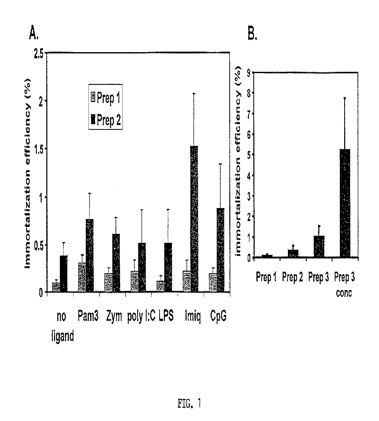
[0169]Toll-Like Receptor (TLR) ligands and EBV concentration did not significantly improve EBV infectivity. Traggiai et al. (2004) reported that addition of at least one TLR ligand (CpG) to cultured memory B cells could enhance EBV infection efficiency. Since naïve B cells express several TLR (Bourke et al., 2003) it was reasonable to assess the effect of several TLR ligands on EBV infection of naïve B cells. Primary B cells were incubated overnight with either Pam3 (Pam3Cys-Ser-(Lys)4) (0.5 μg / mL), zymosan (1 μg / mL), Poly I:C (polyinosinic-polycytidylic acid) (25 μg / mL), LPS (lipopolysaccharide) (5 μg / mL), Imiquimod (1 μg / mL), CpG (10 μg / mL), or no ligand. These are synthetic proteins that mimic common pathogenic antigens. Each of these activates different innate immune pathways in B-cells. Lipopeptide Pam3 (Hamilton-Williams et al., 2005) binds TLR 2 and 1, zymosan (a yeast cell wall component prepared from Saccharomyces cerevisiae) binds TLR 2...

example 3

Materials & Methods for Producing Human B-Cells Secreting Monoclonal Antibodies Reactive with SEB, SEC-2, PLGF, and Ricin B Chain

[0198]Creation of immortalized tonsil repertoires. Generation of concentrated EBV stocks and preparation of B cells from tonsil tissue have been described in Examples 1 and 2. No changes have been made to these methods. For induction of differentiation of EBV immortalized B cells, the inventors used complete RPMI medium (Gibco) supplemented with 10% FBS (Hyclone) containing soluble CD40 ligand (5 ng / ml), BAFF (10 ng / ml), and goat anti-human IgM F(ab′)2 (1.62 ng / ml), as previously described.

[0199]Sample collection for ELISA analysis. Collection and screening of sample culture supernatants for antigen reactivity by ELISA have been modified as follows. Culture supernatants were collected into corresponding wells on a 96-well plate on day 10-14 post-transduction at 100 μl from each well, and aliquots were pooled (30 μl of supernatant from all wells on each pla...


PUM


Abstract

The present invention provides for methods of producing human monoclonal antibodies against a wide variety of antigens including bacterial and viral antigens, as well as tumor antigens, and various autoantigens. Also provided are the antibodies themselves, nucleic acids encoding such antibodies, cells producing such antibodies, and methods of using such antibodies for diagnostic assays and passive immunity against disease states such as infection and cancer.

Description

[0001]This invention was made with government support under grant number 5K01CA095443 awarded by the National Cancer Institute, National Institute of Health. The government has certain rights in the invention.CROSS-REFERENCE TO RELATED APPLICATIONS[0002]This application is a national phase application under 35 U.S.C. §371 of International Application No. PCT / US2008 / 072124, filed Aug. 4, 2008, which claims the priority of U.S. Provisional Patent Application Ser. No. 60 / 953,739, filed Aug. 3, 2007, the entire disclosures of which are specifically incorporated herein by reference.REFERENCE TO A SEQUENCE LISTING SUBMITTED ELECTRONICALLY[0003]The content of the electronically submitted sequence listing (“205350—0002—494617.txt”, 38,573 bytes, created on Nov. 22, 2013) filed with this application is incorporated herein by reference in its entirety.BACKGROUND OF THE INVENTION[0004]I. Field of the Invention[0005]The present invention relates generally to the fields of cell biology and immun...

Claims


Application Information

Patent Timeline
Patent Type & Authority Patents(United States)
IPC IPC(8): C12N5/02A61K35/28A61K39/00
CPCA61K2039/505C07K2317/92C07K16/16C07K16/1018G01N33/6854C07K16/1271C07K16/22C07K2316/96C07K2317/21C12N5/0635C07K16/00C07K16/244C07K2317/75C12N2510/04A61P37/00Y02A50/30
Inventor SUTKOWSKI, NATALIERUBINCHIK, SEMYON
Owner MUSC FOUND FOR RES DEV
Learn More