

Hemostatic sponge
a sponge and hemostatic technology, applied in the field of hemostatic sponges, can solve the problems that the fibrous biomaterials, in particular collagen pads, for wound healing failed to induce hemostasis at conditions with impaired hemostasis, and achieve the effect of reducing the swelling properties and advantageous properties
- Summary
- Abstract
- Description
- Claims
- Application Information
AI Technical Summary
Benefits of technology
Problems solved by technology
Method used
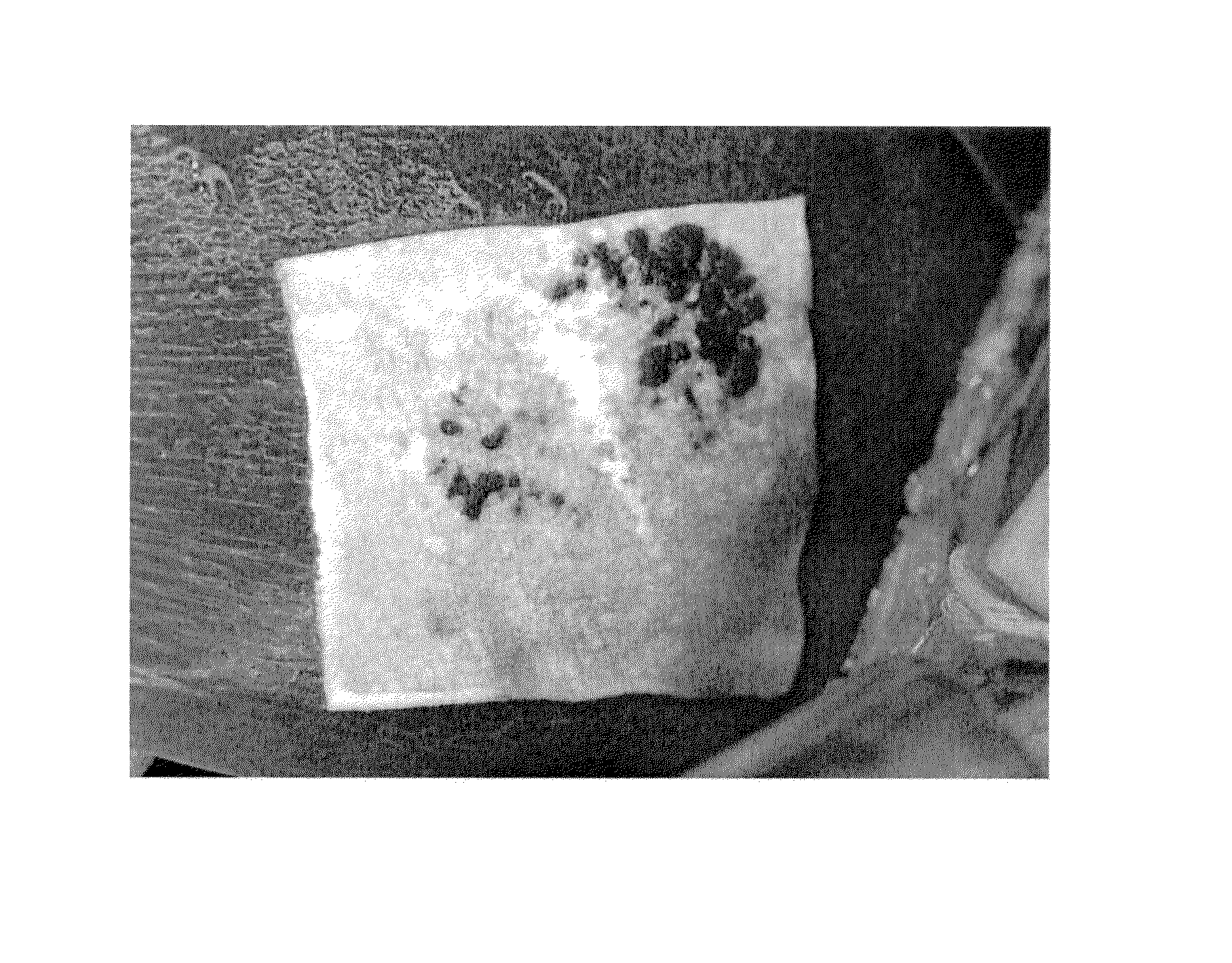
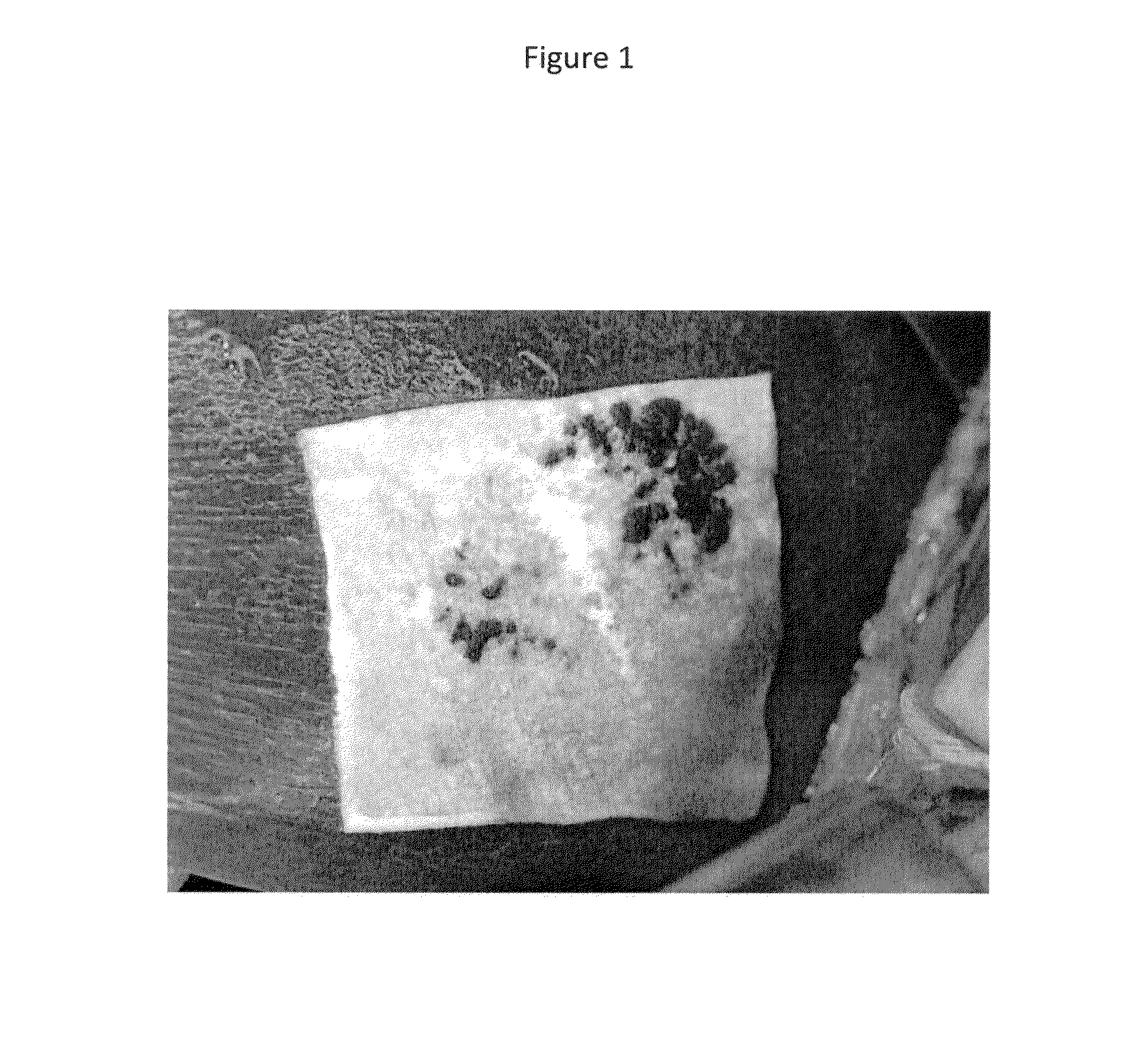
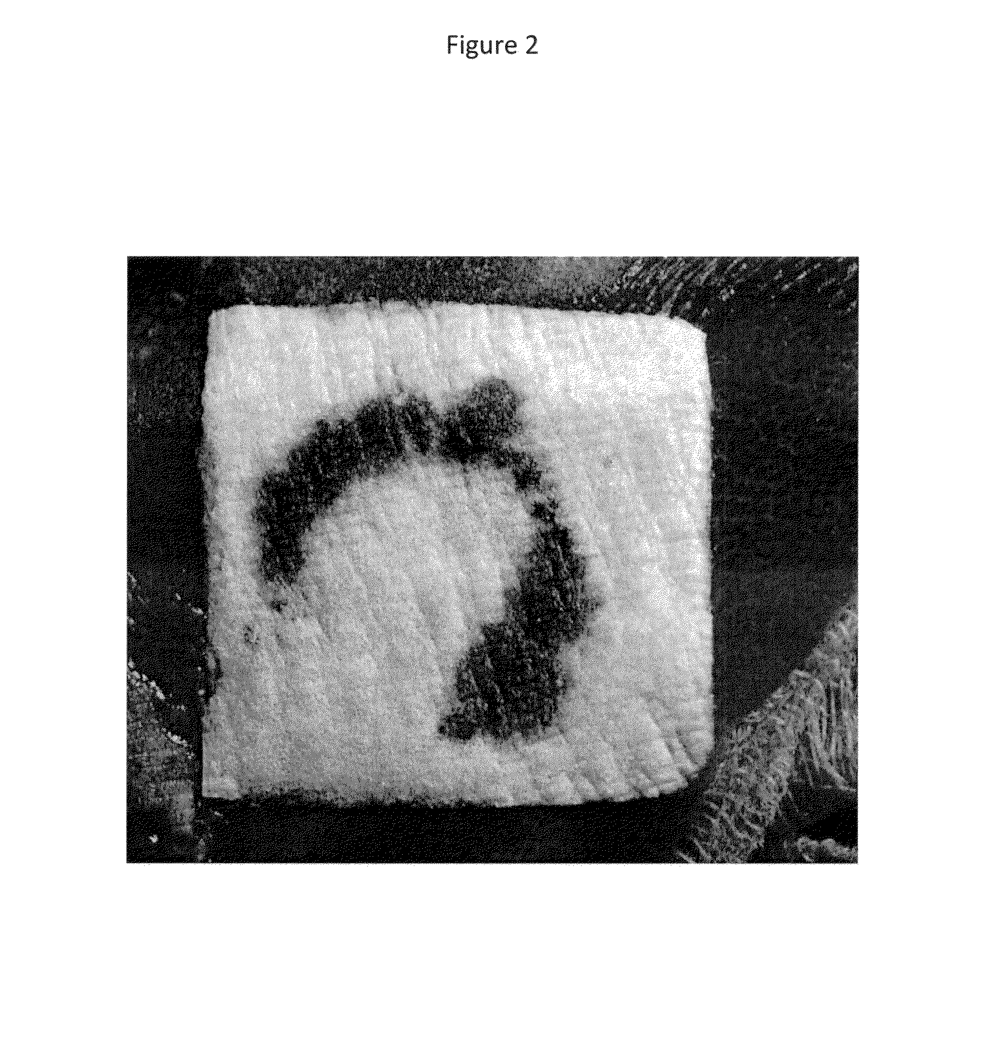
Image

Examples
example 1
Collagen Sponges Treated with Acidic Solution of Two Reactive Pegs
[0068]Aqueous, acidic solutions (pH 3.0, HCl) of COH102 and COH206 with PEG-concentrations (COH102 and COH206 1:1) of 10 mg / cm3, 35 mg / cm3, 70 mg / cm3 and 100 mg / cm3 are prepared and filled into 9×7 cm PET-trays. Commercial available bovine collagen sponges (Matristypt®), 9×7 cm, with the same volume as the previously filled PEG-solution are placed on the top of the solutions. After absorption of the PEG-solution, the collagen materials are lyophilized. After lyophilization the dried sponges may be packed together with desiccants in water vapor impermeable pouches and may be further gamma-sterilized, e.g. with 25 kGray.
example 2
Collagen Sponges Treated with EtOH-Solution of Two Reactive Pegs
[0069]COH102 and COH206 are dissolved in completely dried EtOH. PEG-concentrations (COH102 and COH206 1:1) of 10 mg / cm3, 35 mg / cm3, 70 mg / cm3 and 100 mg / cm3 are prepared and the solutions are filled into 9×7 cm PET-trays. Commercial available bovine collagen sponges (Matristypt®), 9×7 cm, with the same volume as the previously filled PEG-solution are placed on the top of the solutions. After absorption of the PEG-solution the collagen materials are dried in a vacuum chamber.
[0070]Dried sponges may be packed together with desiccants in water vapor impermeable pouches and may be gamma-sterilized, e.g. with 25 kGray.
example 3
Preparation of Collagen- / Reactive PEG Constructs
[0071]22 ml of aqueous, acidic solutions (pH 3.0, HCl) containing various concentrations (2.15 mg / cm3, 4.3 mg / cm3 and 7.2 mg / cm3) of bovine corium collagen and PEG (COH102 and COH206 1:1)-concentrations of 7.2 mg / cm3, 14.3 mg / cm3, 28.6 mg / cm3 and 57.3 mg / cm3 are prepared, filled into PET-trays and lyophilized.
[0072]The dried sponges may be packed together with desiccants in water vapor impermeable pouches and may be gamma-sterilized, e.g. with 25 kGray.
PUM
| Property | Measurement | Unit |
|---|---|---|
| thickness | aaaaa | aaaaa |
| size | aaaaa | aaaaa |
| particle size | aaaaa | aaaaa |
Abstract
Description
Claims
Application Information