Chondrogenic progenitor cells, protocol for derivation of cells and uses thereof

a progenitor cell and chondrocyte technology, applied in the field of cell biology, embryonic stem cells, cell differentiation, can solve the problems of complex development of new transplantation protocols, insufficient treatment of most cases of cartilage degeneration, joint pain and mobility impairment to a degree, etc., and achieve the effect of modulating chondrocyte growth and differentiation

Inactive Publication Date: 2015-05-12
THE UNIV COURT OF THE UNIV OF EDINBURGH
View PDF11 Cites 0 Cited by
  • Summary
  • Abstract
  • Description
  • Claims
  • Application Information

AI Technical Summary

Benefits of technology

[0032]Optionally, replication capacity of the stem cells, such as primate pluripotent stem cells or human embryonic stem cells, can be improved by increasing telomerase activity.

Problems solved by technology

The cartilage tissue performs a structural and mechanical function in skeletal joints and any disease or injury is consequently debilitating for patients.
The degradation leads to joint pain and mobility impairment to a degree that can be disabling.
However, where degradation or injury has already taken place, new therapies are needed to assist in the regenerating of joint cartilage.
Despite the initial success of the clinical methods reported to date, it is clear that current sources of chondrocytes are inadequate to treat most of the instances of cartilage degeneration that present themselves at the clinic.
In addition, problems concerning the use of immunosuppressive drugs have complicated the success of developing new transplantation protocols.
However, the mouse model of embryonic stem cell development does not necessarily yield strategies for differentiation that are applicable to other species (see, e.g. Ginis et al.
However, even with the preparation of chondrocytes derived from undifferentiated progenitor cell populations, such as hES cells, there remains the need to use immunosuppressive drugs in transplantation therapies using chondrocyte cell populations which is unfavourable for the long term success of transplants in patients.

Method used


Image

  • Chondrogenic progenitor cells, protocol for derivation of cells and uses thereof
  • Chondrogenic progenitor cells, protocol for derivation of cells and uses thereof
  • Chondrogenic progenitor cells, protocol for derivation of cells and uses thereof

Examples


example 1

Production of Chondrocyte Progenitor Cells

[0199]The DCCPCs were produced using a protocol as follows. Initially H7 ES cells were grown to 80% confluency using Matrigel® coated flasks and standard ES culture conditions. At this point the media was replaced with chondrogenic media. The cells remained in their original flasks and still on Matrigel® thus reducing cost and handling. Cells were cultured in these conditions for a further 14 days with chondrogenic media replaced 3 times a week. On day 14 the cell layer was washed with PBS and the chondrogenic media replaced with dedifferentiation medium. Again the cells remained in their original flasks. After a further 5 days in culture the cells showed signs of dedifferentiation and were trypsin passaged as single cells.

Materials

[0200]The materials used were: conditioned media, bFGF (Peprotech), tissue culture flasks (Nunc), Matrigel® (BD Biosciences), DMEM (Sigma), and FBS (Gibco).

Protocol

[0201]hESCs were cultured to confluency on Matrig...

example 2

Morphology

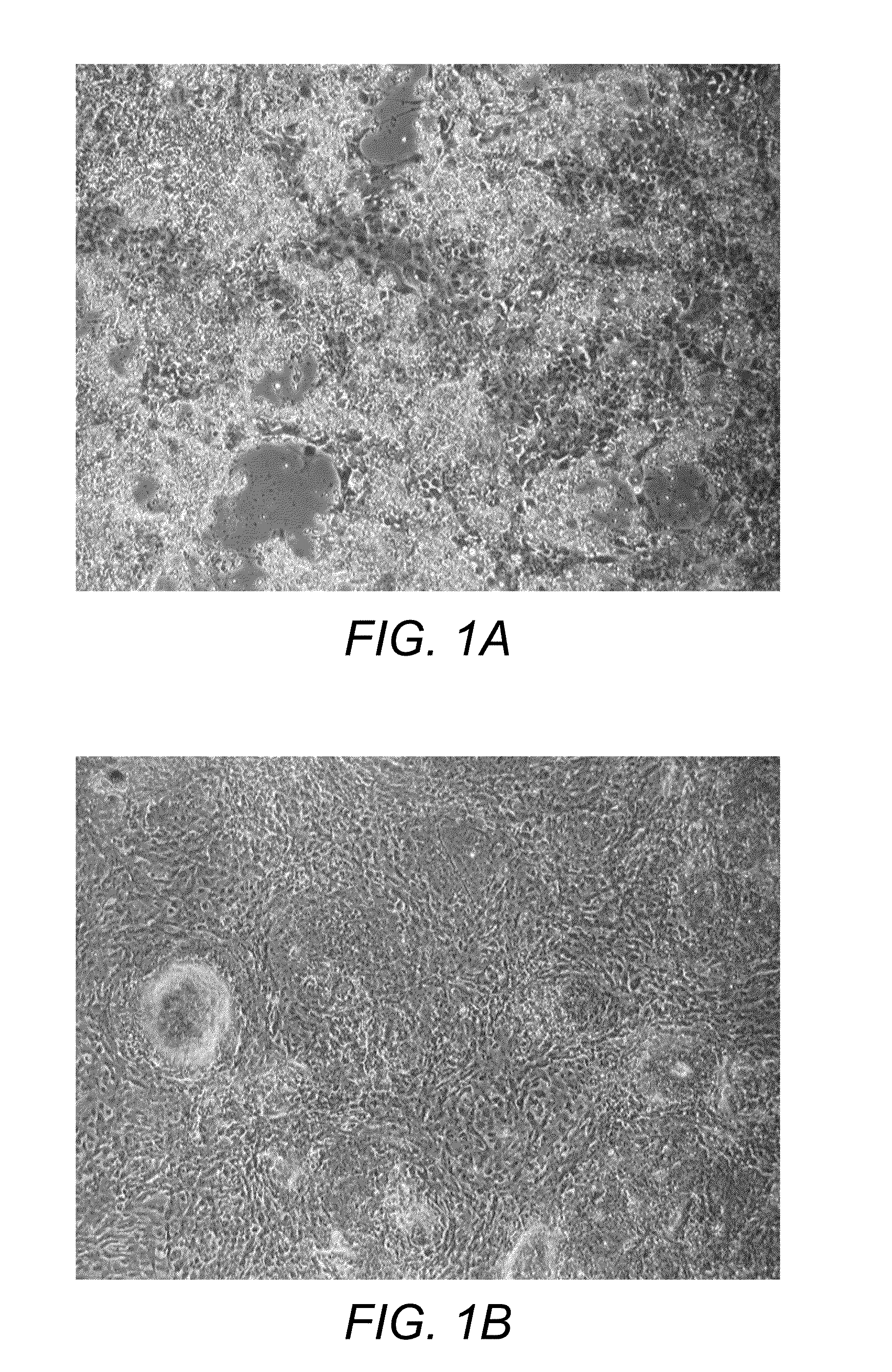
[0207]The morphology of the cells changes dramatically throughout the protocol. After 14 days in our chondrogenic medium the cells show classic chondrocyte morphology with rounding up of the cell body and clustering of cells into dense colonies (FIG. 1). On day 14 the media is changed to dedifferentiation media and at days 15 through 19 in dedifferentiation media, the cells show a fibroblast like morphology and start to repopulate the culture surface

[0208]A high proportion of the original ES cells died and formed rafts of dead cells that appeared as phase bright clumps just above the cell layer. These clumps could be washed off but not without substantial agitation. After 14 days the confluency was as low as 20%. However, the increase in colony density suggested that this decrease in confluency is not directly related to a decrease in cell number.

[0209]After replacement of the chondrogenic medium with de-differentiation media, cell morphology changed dramatically. Cells mi...

example 3

Characterisation of the DCCPCs

[0211]The de-differentiated cells were trypsin passaged and detached from the Matrigel® (and also plastic in later time points) in <5 minutes, as single cells. At this point the suspension of cells was used either for construct formation or the plastic adherent cells seeded for characterisation.

Plastic Adherent Cells:

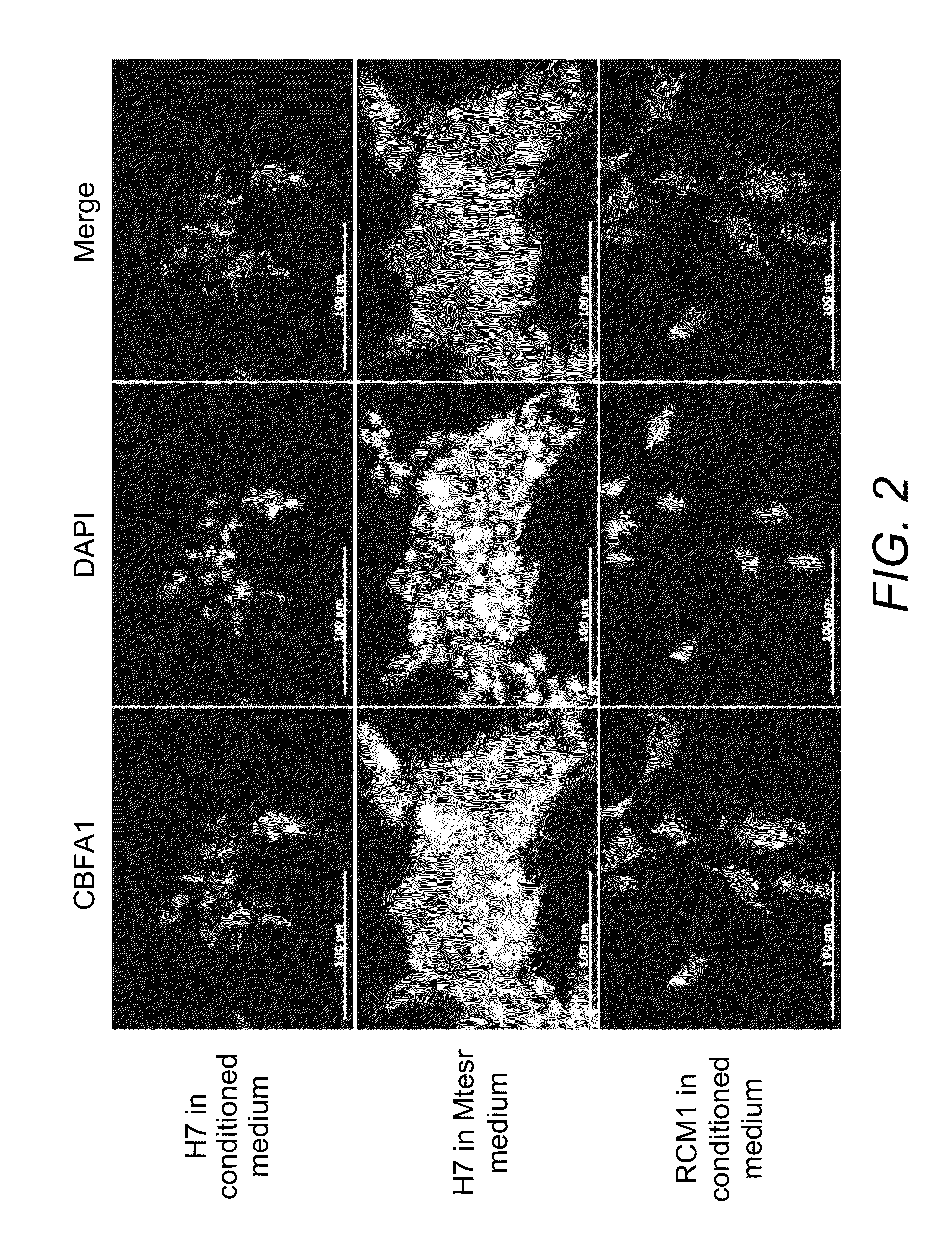
[0212]Plastic adherent DCCPCs maintained in de-differentiation media analysed using FLOW cytometry showed no ES pluripotency markers or MSC markers.

[0213]In agreement with the FLOW data, immunocytochemistry using a different Tra-1-60 antibody showed no evidence of TRA-1-60 protein expression in the DCCPCs. Tra-1-60 staining was not present on the plastic adherent DCCPCs. Undifferentiated H7 ES cells were used as a control for the antibody and showed bright membrane associated staining. After counting 1053 cells no TRA-1-60 positive cells were seen in the DCCPC population. Therefore TRA-1-60 protein expression is undetectable in this populat...


PUM

PropertyMeasurementUnit
surface areaaaaaaaaaaa
timeaaaaaaaaaa
concentrationaaaaaaaaaa

Abstract

The present invention provides an isolated population of chondrocyte precursor cells wherein 1% or less of the cells express Oct4, Nanog and / or TRA-1-60, 7% or less of the cells express no collagen II, collagen X, CD105 or Stro-1 and 85% or more of the cells express CBFA1, methods for preparing such cells and uses of chondrocyte cells derived from said precursor cells.

Description

CROSS-REFERENCE TO RELATED APPLICATIONS[0001]This application claims the benefit of U.S. Provisional Appln. No. 61 / 321,982, filed on Apr. 8, 2010, which is incorporated herein by reference.TECHNICAL FIELD[0002]The present invention relates generally to the fields of cell biology, embryonic stem cells, and cell differentiation. The invention discloses chondrogenic progenitor cells and a method for the preparation of such cells and fully differentiated chondrocytes, including uses of the cells.BACKGROUND[0003]Chondrocytes are specialised cells found in cartilage. Chondrocytes in cartilage produce a large amount of extracellular matrix which is composed of collagen fibers, ground substance, which is rich in proteoglycan, and elastin fibers. The cartilage tissue performs a structural and mechanical function in skeletal joints and any disease or injury is consequently debilitating for patients.[0004]Cartilage degradation is a hallmark of two disease groups: osteoarthritis, a degenerative...

Claims


Application Information

Patent Timeline
Patent Type & Authority Patents(United States)
IPC IPC(8): C12N5/00C12N5/02C12N5/071C12N5/07C12N5/10A61K35/32C12N5/077C12N5/0775
CPCC12N5/0655C12N5/0662A61K35/32C12N2500/25C12N2501/15C12N2501/39C12N2506/02A61P19/00A61P19/02A61P19/04C12N2500/84C12N2500/90C12N2506/03G01N33/5044
Inventor NOBLE, BRENDON STEWARTPIER, DAVID MATTHEW
Owner THE UNIV COURT OF THE UNIV OF EDINBURGH
Why Eureka
Try Eureka