Nano-vehicle derived from tumor tissue, and cancer vaccine using same

a tumor and tumor technology, applied in the field of cancer vaccines, can solve the problems of affecting the production efficiency of cancer vaccines, and affecting the effect of anticancer activity, etc., and achieves the effects of low production yield, difficult separation and preparation, and low production yield

Active Publication Date: 2015-12-29
ROSETTA EXOSOME CO LTD
View PDF13 Cites 1 Cited by
  • Summary
  • Abstract
  • Description
  • Claims
  • Application Information

AI Technical Summary

Benefits of technology

[0016]In spite of reports on using spontaneously shed extracellular vesicles in the development of cancer vaccines, due to their effective anticancer activity, significant limitations have been imposed on the clinical application of the extracellular vesicles because they require in vitro culturing of a patient's cells and are difficult to separate and prepare, in addition to resulting in a low production yield when prepared. In contrast, the tumor tissue-derived nanovesicles according to the present invention can be prepared at high yield while retaining similar properties to those of spontaneously shed extracellular vesicles. Further, the tumor tissue-derived nanovesicles can be diversely modified and can be used as a cancer antigen useful in the development of a cancer vaccine.

Problems solved by technology

However, an effective cure for cancer has not yet been found, and thus, there is a pressing need for novel cancer treatments.
These treatments, however, may provoke significant side effects including the onset of secondary cancer, the metastasis of cancer, immune suppression, and aberrant cellular metabolisms.
Cancer is considerably heterogeneous, and cancer-specific antigens are difficult to define.
Accordingly, a standardized treatment cannot guarantee success in the therapy of cancer.
The use of cancer cells themselves, however, may evoke side effects including the risk of oncogenesis and autoimmune responses.
However, extracellular vesicles are disadvantageous in practical clinical application for the following reasons.
It is difficult to secure extracellular vesicles in a large quantity because they are released in a trace amount.
Intricate separation and purification of extracellular vesicles requires high expense and much time.

Method used


Image

  • Nano-vehicle derived from tumor tissue, and cancer vaccine using same
  • Nano-vehicle derived from tumor tissue, and cancer vaccine using same
  • Nano-vehicle derived from tumor tissue, and cancer vaccine using same

Examples


example 1

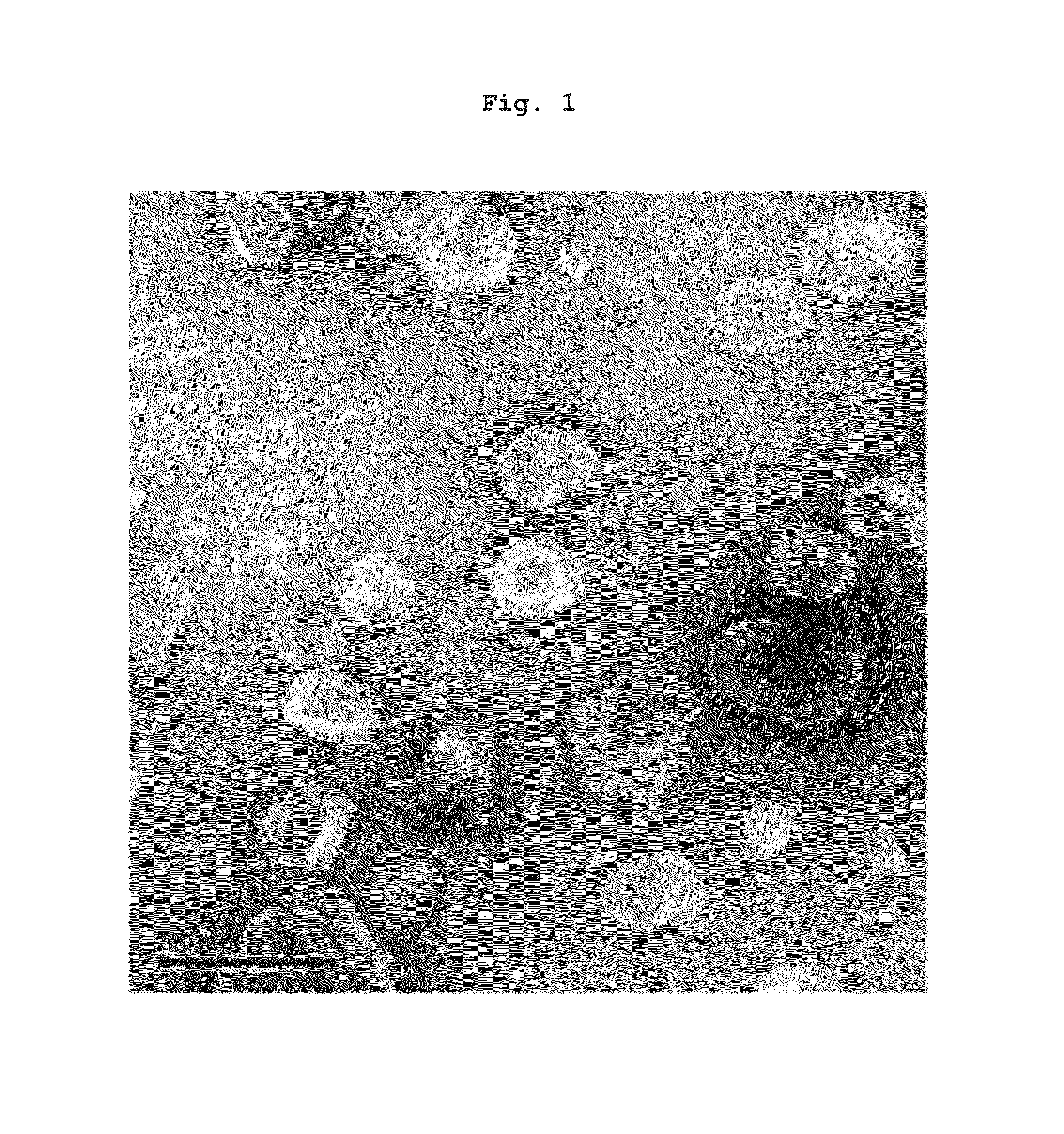
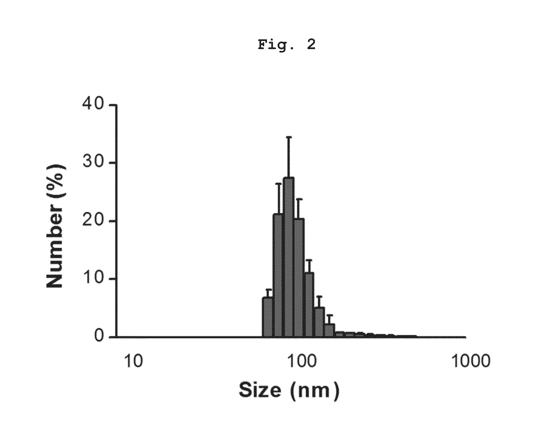
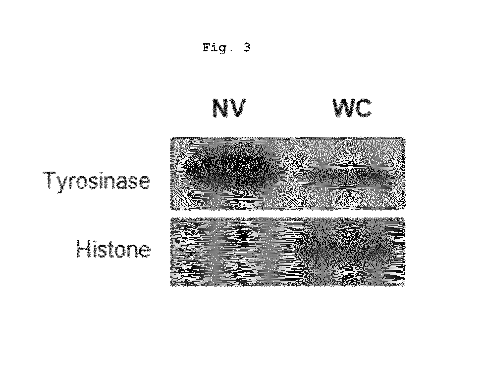
Preparation of Nanovesicles from Melanoma Cells Using Sonication and Their Characterization

[0055]The melanoma cell line (B16BL6) was subcutaneously injected into mice (C57 / BL6, female), cultured for 2˜3 weeks to a mass size of 1.0˜1.5 cm, and then tumor tissues were obtained by surgical excision. The melanoma tissue was ground and passed through a 45 μm filter for homogenization, followed by incubation at 4° C. for 30 min in a hypotonic solution. Then, the filtrate was homogenized with 100 strokes of a homogenizer and the homogenate was adjusted to have a final salt concentration of 150 mM to form vesicles. Centrifugation at 500×g for 10 min removed nucleoproteins and intact cells as a pellet, and the supernatant was sonicated for 30 min in a water bath sonicator to form nano-sized vesicles which were constant in size. Subsequently, cell debris and mitochondria were removed by centrifugation at 10,000×g for 20 min. After being collected, the supernatant was adjusted into a volume of...

example 2

Preparation of Nanovesicles from Colorectal Cancer Cells Using Sonication and Their Characterization

[0057]The colorectal cell line (Colon26) was subcutaneously injected into mice (BALB / C, female), cultured for 2˜3 weeks to a mass size of 1.0˜1.5 cm, and then tumor tissues were obtained by surgical excision. The same procedure as in Example 1 was repeated. That is, the colorectal cancer tissue was ground and filtered for homogenization, followed by incubation at 4° C. for 30 min in a hypotonic solution. Then, the filtrate was homogenized with 100 strokes of a homogenizer and the homogenate was adjusted to have a final salt concentration of 150 mM to form vesicles. Centrifugation at 500×g for 10 min removed nucleoproteins and intact cells as a pellet, and the supernatant was sonicated for 30 min in a water bath sonicator to form nano-sized vesicles which were constant in size. Subsequently, cell debris and mitochondria were removed by centrifugation at 10,000×g for 20 min. After being...

example 3

Preparation of Nanovesicles from Melanoma Cells Using Extrusion and Their Characterization

[0058]The melanoma cell line (B16BL6) was subcutaneously injected into mice (C57 / BL6, female), cultured for 2˜3 weeks to a mass size of 1.0˜1.5 cm, and then tumor tissues were obtained by surgical excision. The melanoma tissue was ground and passed through a 45 μm filter for homogenization, followed by incubation at 4° C. for 30 min in a hypotonic solution. Then, the filtrate was homogenized with 100 strokes of a homogenizer and the homogenate was adjusted to have a final salt concentration of 150 mM to form vesicles. Centrifugation at 500×g for 10 min removed nucleoproteins and intact cells as a pellet, and the supernatant was rendered to pass three times through a membrane filter with a pore size of 1 μm and then three times through a membrane filter with a pore size of 0.4 μm. After being collected, the filtrate was adjusted into a volume of 10 ml, placed on a sucrose cushion comprising 0.1 ...


PUM

PropertyMeasurementUnit
sizeaaaaaaaaaa
sizeaaaaaaaaaa
volumeaaaaaaaaaa

Abstract

The present invention relates to a cancer vaccine, and specifically, to a pharmaceutical composition for treating cancer containing a nano-vehicle derived from tumor tissue, and a method for treating cancer using the nano-vehicle derived from tumor tissue, and the like. According to the present invention, a nano-vehicle antigen derived from tumor tissue indicates a high yield while having properties similar to those of an exocellular vehicle, and can be variously modified, and is thus expected to be very useful for developing the cancer vaccine.

Description

CROSS-REFERENCE TO RELATED APPLICATIONS[0001]This application is a 371 of PCT / KR2012 / 004162 filed on May 25, 2012, which claims the benefit of Korean Patent Application No. 10-2011-0050854 filed on May 27, 2011, the contents of each of which are incorporated herein by reference.TECHNICAL FIELD[0002]The present invention relates to a cancer vaccine. More particularly, the present invention relates to a pharmaceutical composition for the treatment of cancer, comprising nanovesicles derived from a tumor tissue, and a method for treating cancer using the tumor tissue-derived nanovesicles.BACKGROUND ART[0003]Cancer is a broad group of various diseases, all involving unregulated cell growth without mortality, as a result of the abnormal differentiation or proliferation induced by the modification of the structural and functional unit cells. In cancer, cells invade nearby parts of the body, and may also spread to more distant parts of the body through the lymphatic system or bloodstream, a...

Claims


Application Information

Patent Timeline
Patent Type & Authority Patents(United States)
IPC IPC(8): A61K9/133A61K47/00A61K47/36A61K47/40A61K39/00A61K9/127A61K47/10A61K38/17
CPCA61K39/0011A61K9/127A61K47/40A61K47/10A61K2039/55561A61K38/1709A61P35/00A61K47/6951
Inventor GHO, YONG SONGKIM, YOON KEUNLEE, EUN YEONGJANG, SU CHUL
Owner ROSETTA EXOSOME CO LTD
Why Eureka
Try Eureka