Method of using microfluidic chip for nucleic acid hybridization
a microfluidic chip and nucleic acid technology, applied in the field of nucleic acid hybridization, can solve the problem that the nucleic acid of unhybridized test macromolecular nucleic acids cannot be displayed on the porous substrate, and achieve the effect of rapid nucleic acid hybridization
- Summary
- Abstract
- Description
- Claims
- Application Information
AI Technical Summary
Benefits of technology
Problems solved by technology
Method used
Image
Examples
example 1
Analysis of Elution of Test Double Helix DNA / Syber and Probe / HEX Using SDS
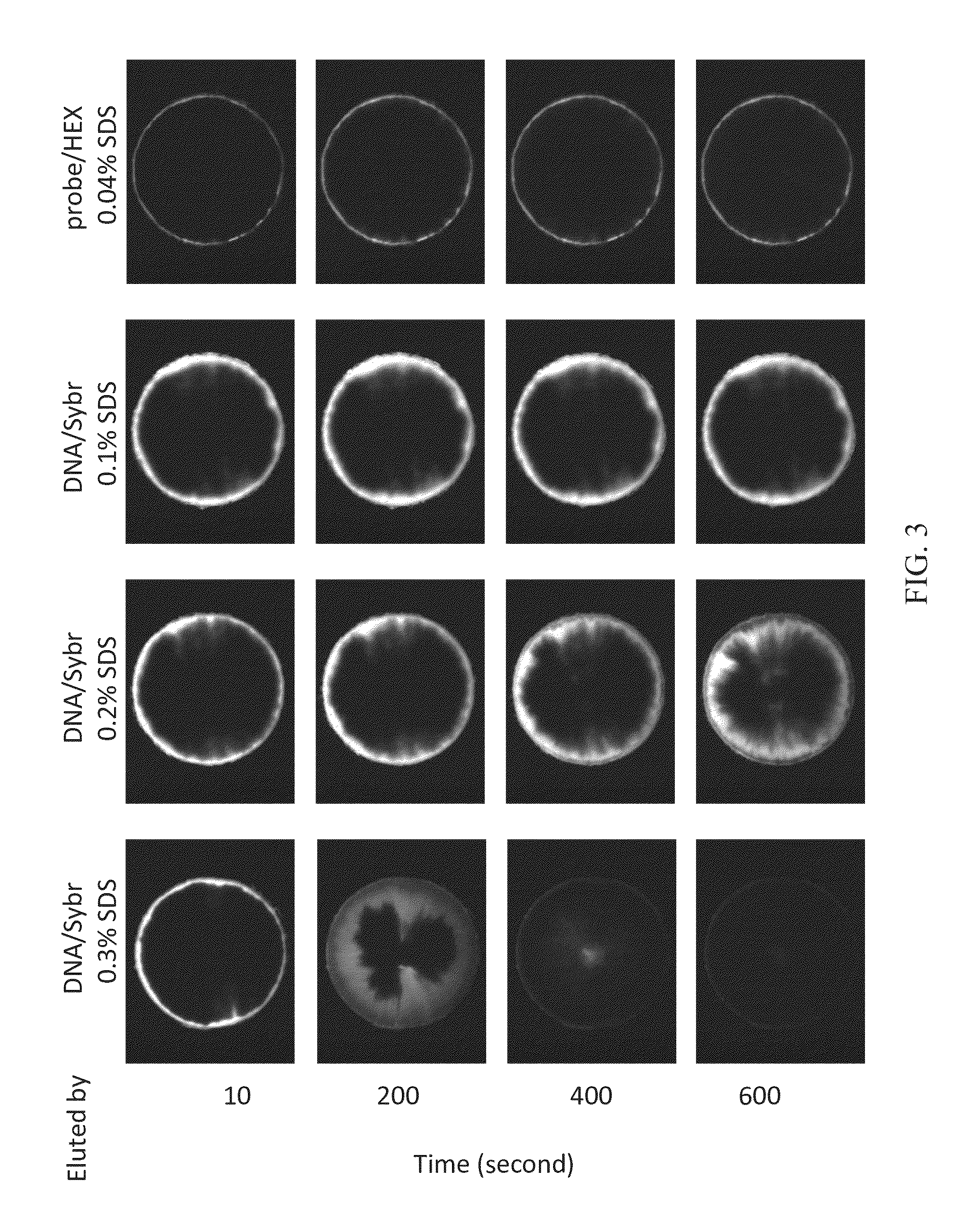
[0032]FIG. 3 clearly illustrates the results upon the mixture of test double helix DNA, which is bound to SYBR® Green I stain and abbreviated as DNA / Sybr, and the probe / HEX were injected, and different concentrations of SDS were used for elution. The concentrations of SDS are 0.04% (w / v), 0.1% (w / v), 0.2% (w / v), and 0.3% (w / v). As shown in FIG. 3, test macromolecular 16S rDNA (approximately 1540 bp in length) began to move towards the center of the substrate after eluting for 200 seconds using 0.1% (w / v) SDS; the test macromolecular DNA began to move significantly towards the center of the substrate after eluting for 400 seconds using 0.2% (w / v) SDS; almost all test macromolecular DNA move to the center of the substrate after eluting for 400 seconds using 0.3% (w / v) SDS. Meanwhile, small molecular probe / HEX were washed away immediately by the elution of 0.04% (w / v) SDS since they were not hybridized with the t...
example 2
Analysis of Elution of Test Double Helix DNA / Syber and Probe / HEX Using Sarcosine
[0033]FIG. 4 clearly illustrates the results upon the mixture of test double helix DNA, which is bound to SYBR® Green I stain and abbreviated as DNA / Sybr, and the probe / HEX were injected and different concentrations of sarcosine were used for elution. The concentrations of sarcosine are 0.2% (w / v), 0.3% (w / v), and 0.4% (w / v). As shown in FIG. 4, a test macromolecular DNA began to move towards the center of the substrate after eluting for 600 seconds using 0.3% (w / v) sarcosine; the macromolecular DNA began to move significantly towards the center of the substrate after eluting for 400 seconds using 0.4% (w / v) sarcosine and almost all test macromolecular DNA were washed away and were discharged from the substrate of the chip via the outlet 12 after eluting for 600 seconds using 0.4% (w / v) sarcosine. On the other hand, small molecular probe / HEX were washed away immediately by the elution of 0.2% (w / v) or 0....
example 3
Analysis of Elution of Probe / HEX Using a Mixture of SDS and SSC
[0034]FIG. 5 clearly illustrates the results upon probe / HEX were injected and solely 0.1% (w / v) SDS or a mixture of 0.1% (w / v) SDS and 0.1× SSC were used for elution. As shown in FIG. 5, during the process of elution using solely anionic surfactant, SDS, except a few immobile small molecular probes trapped in the substrate, free probes were coated by SDS to form micelles and migrated rapidly towards the center of the substrate. During the movement, the micelles coupled with the positive charges on the substrate and became immobile. The immobilization of micelles became significant from after 300 seconds since the florescence light gradually faded from the outside towards the inside of substrate. Comparing with the movement of probes during the elution by a mixture of SDS and 0.1× SSC, it was found that after 200 seconds, the Cl− and citrate− competed with the probe / DNA for the positive charges on the substrate, which eff...
PUM
| Property | Measurement | Unit |
|---|---|---|
| reaction time | aaaaa | aaaaa |
| diameter | aaaaa | aaaaa |
| diameter | aaaaa | aaaaa |
Abstract
Description
Claims
Application Information
 - R&D
- Intellectual Property
- Life Sciences
- Materials
- Tech Scout
- Unparalleled Data Quality
- Higher Quality Content
- 60% Fewer Hallucinations
Browse by: Latest US Patents, China's latest patents, Technical Efficacy Thesaurus, Application Domain, Technology Topic, Popular Technical Reports.
© 2025 PatSnap. All rights reserved.Legal|Privacy policy|Modern Slavery Act Transparency Statement|Sitemap|About US| Contact US: help@patsnap.com


