Apparatus and method for assessing tissue composition

a tissue composition and monitoring apparatus technology, applied in the field of tissue composition monitoring apparatus, can solve the problems of even ultrasound images, difficult to quantify optical sensing data in a beating heart, and significant errors in spectrophotometer readings

Active Publication Date: 2016-12-27
LIM BERNARD BOON CHYE
View PDF33 Cites 12 Cited by
  • Summary
  • Abstract
  • Description
  • Claims
  • Application Information

AI Technical Summary

Benefits of technology

[0015]In one aspect of the present invention, a tissue composition monitoring apparatus, an ablation lesion monitoring method and an ablation lesion determination automated algorithm incorporates Arrhenius's model for thermal denaturation kinetics for determining the characteristics of the ablation lesion. The apparatus includes a basket catheter configuration for achieving stability and consistent contact pressure during ablation for collection of optical spectra. The basket catheter incorporates a linear portion which “hugs” the conical portion of the pulmonary vein during atrial fibrillation ablation and allows a variable spatial offset of between 10 mm to 20 mm between the illuminating optic fiber and the receiving optic fiber.

Problems solved by technology

To quantify optical sensing data in a beating heart is challenging.
This movement can lead to significant error in spectrophotometer readings and even ultrasound images.
Harks et al., (WO 2012 / 049621 A1), hereinafter “Harks,” incorporates optic fiber within a standard ablation catheter that is subject to cardiac motion which can lead to significant errors in spectrophotometer readings and is subject to different contact pressures which can lead to significant errors in spectrophotometer readings.
With past ablation systems such as those used for atrial fibrillation and ventricular tachycardia, there is no method of checking for transmurality so that there is a significant variation in power delivery and duration of power delivery between different operators.
For ventricular tachycardia, the recurrence rate is even higher because ventricular tissue is thicker and so it is harder to achieve transmurality.
Studies have shown that when inpatients come back with recurrence of atrial fibrillation, the pulmonary veins typically have reconnected, proving the inability of current ablation systems to achieve transmurality.
Histology studies have shown that a rim of unablated tissue as little as 1.4 mm can lead to reconnection of pulmonary veins and lead to recurrence of atrial fibrillation.
As such, the operator is not able to discern when the nerves have been ablated.
As such there have been reports of thrombus formation and even dissection of the renal artery, due perhaps to delivery too much power for too long.
The geometry of standard ablation catheters does not permit a wide spatial offset between illuminating and receiving optic fibers positioned at the distal tip that is required to truly achieve analysis of transmurality for the reason that typically, only between 3 to 4 mm of the distal tip of the catheter is in contact with the muscle tissue.
It is difficult to ensure a constant end-on or side-on contact of standard ablation catheters with the tissue surface.

Method used


Image

  • Apparatus and method for assessing tissue composition
  • Apparatus and method for assessing tissue composition
  • Apparatus and method for assessing tissue composition

Examples


Embodiment Construction

Reference Listing

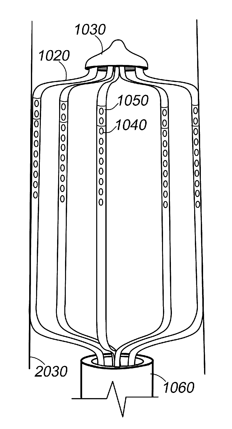
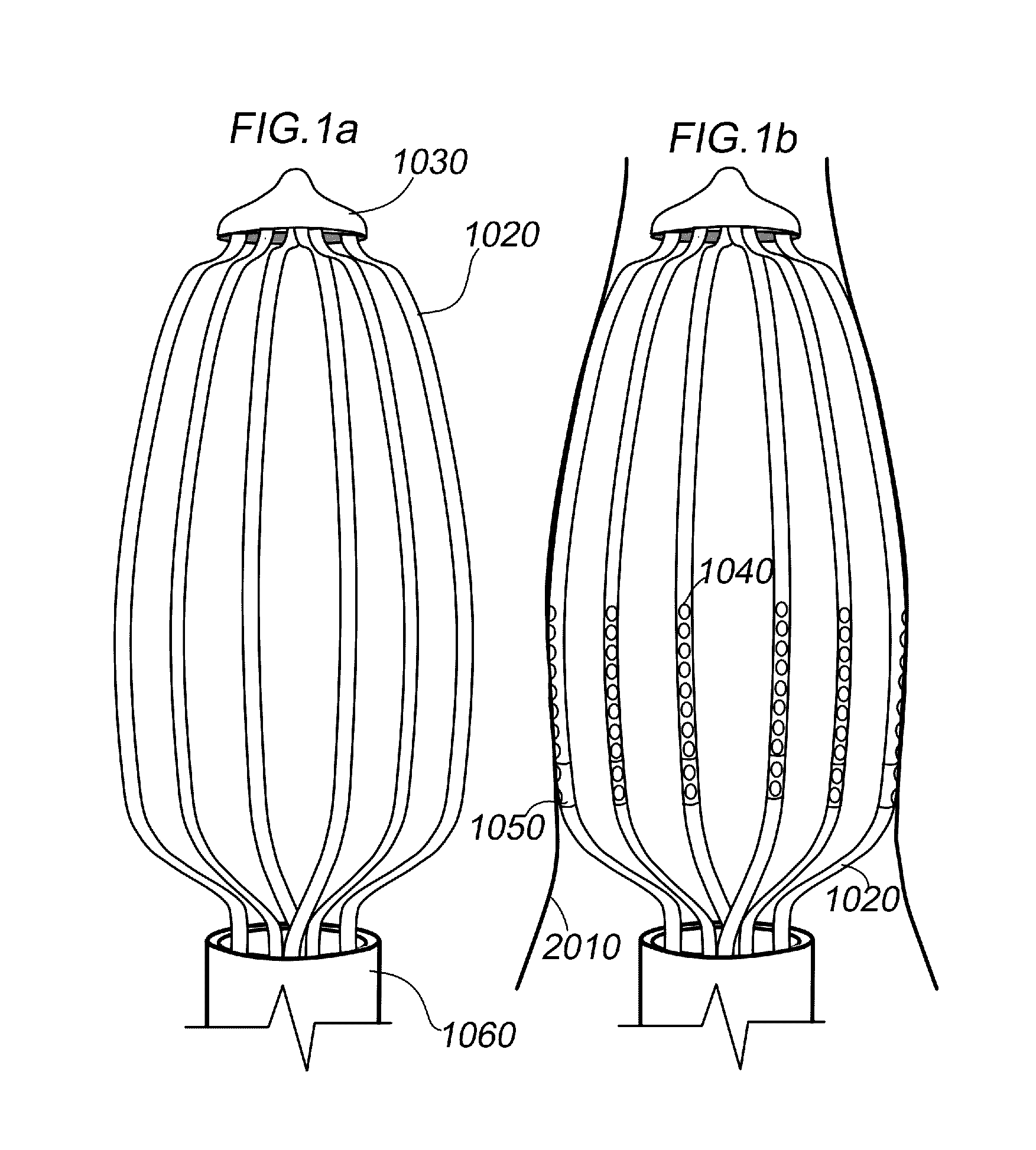
[0047]1000 catheter[0048]1010 optical sensor[0049]1020 spline / arm[0050]1024 photon path[0051]1025 optic fiber[0052]1027 receiving fiber[0053]1028 illuminating fiber[0054]1029 mirror[0055]1030 cap[0056]1040 aperture[0057]1050 electrode[0058]1051 wire to electrodes[0059]1060 sheath[0060]1070 basket[0061]1080 MEMS gyroscope[0062]2000 pulmonary vein[0063]2010 opening pulmonary vein[0064]2020 renal artery[0065]2030 opening renal artery[0066]3000 ablated tissue

[0067]The present invention includes a catheter including optical sensors for generating optical sensing data that are indicative of the optical property of the tissue. FIG. 1 shows a preferred embodiment according to the present invention for determining the properties of a tissue. The distal end of catheter 1000 expands into a basket 1070 configuration with a plurality of arms or splines 1020.

[0068]By way of non-limiting examples shown in FIG. 4, the basket can have a plurality of shapes. In a preferred embodiment...


PUM


Abstract

A tissue composition monitoring apparatus that includes a catheter with an optical sensor for tissue composition monitoring and ablation lesion determination via an automated algorithm incorporating Arrhenius model thermal denaturation kinetics for determining the characteristics of tissue for example the transmurality of the ablation lesion.

Description

[0001]This Application Claims the benefit of U.S. Provisional Patent Application No. 61 / 673,025 filed Jul. 18, 2012 and titled SYSTEMS AND METHODS FOR IMPROVING EFFICACY AND SAFETY IN ABLATION PROCEDURESFIELD OF THE INVENTION[0002]The invention relates to a tissue composition monitoring apparatus, a tissue composition monitoring method and an ablation lesion determination automated algorithm incorporating Arrhenius model thermal denaturation kinetics for determining the characteristics of tissue for example the transmurality of the ablation lesion.BACKGROUND OF THE INVENTION[0003]Methods incorporating diffuse reflectance spectroscopy (DRS), into standard ablation catheters suffer from instability of collection of optical spectra due to motion of the catheter and inconsistent contact pressure. Furthermore, DRS deployed with a small offset between illuminating and collecting fibers have only small penetration depths. Standard ablation catheters cannot achieve a sufficient spatial offs...

Claims


Application Information

Patent Timeline
Patent Type & Authority Patents(United States)
IPC IPC(8): A61B5/00
CPCA61B5/0075A61B19/5244A61B5/0073A61B5/0044A61B5/0084A61B5/6858A61B18/1492A61B2017/00057A61B2018/00267A61B2018/00357A61B2018/00404A61B2018/00434A61B2018/00511A61B2018/00577A61B2018/00642A61B2018/00773A61B2034/2048A61B2090/3966
Inventor LIM, BERNARD BOON CHYE
Owner LIM BERNARD BOON CHYE
Why Eureka
Try Eureka