Cell attachment coatings and methods using phosphorous-containing photoreagent

a technology of phosphorous-containing photoreagents and cell attachment coatings, which is applied in the field of article-based cell attachment coatings, can solve the problems of ineffective surface, inability to provide the intended function of the surface, and difficulty in the technical process of mimicking the natural function of collagen on a synthetic surface, so as to improve the number of proliferating cells, improve the cell attachment surface, and enhance the effect of cell attachmen

Active Publication Date: 2017-04-25
SURMODICS INC
View PDF37 Cites 0 Cited by
  • Summary
  • Abstract
  • Description
  • Claims
  • Application Information

AI Technical Summary

Benefits of technology

[0007]The present invention is directed to articles having biocompatible cell attachment coatings, and methods for forming these coatings. The coatings have an arrangement of coating components including a phosphorous-containing component with photoreactive groups, and a cell attachment component that is a cell attachment molecule comprising amino acids. The cell attachment molecule comprises an extracellular matrix (ECM) protein, or a peptide that includes an active portion of an ECM protein. The phosphorous-containing component is bonded in the coating using the photoreactive groups, resulting in the presentation of chemical groups which provide a distinct and improved cell attachment surface. Generally, the coatings promote enhanced attachment of cells on the coated surface of the article. In turn, this increases the number of proliferating cells on the device surface. In the body, these improved functional surfaces can enhance generation of tissue in association with the coated surface.
[0008]In some aspects, the coatings are formed on implantable medical articles, and these articles can be used in methods for the treatment of a medical condition. In the body this can promote the formation of tissue in association with the article. The enhanced cell attachment and proliferation improves integration of the implant in the body, and makes the implant more effective for medical use. The invention also contemplates the use of these coatings on cell culture articles and in vitro methods for enhancing cell attachment.
[0010]The coating can be formed by a method in which a first composition including the first component comprising the phosphorous-containing group and the bonding group is applied to a device surface. A second composition including the cell attachment molecule comprising amino acids, which is an ECM protein, or a peptide that includes an active portion of an ECM protein, is applied after the first composition is applied. The method also includes a step of irradiating the coating which can be performed after the first composition is applied, after the second composition is applied, or both. Irradiation of the bonding group causes its activation and covalent bonding to a target moiety and immobilization in the coating. The target moiety that reacts with the bonding group can be a component of the device surface, another first component, or the component selected from the ECM protein or peptide that includes an active portion of an ECM protein. The presentation of components in the coating achieved by irradiation and bonding using the bonding groups provides a particularly favorable presentation of coating components shown herein to enhance attachment of cells to the device surface.
[0011]Cell attachment studies associated with the invention revealed that the inventive coatings promoted a greater level of cell attachment to the coated surfaces over coatings using collagen alone, or the phosphorous-containing reagent alone. The enhanced cell attachment results were rather surprising considering that, in theory, the coating process should result in the phosphorous-containing reagent being buried under the collagen layer.
[0012]The coatings of the present invention can be formed on implantable medical articles such as, but not limited to, hernia meshes, aneurysm devices, and prosthetic devices such as coronary stents. Following implantation of the coated article in the body, the coating promotes an increased level of attachment of cells, such as endothelial cells and / or fibroblasts. Over time, tissue is formed on or around the coated implantable article. The natural tissue response can enhance integration, function, and lifetime of the implanted article.

Problems solved by technology

However, the process of mimicking the natural function of collagen on a synthetic surface is technically challenging.
Preparation of collagen-containing coatings can often result in surfaces that do not provide the intended function following implantation.
In coatings wherein collagen is not properly immobilized, collagen can leach out or be released from the surface, rendering the surface ineffective.
Further, even if collagen is successfully immobilized in a coating, the coating may have the ability to attract cells to a certain extent, but not in a manner that provides for subsequent proliferation of the cells, which is important for tissue formation.

Method used


Image

  • Cell attachment coatings and methods using phosphorous-containing photoreagent
  • Cell attachment coatings and methods using phosphorous-containing photoreagent
  • Cell attachment coatings and methods using phosphorous-containing photoreagent

Examples


example 1

Synthesis of Photocollagen (COL1)

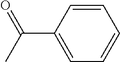
[0124]Bovine skin collagen (Semed S Powder) was purchased form Kensey Nash Corporation (Exton, Pa.). This collagen has the proportions of type I collagen (95%) and type III collagen (5%) that are usual for skin-derived collagens. Type 1 collagen was photoderivatized by the addition of (benzoylbenzoic acid)-(epsilon aminocaproic acid)-(N-oxysuccinimide)(BBA-EAC-NOS). BBA-EAC-NOS has a benzophenone photoactivatible group on one end (benzoyl benzoic acid, BBA), a spacer in the middle (epsilon aminocaproic acid, EAC), and an amine reactive thermochemical coupling group on the other end (N-oxysuccinimide, “NOS”). BBA-EAC was synthesized from 4-benzoylbenzoyl chloride and 6-aminocaproic acid. Then the NOS ester of BBA-EAC was synthesized by esterifying the carboxy group of BBA-EAC by carbodiimide activation with N-hydroxysuccimide to yield BBA-EAC-NOS. See U.S. Pat. No. 5,744,515 (columns 13 and 14), and U.S. Pat. No. 7,220,276. Atelocollagen (Biom'Up, Sai...

example 2

Synthesis of Phosphorous-Containing Crosslinker 1

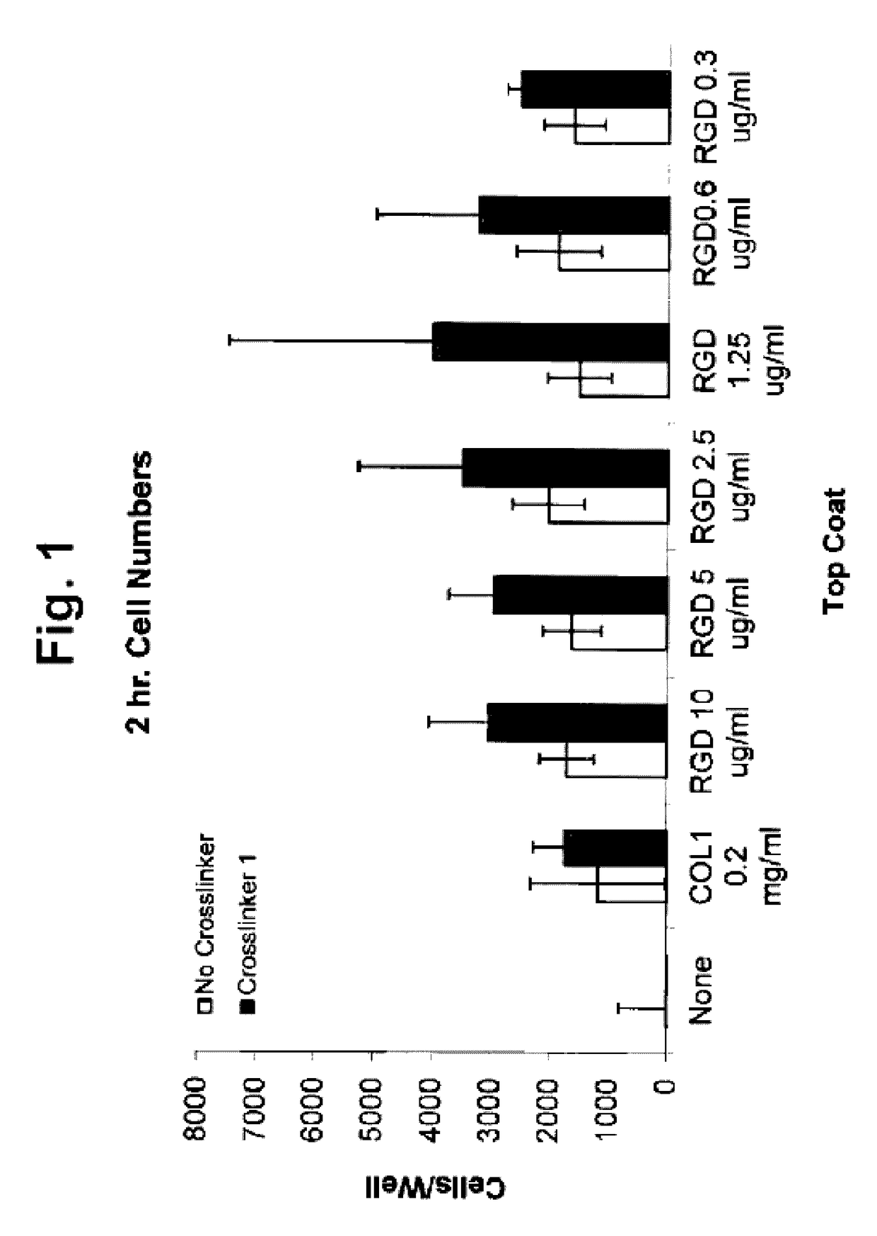
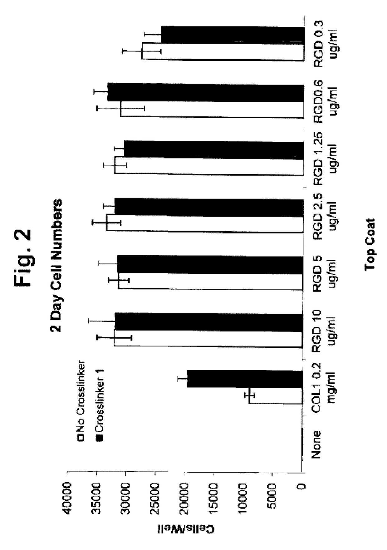
[0125]Low molecular weight compounds that include photoreactive groups and phosphorous-containing groups were used to form the coatings of the invention. Such compounds include bis(4-benzoylphenyl)hydrogen phosphate, and alkaline salts thereof and tris(4-benzyolphenyl) phosphate. These phosphorous-containing crosslinkers can be synthesized according to US Patent Publication No. 2012 / 00463384 (Kurdyumov, at al.).

example 3

Synthesis of Photopolymer Peptides

[0126]Photopolymer peptides are prepared as described in U.S. Pat. No. 6,514,734 (Clapper et al.)


PUM

PropertyMeasurementUnit
thicknessaaaaaaaaaa
diameteraaaaaaaaaa
thickaaaaaaaaaa

Abstract

Cell attachment coatings for articles such as implantable medical devices and cell culture vessels are disclosed. The coatings include an intermediate coater layer which includes a phosphorous-containing component that is bonded in the coating by reacted photoreactive functional groups. The coating also include a second coated layer including an immobilized ECM protein or peptide that includes an active portion of an ECM protein that is able to serve as an outer layer to contact cells during use. The coatings promoted enhanced cell binding and growth.

Description

CROSS-REFERENCE TO RELATED APPLICATION[0001]The present non-provisional Application claims the benefit of commonly owned provisional Application having Ser. No. 61 / 666,516, filed on Jun. 29, 2012, entitled CELL ATTACHMENT COATINGS AND METHODS USING PHOSPHOROUS-CONTAINING CROSSLINKER, which Application is incorporated herein by reference in its entirety. Also, the entire contents of the ASCII text file entitled “SRM0141_Sequence_Listing_2_ST25.txt” created on May 5, 2014, having a size of 178 kilobytes is incorporated herein by reference.FIELD OF THE INVENTION[0002]The invention relates to cell attachment coatings for articles and methods for enhancing cell attachment to an article surface.BACKGROUND OF THE INVENTION[0003]Thin surface coatings on implantable medical articles have proved to be valuable in cases where it is desired to provide the article surface with a property that is not present on the uncoated surface. Polymeric coatings have been used to improve the wettability and...

Claims


Application Information

Patent Timeline
Patent Type & Authority Patents(United States)
IPC IPC(8): C12N11/06A61K38/08A61L31/10A61L31/16
CPCC12N11/06A61L31/10A61L31/16C08L71/00C08L89/06C12N2303/00A61L2300/25A61L2300/252A61L2300/64A61L2420/08
Inventor MCGONIGLE, JOSEPH S.
Owner SURMODICS INC
Why Eureka
Try Eureka