Drainage devices and methods for use

a technology of lymphatic drainage and draining device, which is applied in the direction of catheters, blood vessels, angiography, etc., can solve the problems of reducing cardiac performance, affecting the accuracy of immune suppression, so as to reduce the infection rate, the effect of safe and minimally invasive methods and accurate indication of the level of immune suppression

Active Publication Date: 2017-06-20
LXS LLC +1
View PDF16 Cites 30 Cited by
  • Summary
  • Abstract
  • Description
  • Claims
  • Application Information

AI Technical Summary

Benefits of technology

[0018]The device can allow fluid communication between a lymphatic vessel and an external reservoir. For example, the device can open the wall of (i.e., access) the thoracic duct. The device can drain fluid from the lymphatic vessel. For example, the cannula can drain fluid from the thoracic duct. The device can drain lymphatic fluid from the lymphatic system, for example, when the patient becomes fluid-overloaded or suffers from congestive heart failure. The device can help prevent and / or treat congestive heart failure and pulmonary edema, for example, by drainage of the lymphatic system.
[0024]Periodic monitoring of lymphocyte antigen, for example, after solid organ transplant, can provide an early warning of graft rejection and a more accurate indication of the level of immune suppression. For example, lymph fluid can be withdrawn through the device and analyzed after a high risk graft or transplant procedure. For example, the device can be implanted and lymph fluid can be removed through the device and analyzed for graft rejection after a heart, lung, kidney, skin, liver, or small intestine transplant or graft. The device can provide a safe and minimally invasive method of lymphatic access, which could be provided with a transcutaneous catheter. Routine drainage of sensitized lymphocyte subpopulations could help prevent or rescue an acute episode of rejection.
[0025]As a clinical modality, catheter drainage of the thoracic duct through the device can remove the lymph fluid to deplete large numbers of CD-4 cells (for example, CD-4+ T cells). Combining drainage with conventional pharmacological therapy to decrease the infection rate in remaining CD-4 cells can promote a shift towards a virus-free state and represents an intriguing alternative to current treatment for HIV or other infectious diseases with similar modalities.
[0026]In accordance with another embodiment, a method is provided for reducing interstitial volume in a patient using a device comprising a tubular member that includes placing a distal end of the tubular member into a patient's venous system such that the distal end is located adjacent the thoracic duct; draining fluid from the lymphatic system via the thoracic duct through a lumen of the tubular member to reduce fluid volume within the patient's body; and discontinuing draining fluid when the fluid volume is sufficiently reduced.
[0028]In accordance with yet another embodiment, a method is provided for decreasing lymphocyte numbers in a patient affected by autoimmune or infectious disease using a device comprising a tubular member that includes placing a distal end of the tubular member into a patient's venous system such that the distal end is located adjacent the thoracic duct; and draining fluid from the lymphatic system via the thoracic duct through a lumen of the tubular member.

Problems solved by technology

Volume overload in patients with heart failure is an acute manifestation of a chronic decrease in cardiac performance such that the heart is unable to pump and circulate the volume of blood returning from the venous system.
A common and potentially life threatening consequence of decompensated heart failure is pulmonary edema, which presents as shortness of breath and poor oxygenation, both indicative of fluid accumulation in lung tissues and small airways.
If left untreated, patients are at risk for hypoxic arrest and death.
The sudden onset of volume overload is due to either a miscalculation in salt and fluid intake or poor medication compliance on the part of the patient, or a result of a worsening heart condition, renal impairment, or resistance to diuretic medications.
It is suggested that the high readmission rate is due to inadequate volume reduction during their previous admission.
Furthermore, approximately 30% of chronic heart failure patients on diuretics will develop resistance or renal failure as a result of their medication and require more invasive and higher risk therapies to control their volume status.
In this case, the lymphatic system is limited by volume capacity and higher than expected flow pressures generated by the venous system into which the thoracic duct is draining.
Interstitial fluid accumulation can become a critical issue in any post-operative patient and is especially concerning after heart surgery, when it is important to limit cardiac stress.

Method used


Image

  • Drainage devices and methods for use
  • Drainage devices and methods for use
  • Drainage devices and methods for use

Examples


Embodiment Construction

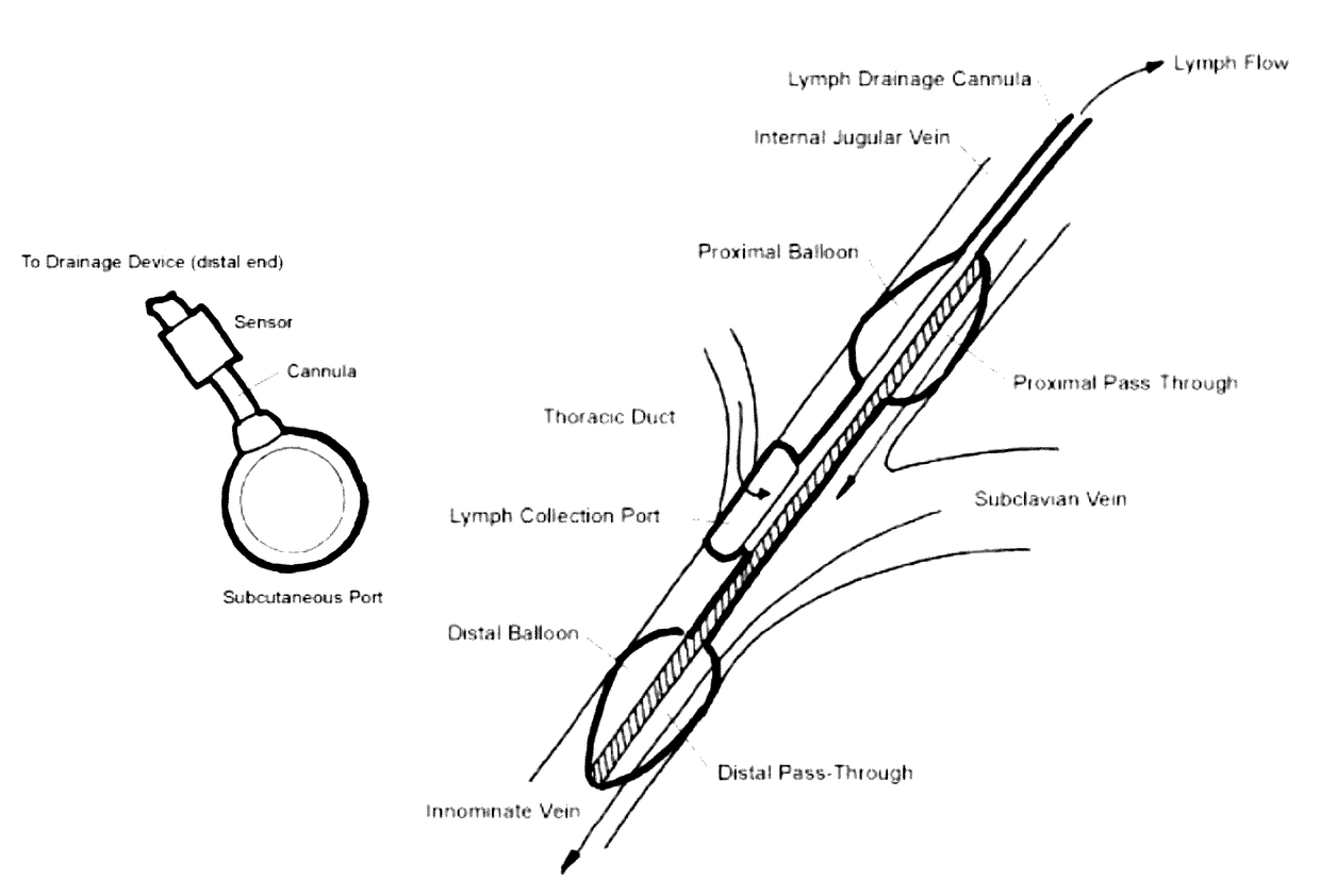
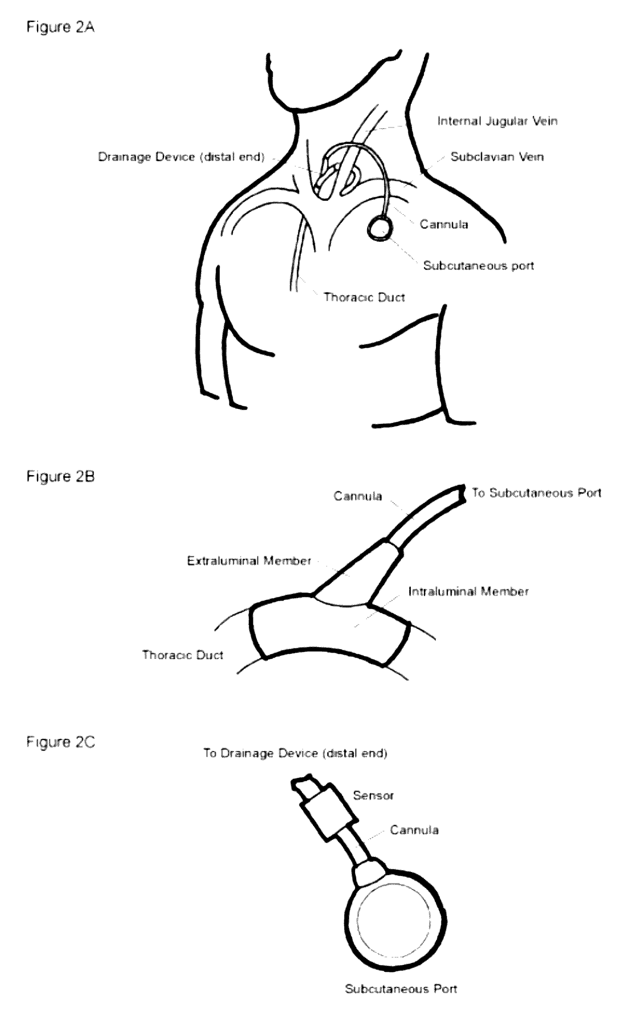
[0056]The devices and methods disclosed herein enable removal of fluid from a vessel within the body. For example, the device can be placed in a lymph or blood vessel to remove lymph fluid from the body. The removal of lymph fluid can reduce the hemostatic pressure in the lymphatic system, reducing symptoms of congestive heart failure.

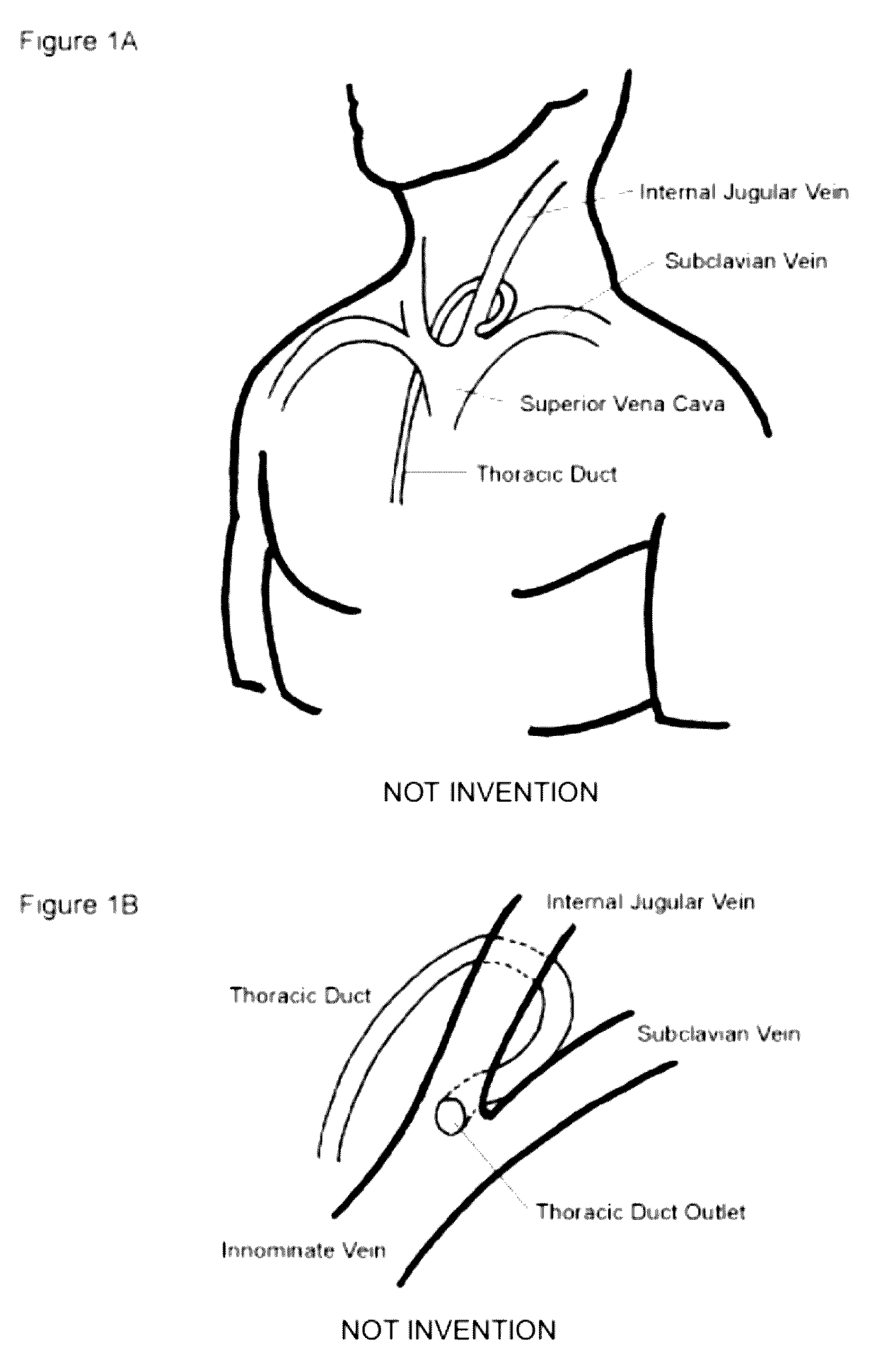
[0057]The device can access the lumen of a body vessel, for example, a lymphatic vessel and / or blood vessel. Fluid can drain from the vessel into the device. For example, the device can be in fluid communication with the thoracic duct or central veins (e.g., subclavian vein, internal jugular vein, superior vena cava, and innominate vein) shown in FIG. 1A and FIG. 1B. The device can reduce the pressure within the lumen by draining and removing excess fluid. The excess fluid can be stored in the device. The excess fluid can flow through a port and removed percutaneously. The device could treat pulmonary edema, for example, by reducing the pressure in the...


PUM


Abstract

Devices and methods for draining excess lymph fluid are disclosed. The device can be fixed to the blood vessel adjacent to the thoracic duct. The device can have a port for withdrawing lymph fluid exiting the thoracic duct. The device can have a cannula and / or subcutaneous port to draw the lymph fluid away from the thoracic duct and reduce hemostatic pressure in the lymphatic system.

Description

CROSS REFERENCE TO RELATED APPLICATIONS[0001]This application is a continuation-in-part of co-pending International Application No. PCT / US2010 / 000073, filed Jan. 12, 2010, which claims priority to U.S. Provisional Application No. 61 / 204,893, filed 12 Jan. 2009. The entire disclosures of these applications are incorporated by reference herein.BACKGROUND OF THE INVENTION[0002]1. Field of the Invention[0003]The present device and method relate generally to accessing, draining, and monitoring the lymphatic system. This device can be used to treat volume overload in heart failure, pulmonary edema, after surgical procedures and in other disease states in which interstitial volume removal or lymphatic sampling are of use.[0004]2. Description of the Related Art[0005]Congestive heart failure affects 5.5 million Americans and is a leading cause of death in the U.S. Each year, over one million patients are hospitalized for heart failure and 90% of these are diagnosed with volume overload. Volu...

Claims


Application Information

Patent Timeline
Patent Type & Authority Patents(United States)
IPC IPC(8): A61M39/02A61B5/03A61B5/0215A61B5/145A61B5/00A61M25/01A61M27/00A61F2/82A61F2/06A61M25/00
CPCA61M39/0247A61B5/0215A61B5/036A61B5/14539A61B5/41A61B5/413A61M25/01A61B5/418A61F2/064A61F2/82A61F2002/061A61M27/002A61M2025/0057A61M2025/0197A61M2039/0258A61M2039/0276A61M2039/0291A61M2039/0297A61M2202/0405A61M2230/208
Inventor CALLAGHAN, MATTHEW J.FAULKNER, JOELLE A.CHAO, KEVIN ZI JUNROOP, JOHN AVI
Owner LXS LLC
Why Eureka
Try Eureka