
System and method for imaging of the vascular components with temporal information and suppressed blood pools using magnetic resonance imaging

a technology of temporal information and imaging of vascular components, applied in the field of system and method for performing magnetic resonance imaging, can solve the problems of reducing clinical utility, major source of mortality and morbidity, and difficult to predict which patients will experience devastating stroke, and achieve the effect of facilitating faster clinical adoption

Active Publication Date: 2018-05-15
NORTHSHORE UNIV HEALTHSYST
View PDF5 Cites 0 Cited by
  • Summary
  • Abstract
  • Description
  • Claims
  • Application Information

AI Technical Summary

Benefits of technology

The present invention provides a way to get four-dimensional, dark blood images of vascular or cardiac structures using magnetic resonance imaging. This technique can be used on humans and can help to image the carotid arteries. This new method can be compared to existing techniques to make it easier to adopt in clinical practice.

Problems solved by technology

Carotid artery atherosclerosis is a main cause of stroke and a major source of mortality and morbidity.
Despite the relatively high prevalence of atherosclerosis at the carotid bifurcation, it remains difficult to predict which patients will experience devastating stroke.
These magnetization preparations, however, each suffer from limitations that reduce the clinical utility.
Given these constraints and the general requirement of high spatial resolution imaging to adequately define the arterial wall, reported dark blood arterial wall MR protocols have largely been limited to static imaging protocols that, by definition and design, lack temporal resolution.
These static imaging protocols, which may be in either two or three spatial dimensions, do not have the ability to display vascular pulsation occurring over the cardiac cycle, which limits the clinical usefulness of the information collected in the imaging process.
However, this imaging technique provides only a limited extent of vascular coverage, cannot provide contiguous data with fine isotropic spatial resolution, and has not been validated against other measures of arterial distension.
Unfortunately, there is no MRI method that provides large coverage dark-blood display of the carotid arteries with submillimeter isotropic spatial resolution over the cardiac cycle.

Method used


Image

  • System and method for imaging of the vascular components with temporal information and suppressed blood pools using magnetic resonance imaging
  • System and method for imaging of the vascular components with temporal information and suppressed blood pools using magnetic resonance imaging
  • System and method for imaging of the vascular components with temporal information and suppressed blood pools using magnetic resonance imaging

Examples


Embodiment Construction

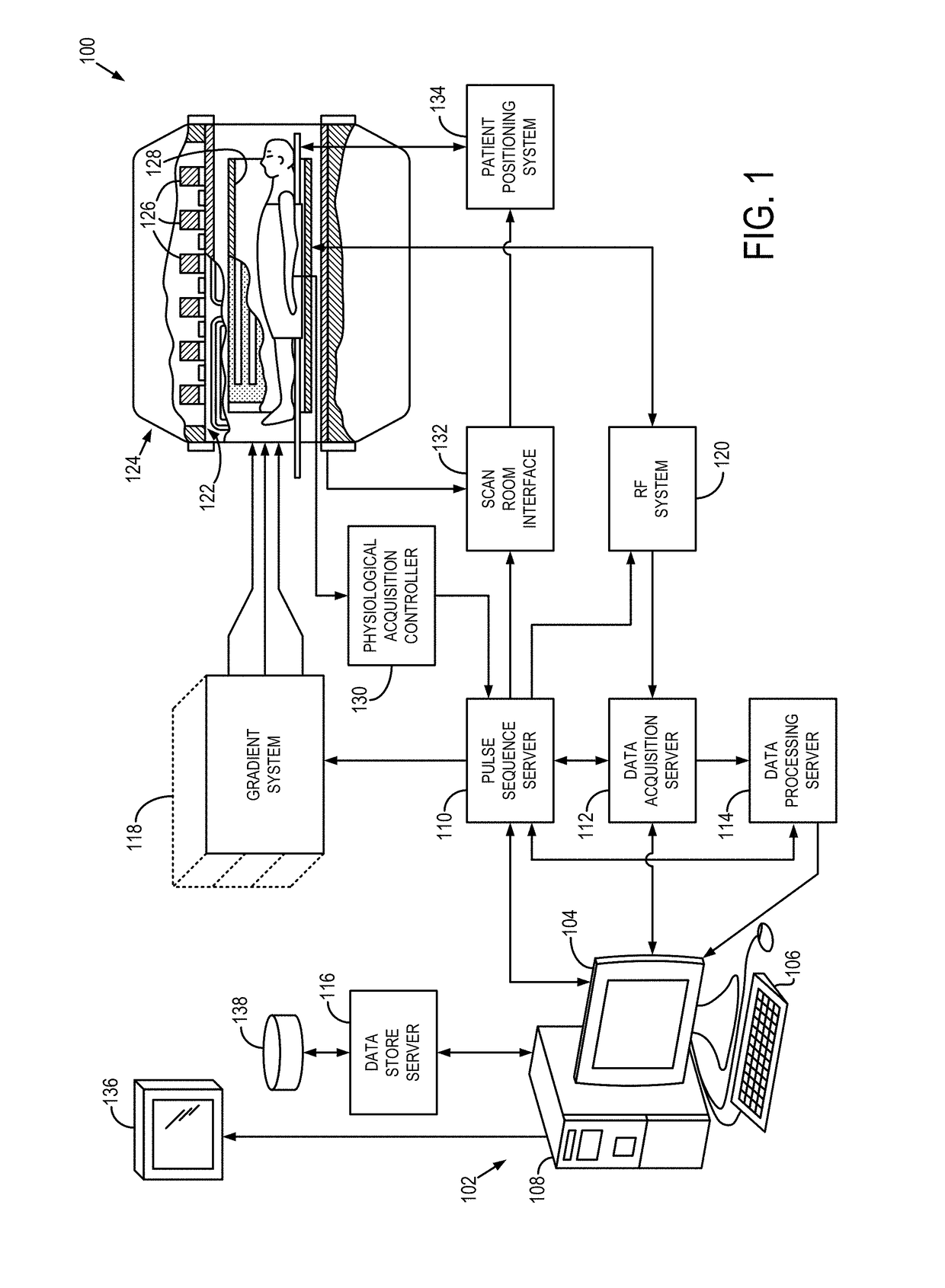
[0024]Referring particularly to FIG. 1, an example of a magnetic resonance imaging (“MRI”) system 100 is illustrated. The workstation 102 includes a processor 108, such as a commercially available programmable machine running a commercially available operating system. The workstation 102 provides the operator interface that enables scan prescriptions to be entered into the MRI system 100. The workstation 102 is coupled to four servers: a pulse sequence server 110; a data acquisition server 112; a data processing server 114; and a data store server 116. The workstation 102 and each server 110, 112, 114, and 116 are connected to communicate with each other.

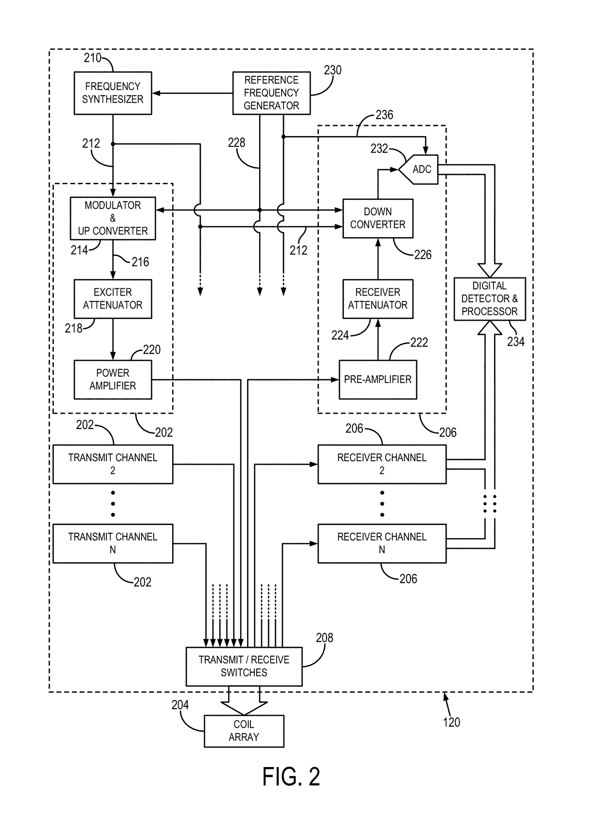
[0025]The pulse sequence server 110 functions in response to instructions downloaded from the workstation 102 to operate a gradient system 118 and a radiofrequency (“RF”) system 120. Gradient waveforms necessary to perform the prescribed scan are produced and applied to the gradient system 118, which excites gradient coils in an ass...


PUM


Abstract

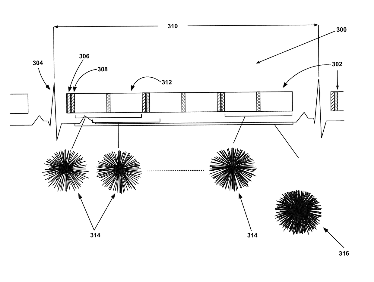
A system and method is provided that includes a) monitoring a cardiac cycle of the subject to identify a predetermined point and, b) upon identifying the predetermined point, performing the steps of i) performing at least one of a desired number of magnetization suppressing preparations to suppress signal from blood flow through at least the region of interest, ii) acquiring a first set of imaging data from the region of interest, and iii) repeating step i) and step ii) to acquire at least a second set of imaging data from the region of interest. The method further includes c) repeating step b) a predetermined number of times over a series of cardiac cycles to acquire respective sets of medical imaging data of the region of interest and d) reconstructing first set of imaging data and the second set of imaging data into a time-resolved series of images.

Description

CROSS-REFERENCE TO RELATED APPLICATIONS[0001]N / AFIELD OF THE INVENTION[0002]The invention relates to a system and method for performing magnetic resonance imaging and, more particularly, to a system and method for acquiring images using magnetic resonance imaging that allow clinical review of a subject's vascular components, including the blood vessels and the heart, over time and without signal from blood pools within the vessels or heart obscuring the vascular components.BACKGROUND OF THE INVENTION[0003]When a substance such as human tissue is subjected to a uniform magnetic field (polarizing field B0), the individual magnetic moments of the nuclear spins in the tissue attempt to align with this polarizing field, but precess about it in random order at their characteristic Larmor frequency. Usually the nuclear spins are comprised of hydrogen atoms, but other NMR active nuclei are occasionally used. A net magnetic moment Mz is produced in the direction of the polarizing field, but ...

Claims


Application Information

Patent Timeline
Patent Type & Authority Patents(United States)
IPC IPC(8): A61B5/055G01R33/563G01R33/56A61B5/00A61B5/026A61B5/0452
CPCA61B5/055A61B5/0044A61B5/0263G01R33/563G01R33/5607G01R33/5635A61B5/7285A61B5/0452A61B5/349
Inventor KOKTZOGLOU, IOANNIS
Owner NORTHSHORE UNIV HEALTHSYST
Learn More