Immuno imaging agent for use with antibody-drug conjugate therapy
a conjugate therapy and immuno-imaging technology, applied in the field of companion diagnostic antibodylike binding protein, can solve the problems of lack of clinical benefit, lack of safety, and many initial agents that do not reach the market, and achieve the effect of facilitating patient stratification and early evaluation
- Summary
- Abstract
- Description
- Claims
- Application Information
AI Technical Summary
Benefits of technology
Problems solved by technology
Method used
Image
Examples
example 1
Radiolabeling and In Vitro Characterization of Humanized DS6 Monoclonal Antibody and DS6-DM4
[0195]A clinically useful molecular imaging agent produces high contrast images within a reasonable amount of time following administration, demonstrates tumor penetration, has faster clearance kinetics and exhibits an excellent tumor to blood ratio. A caveat of using intact native antibodies in bioimaging is inherently slow kinetics of tumor uptake and blood clearance; they also tend to have high background and require a long time (hours to days) to obtain clear images of the tumor.
[0196]It has been previously shown that a radiolabeled tracer combined with bio-imaging can translate from a preclinical tumor model to a clinical setting (Williams et al., 2008, Cancer Biother and Radiopharm 23(6):797; Liu et al., (2009) Mol Imaging Biol 12:530). We further demonstrated that a radiolabeled DS6 antibody and DS6-DM4 effectively targeted WISH xenograft tumors in mice. Based on these results, three e...
example 2
Expression of DS6 Engineered B-Fab
[0197]The expression plasmids encoding the heavy and light chains of the corresponding constructs were propagated in E. coli DH5a cells. Plasmids used for transfection were prepared from E. coli using the Qiagen EndoFree Plasmid Mega Kit (polypeptide and DNA sequences for the B-Fab antibody-like binding protein are in Table 2).
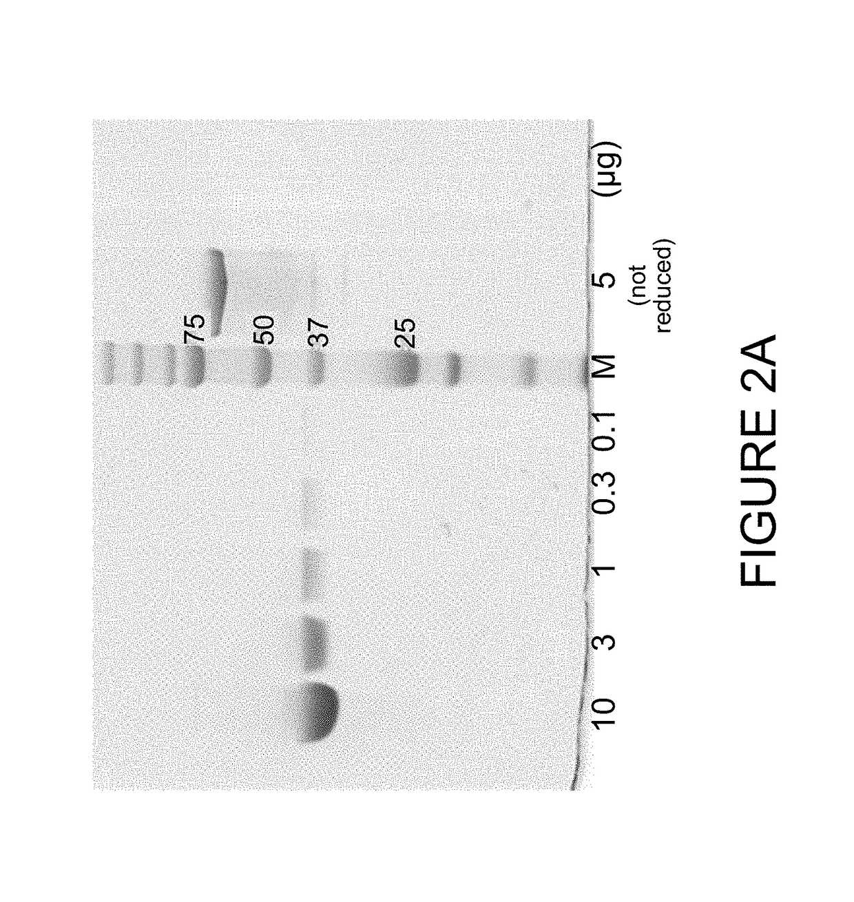
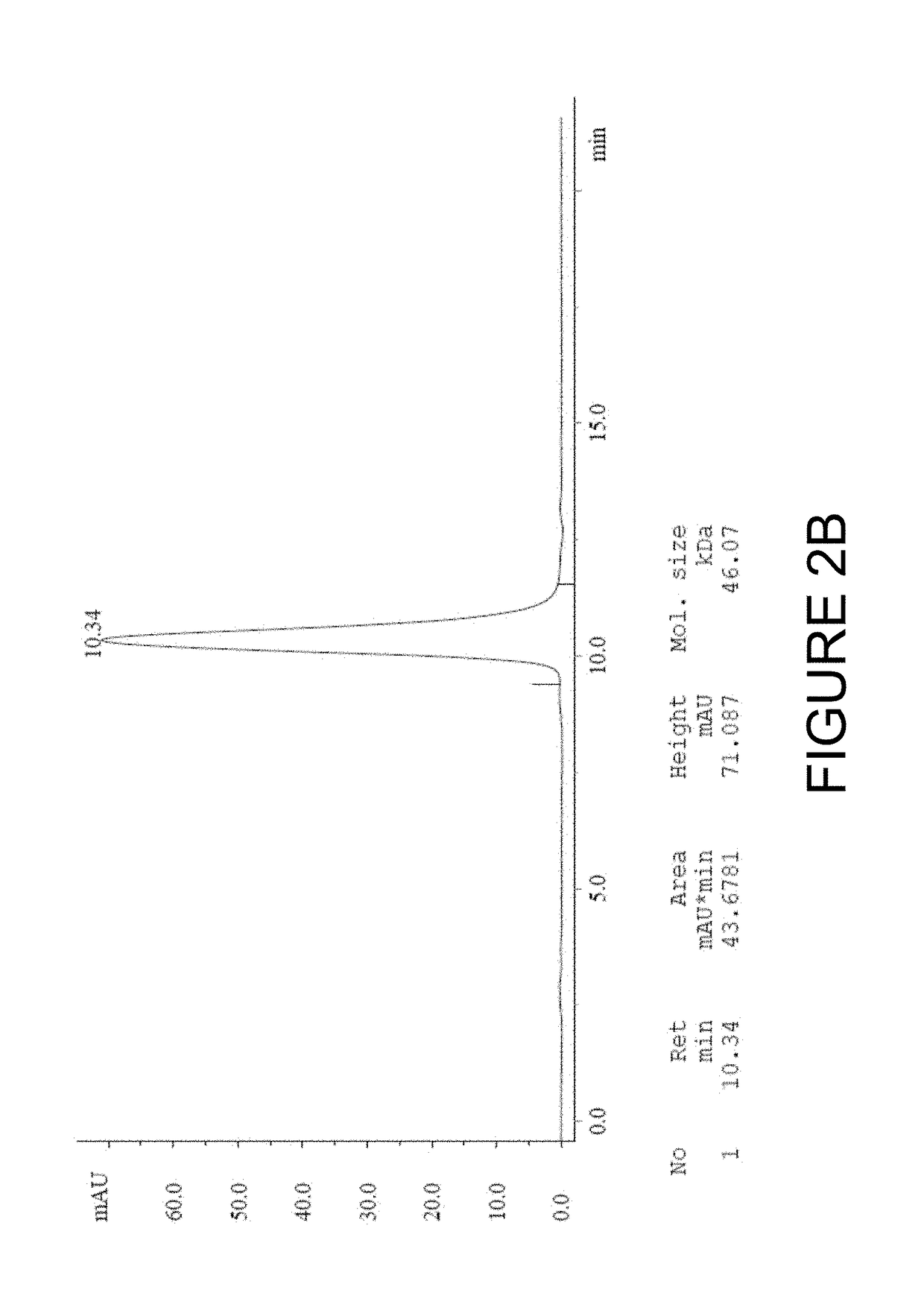
[0198]The DS6 antibody-like binding protein B-Fab was produced by transient transfection of HEK293 cells in FreeStyle F17 medium (Invitrogen). After 7 days after transfection, supernatant was harvested, and subjected to centrifugation and 0.2 μm filtration to remove particles. Purification was carried out by capture on KappaSelect (GE Healthcare). After binding the KappaSelect column, the column was washed with PBS or HEPES buffer (50 mM HEPES, 150 mM NaCl, pH 7.2, FP12016) followed by elution with 0.1M Glycin (pH 2.5) and neutralization with 1M Tris / HCl pH 9. The purification polishing step consisted of size exclusion chromat...
example 3
Development of Engineered Antibody-Like Binding Proteins Based on the DS6 Antibody
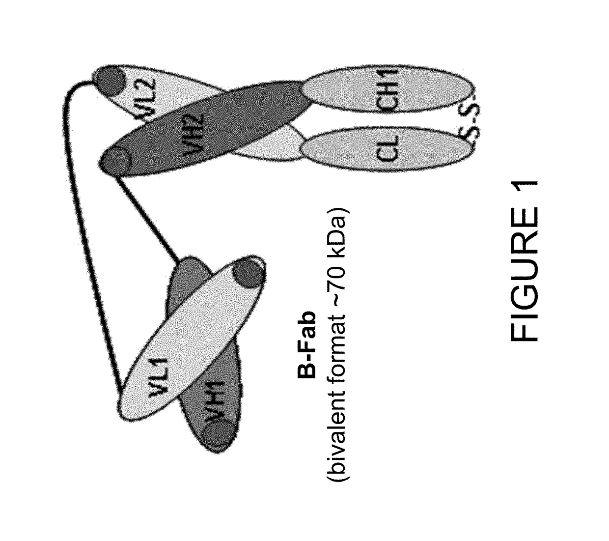
[0201]In order to develop an imaging based companion diagnostic antibody-like binding protein for huDS6-DM4, three engineered antibody-like binding proteins (B-Fab, see FIG. 1) based on CA6 binding sequences from the humanized DS6 monoclonal antibody were created. Each of the three engineered antibody-like binding proteins was purified and tested as described below; the results are shown in Table 3. Desirable characteristics include >95% purity by HPLC or SDS-PAGE, ˜10 mg / mL concentration, a stable peptide, binding similar to the DS6 antibody, and to determine whether the presence of a poly-histidine (6x-His) tag affects PET tracers. Additional selection criteria includes: a tumor signal of >5% ID / g; a tumor to muscle ratio of 3:1; efficient renal clearance; and a statistically significant difference (p<0.05) between unblocked and blocked uptake (see Table 4 for summary of quality criteria for radiolab...
PUM
| Property | Measurement | Unit |
|---|---|---|
| molecular weight | aaaaa | aaaaa |
| molecular weight | aaaaa | aaaaa |
| dissociation constant | aaaaa | aaaaa |
Abstract
Description
Claims
Application Information
 - R&D
- Intellectual Property
- Life Sciences
- Materials
- Tech Scout
- Unparalleled Data Quality
- Higher Quality Content
- 60% Fewer Hallucinations
Browse by: Latest US Patents, China's latest patents, Technical Efficacy Thesaurus, Application Domain, Technology Topic, Popular Technical Reports.
© 2025 PatSnap. All rights reserved.Legal|Privacy policy|Modern Slavery Act Transparency Statement|Sitemap|About US| Contact US: help@patsnap.com


