Method for detecting COL7A1 gene mutation and uses thereof

A genetic and functional technology, applied in the field of genetic testing, can solve problems such as indistinguishability

Inactive Publication Date: 2008-06-18
HUAZHONG UNIV OF SCI & TECH
View PDF0 Cites 18 Cited by
  • Summary
  • Abstract
  • Description
  • Claims
  • Application Information

AI Technical Summary

Problems solved by technology

Family history is important, but it is difficult to distinguish the two due to the rare occurrence of large families or the occurrence of spontaneously mutated (de novel) DDEB

Method used


Image

  • Method for detecting COL7A1 gene mutation and uses thereof
  • Method for detecting COL7A1 gene mutation and uses thereof

Examples


Embodiment 1

[0043] Next, the COL7A1 gene mutation was detected by gene sequencing.

[0044] 1. PCR amplification of COL7A1 gene coding region

[0045] Objects: The families with dystrophic epidermolysis bullosa collected in outpatient clinics included 9 patients, 11 healthy people and 200 normal controls. Detailed physical examination was performed on all participants, and 5ml blood samples were collected from each participant after signing the informed consent form.

[0046] Genomic DNA extraction: using phenol chloroform extraction method.

[0047] 1. Anticoagulant blood was diluted 1-fold with PBS.

[0048] 2. Add 2 times the volume of lymphatic separation solution (18°C-28°C) into the centrifuge tube, spread a layer of 1 times the volume of diluted blood on top, and centrifuge at 1000*g for 20 minutes at room temperature.

[0049] 3. Discard the supernatant, carefully suck out the nucleated cell layer in the middle, transfer to a 5ml Ep tube, 5000g, 10 minutes, and then wash once w...

Embodiment 2

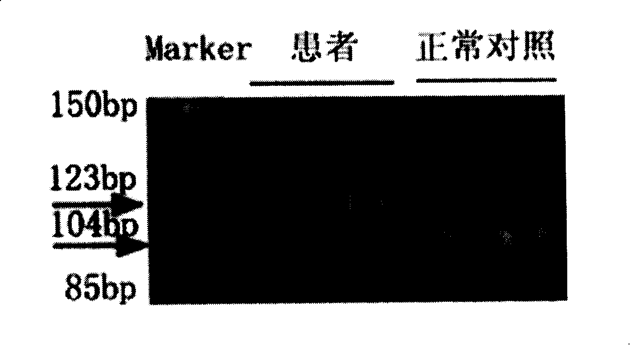
[0096] Next, the change of said base is detected by restriction fragment length polymorphism.

[0097] 1. PCR amplification of intron 86 / exon 87 of COL7A1 gene

[0098] 1. Use primers p87F and p87R and other reagents to PCR amplify 86 / exon 87 of the sample DNA. For the steps, see the first part of Example 1.

[0099] 2. After the PCR product was diluted 200 times, take 1 μl as a template for the second round of PCR. The primers are as follows: p87Fa: 5'-TCCCACGGGGGCCCCTGGACAG-3' and p87Ra: 5'-TCCCCCGCCCCCACCCTGCCA-3'. For other reagents and steps, see Example 1 first part.

[0100] 3. For the purification of PCR products, refer to steps 1-5 in the second part of Example 1 for the steps.

[0101] 4. Add 1 U of Bsl I (NEB Company) to 20 μl of PCR product, 2.5 μl of 10*buffer, add 25 μl of water to make up, and incubate at 55°C for 2 hours.

[0102] 5. Use 1.5% agarose gel for electrophoresis, 1*TAE electrophoresis buffer, voltage 5V / cm, electrophoresis for 30 minutes, observe u...

Embodiment 3

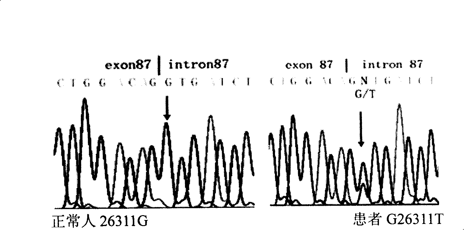
[0104] Kit for G26311T point mutation of the causative gene COL7A1 of dystrophic epidermolysis bullosa and its application

[0105] 1 kit contains:

[0106] Primers for amplification: p87F: TGGGCCTGAAATATGAGGAG

[0107] P87R: TAGGCCACTGGAGAGACAGG

[0108] Taq enzyme 5u / ul for PCR amplification

[0109] 10* buffer (containing 15ml MgCl 2 . )

[0110] dNTP2mM

[0111] Big Dyemix

[0112] 2 How to use:

[0113] It mainly includes the following steps:

[0114] A. extract DNA, utilize above-mentioned primer, carry out PCR reaction;

[0115] B: The PCR reaction product is directly sequenced after purification, and the obtained sequence is compared with the standard sequence in Genebank to determine the existence of the mutation site.

[0116] The specific method is referring to embodiment 1.

[0117] The base change can also be detected by specific hybridization of the COL7A1 gene isolated from the tissue with the COL7A1 allele.


PUM


Abstract

The invention discloses a method of detecting 26311 Mutation of COL7A1 gene and a usage thereof. The method adopts primers of p87F (TGGGCCTGAAATATGAGGAG) and P87R (TAGGCCACTGGAGAGACAGG) to PCR-amplify COL7A1 gene including the section of 26251-26350, implements the sequence after the purification of products, or uses BslI for the endonuclease detection after using p87Fa (TCCCACGGGGGCCCCTGGACAG) and P87Ra (TCCCCCGCCCCCACCCT GCCA) for the nest amplification of the PCR products. The usage is the application in preparations or gene chips for detecting the diagnosis of dominant dystrophic epidemolysis bullosa genealogy. The invention provides a novel way for the prenatal diagnosis of the bullosa and provides a basis for the gene therapy.

Description

technical field [0001] The invention belongs to the technical field of gene detection, and in particular relates to a method for detecting COL7A1 gene mutation, especially a method for detecting COL7A1 gene mutation G26311T. Application on gene chips or preparations for resolving diseases. Background technique [0002] Hereditary epidermolysis bullosa is composed of a group of diseases related to clinical manifestations. According to the standard of electron microscopy, the position of the blister on the skin can be used as the standard, and it can be divided into epidermolysis bullosa simplex and junctional type. Epidermolysis bullosa and dystrophic epidermolysis bullosa. [0003] Type VII collagen was first isolated by Bebtz et al., and was named long-chain collagen (LC) at that time. Ryynanen et al found that type VII collagen mRNA was expressed in keratinocytes and oral epidermal carcinoma cell lines by Northern hybridization. Indirect immunofluorescence showed that t...

Claims


Application Information

Patent Timeline
Patent Type & Authority Applications(China)
IPC IPC(8): C12Q1/68
Inventor 任翔王擎刘木根刘静宇姚淇蔡洲代小华
Owner HUAZHONG UNIV OF SCI & TECH
Why Eureka
Try Eureka