Agent and method for testing reticulocyte

A technology for reticulocytes and detection reagents, which is applied in biological testing, biochemical equipment and methods, material testing products, etc., can solve the problems of high fluorescence background, errors, and unfavorable standard marking of blood analyzers, and achieves good repeatability, Good consistency and low non-specific fluorescence background

Active Publication Date: 2008-07-30
SHENZHEN MINDRAY BIO MEDICAL ELECTRONICS CO LTD +1
View PDF13 Cites 21 Cited by
  • Summary
  • Abstract
  • Description
  • Claims
  • Application Information

AI Technical Summary

Problems solved by technology

[0019] SYSMEX US5821127, CN1172953A, and CN1154966A describe a fluorescent dye used for automatic analysis of reticulocytes. The fluorescent dye has significant non-specific binding to red blood cells, not only binding to RNA, but also binding to other particles in the cell, such as mitochondria, ribosomes, etc., causing Defects with high fluorescence background, and require an incubation temperature of 40°C
[0020] The fluorescent dye described in US200203758 is used to label reticulocytes and nucleated red blood cells, but it needs to be incubated for up to 1 minute, and the labeling time changes with the ambient temperature, which is not conducive to the specification in blood analyzers mark, resulting in certain errors

Method used


Image

  • Agent and method for testing reticulocyte
  • Agent and method for testing reticulocyte
  • Agent and method for testing reticulocyte

Examples


Embodiment 1

[0073] Configure the reticulocyte detection reagent composed as follows.

[0074] Fluorescent dye (III) 7mg

[0075] NaH 2 PO 4 .H 2 O 53.8mg

[0076] Na 2 HPO 4 .7H 2 O 163.4mg

[0077] Cocamidopropyl Betaine 100mg

[0078] Delicate water 1L

[0079] (adjust pH to 7)

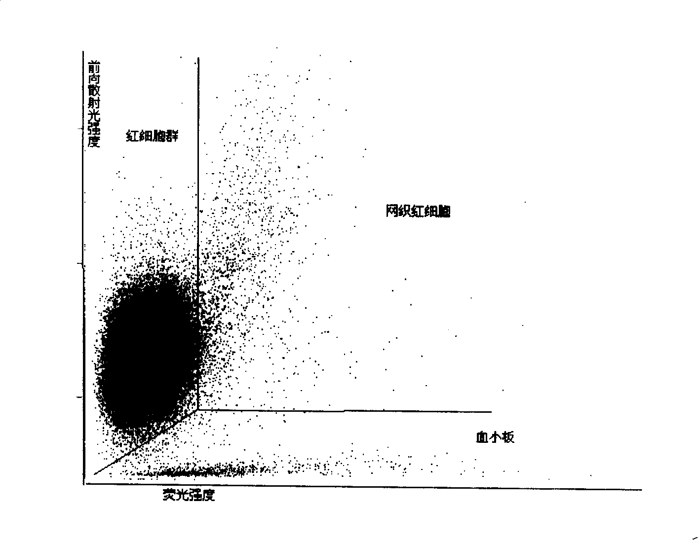
[0080] 4 microliters of anticoagulant-treated blood were added to 1 milliliter of the reagent, and left at a constant temperature at 40° C. for 30 seconds, or incubated in an incubation tank for 30 seconds, to form a measurement sample. The sample for measurement is detected by a detection instrument with a red semiconductor laser, the excitation wavelength is 633nm-635nm, and the power is 5mW. By measuring the forward low-angle scattered light intensity and fluorescence intensity, the following figure 1 The scatter plot shown, where the proportion of reticulocytes (RET) in the total erythrocytes is 1.25%.

[0081] In addition, the same blood sample was detected by traditional manual microscopy met...

Embodiment 2

[0084] Configure the reticulocyte detection reagent composed as follows.

[0085] Fluorescent dye (III) 7mg

[0086] NaH 2 PO 4 .H 2 O 53.8mg

[0087] Na 2 HPO 4 .7H 2 O 163.4mg

[0088] Cocamidopropyl Betaine 100mg

[0089] Delicate water 1L

[0090] (adjust pH to 7)

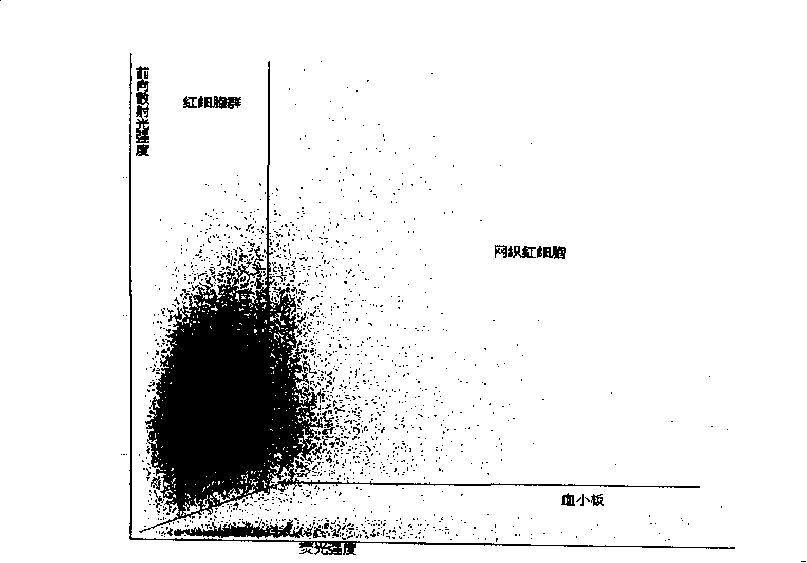
[0091] Add 4 microliters of anticoagulant-treated blood to 1 milliliter of the reagent, place it at a constant temperature at 40° C. for 30 seconds, or incubate in an incubation pool for 30 seconds to form a test sample. The sample for measurement is detected by a detection instrument with a red semiconductor laser, the excitation wavelength is 633nm-635nm, and the power is 5mW. By measuring the forward low-angle scattered light intensity and fluorescence intensity, the following figure 2 The scatter plot shown. Among them, reticulocytes (RET) accounted for 5.08% of the total red blood cells.

[0092] In addition, the same blood sample was tested by traditional manual microscopy, and the result w...

Embodiment 3

[0096] A reagent for measuring reticulocytes with the following composition was prepared.

[0097] Fluorescent dye (IV) 7mg

[0098] Disodium hydrogen phosphate 53.8mg

[0099] Sodium dihydrogen phosphate 163.4mg

[0100] Cocamidopropyl Betaine 100mg

[0101] Delicate water 1L

[0102] (adjust pH to 7)

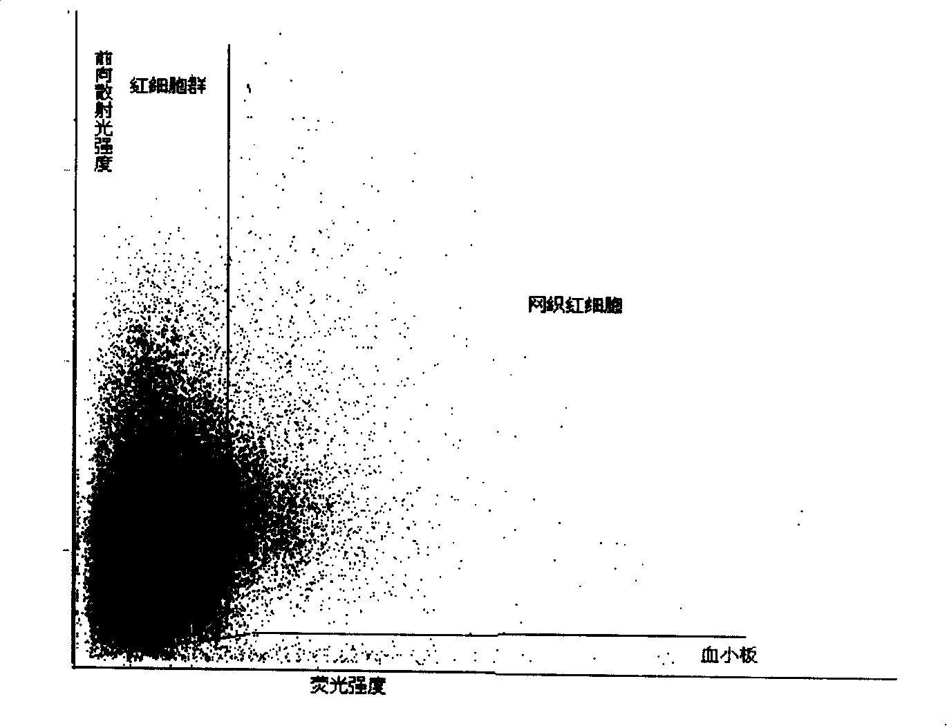
[0103] Add 4 microliters of anticoagulant-treated blood to 1 milliliter of the reagent, place it at a constant temperature at 40° C. for 30 seconds, or incubate in an incubation pool for 30 seconds to form a test sample. The sample for measurement is detected by a detection instrument with a red semiconductor laser, the excitation wavelength is 633nm-635nm, and the power is 5mW. By measuring the forward low-angle scattered light intensity and fluorescence intensity, the following image 3 The scatter plot shown. Among them, reticulocytes (RET) accounted for 5.35% of the total red blood cells.

[0104] In addition, the same blood sample was tested by traditional manual ...


PUM

PropertyMeasurementUnit
concentrationaaaaaaaaaa
wavelengthaaaaaaaaaa

Abstract

The invention discloses a reticular cell detect reagent and the testing method thereof, and the reagent and the method can be applied to an apparatus based on the streaming technical principle to count accurately the analyzing reticular cell in blood specimen. The reagent and the method of the invention adopt a novel red fluorescence excitation dye which is used for marking RNA in the reticular cell, and the rapid sign of RNA in the reticular cell can be realized under the environmental temperature through a matched sphericize reagent system.

Description

technical field [0001] The invention relates to a cell detection reagent and method, in particular to a detection reagent and detection method for reticulocytes in blood. Background technique [0002] Reticulocytes are immature red blood cells between late immature red blood cells and mature red blood cells. The reticulocyte count is an important index to evaluate bone marrow hematopoietic function and erythropoietic activity. [0003] Under normal physiological conditions, it takes 4 days for the bone marrow to transform from primitive cells to reticulocytes, and then the reticulocytes mature in the bone marrow for 48 hours and enter the peripheral blood, and the reticulocytes in the blood continue to mature for 48 hours to become mature red blood cells. The number of reticulocytes in peripheral blood is of great significance for evaluating bone marrow erythroid hematopoiesis and the transfer rate of reticulocytes from bone marrow to peripheral blood [0004] Counting ret...

Claims


Application Information

Patent Timeline
Patent Type & Authority Applications(China)
IPC IPC(8): G01N21/64G01N33/52C12Q1/02C07D209/14C07D401/06
Inventor 徐兵匡玉吉
Owner SHENZHEN MINDRAY BIO MEDICAL ELECTRONICS CO LTD
Why Eureka
Try Eureka