
Immune tissue chemical diagnostic kit used for pathological diagnosis of tumour

An immunohistochemistry and diagnostic kit technology, applied in the field of bioengineering, which can solve the problems of poor repeatability, unsatisfactory results, and large differences between antibody batches

Active Publication Date: 2009-12-09
FOURTH MILITARY MEDICAL UNIVERSITY
View PDF1 Cites 31 Cited by
  • Summary
  • Abstract
  • Description
  • Claims
  • Application Information

AI Technical Summary

Problems solved by technology

There is no formal batch number, and the antibodies used in pathological diagnosis have large differences between antibody batches, poor reproducibility, and unsatisfactory results

Method used


Image

  • Immune tissue chemical diagnostic kit used for pathological diagnosis of tumour
  • Immune tissue chemical diagnostic kit used for pathological diagnosis of tumour
  • Immune tissue chemical diagnostic kit used for pathological diagnosis of tumour

Examples


specific Embodiment

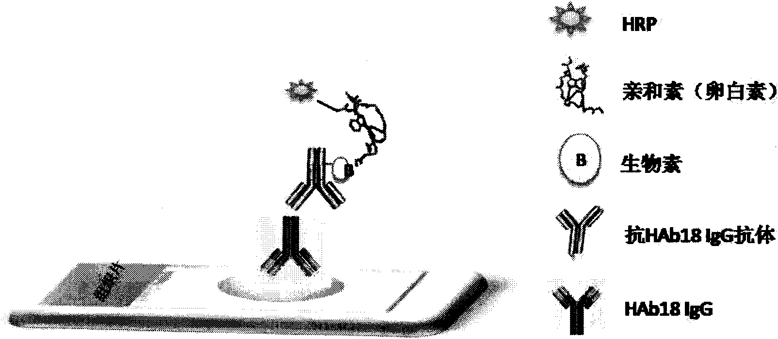
[0047] The experimental method of the HAb18G / CD147 cancer marker immunohistochemical diagnostic kit is as follows, and the following examples refer to this method, referred to as "experimental method" for short:

[0048] 1. Sections of paraffin specimens of clinical tissue cases were dewaxed in xylene for 20 minutes, three times;

[0049] 2. 100% alcohol to remove xylene, 5min, twice; 95%-75% alcohol, 2min / time;

[0050] 3. 3% H2O2 to block endogenous peroxidase, 22-28°C, 10 minutes;

[0051] 4. Soak in PBS buffer for 3 minutes, 3 times;

[0052] 5. 5% normal sheep serum to block non-specific sites in the tissue, 22-28°C, 20 minutes;

[0053] 6. HAb18 monoclonal antibody (30ug / mL), 37°C, 120 minutes;

[0054] 7. Soak in PBS buffer for 5 minutes, 3 times;

[0055] 8. Avidin-labeled anti-specific anti-antibody, 22-28°C, 15 minutes;

[0056] 9. Soak in PBS buffer for 5 minutes, 3 times;

[0057] 10. Horseradish peroxidase-labeled biotin, 22-28°C, 20 minutes;

[0058] 11. S...

Embodiment 1

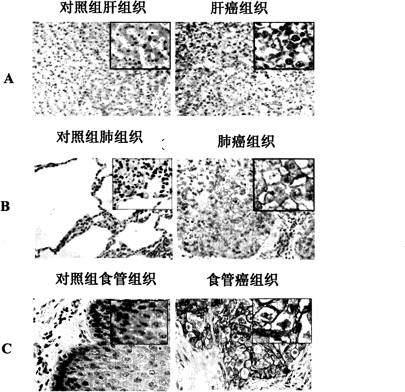
[0065] The HAb18G / CD147 cancer marker immunohistochemical diagnostic kit for auxiliary diagnosis or differential diagnosis of cancer is expressed in liver tissue. The specific test process is as follows:

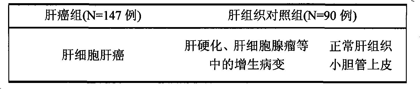
[0066] 1. Sample selection: (liver cancer, cases determined according to the gold standard HE staining)

[0067] Table 1 Case selection of liver cancer and liver tissue

[0068]

[0069] 2. Test operation: see "Experimental Method" for details.

[0070] 3. Test results: (see attached figure 2 A)

[0071] Table 2 Staining results of HAb18G / CD147 cancer marker diagnostic kit in liver cancer and liver tissue

[0072]

Embodiment 2

[0074] HAb18G / CD147 cancer marker immunohistochemical diagnostic kit for auxiliary diagnosis or differential diagnosis of cancer expressed in lung tissue The specific test process is as follows:

[0075] 1. Sample selection: lung cancer, according to the results of the gold standard HE staining method to determine the case

[0076] Table 3: Case selection for lung cancer and lung tissue

[0077]

[0078] 2. Test operation: see "Experimental Method" for details.

[0079] 3. Test results: (see attached figure 2 B)

[0080] Table 4: Staining results of HAb18G / CD147 cancer marker diagnostic kit in lung cancer and lung tissue

[0081]


PUM


Abstract

The invention discloses an immune tissue chemical diagnostic kit of HAb18G / CD147 cancer landmark used for pathological diagnosis of tumour, including a blocking agent capable of removing endogenous enzyme in a human tissue slice, a sealing agent used for sealing cross protein reaction in the human tissue slice, a first antibody capable of combining with the HAb18G / CD147 cancer landmark in human tumour tissue, a biotin marked secondary antibody capable of combining with the first antibody, and an enzyme marked avidin, chromogenic reagent and a phosphate buffer system used for combining with the secondary antibody. The kit has the characteristics of good specificity, high sensibility, stability and universality, is mainly used for pathologic diagnosis of epithelium-derived malignant tumour (such as liver cancer, lung cancer, esophageal cancer, gastric cancer, colorectal cancer, breast cancer, cervical cancer, ovarian cancer and the like), and is beneficial to warning screen, early diagnosis, etiologic diagnosis, treatment reaction and curative effect monitoring of tumour, recrudescence and transfer and prognosis judgement of tumour.

Description

technical field [0001] The invention belongs to the field of bioengineering, and relates to a kit for clinical diagnosis, in particular to a kit for immunohistochemical diagnosis of HAb18G / CD147 cancer markers for pathological diagnosis of tumors. [0002] HAb18G / CD147 cancer marker is a novel, broad-spectrum and highly specific cancer marker. Using a specific antibody against HAb18G / CD147 cancer markers, combined with a general immunohistochemical method, HAb18G / CD147 cancer marker immunohistochemical diagnostic kit is applied to various epithelial malignant tumor histopathological specimens for tumor pathological diagnosis, which is helpful for tumor early warning screening, early diagnosis, qualitative diagnosis, treatment response and curative effect monitoring, tumor recurrence, metastasis and prognosis. Background technique [0003] In recent years, the incidence of various tumors has risen sharply, and the medical expenses of tumors and the death of cancer patients ...

Claims


Application Information

Patent Timeline
IPC IPC(8): G01N33/577G01N33/574G01N1/30
Inventor 米力陈小春张阳陈志南
Owner FOURTH MILITARY MEDICAL UNIVERSITY
Features
  • Generate Ideas
  • Intellectual Property
  • Life Sciences
  • Materials
  • Tech Scout
  • Unparalleled Data Quality
  • Higher Quality Content
  • 60% Fewer Hallucinations
Learn More