Methods of evaluating cells and cell cultures

A technology for cell culture and culture, applied in the field of determining the composition of cell culture

Active Publication Date: 2010-03-24
GENZYME CORP
View PDF2 Cites 4 Cited by
  • Summary
  • Abstract
  • Description
  • Claims
  • Application Information

AI Technical Summary

Problems solved by technology

Thus, the expression levels of such fibroblast markers may not necessarily indicate whether the ce

Method used


Image

  • Methods of evaluating cells and cell cultures
  • Methods of evaluating cells and cell cultures
  • Methods of evaluating cells and cell cultures

Examples


Embodiment 1

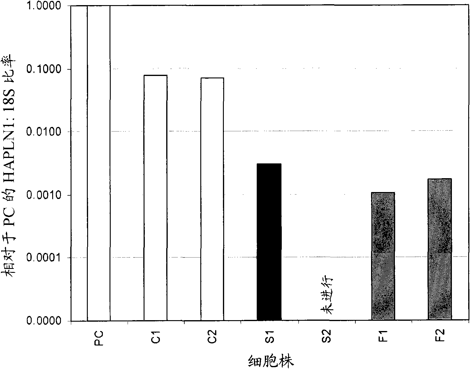
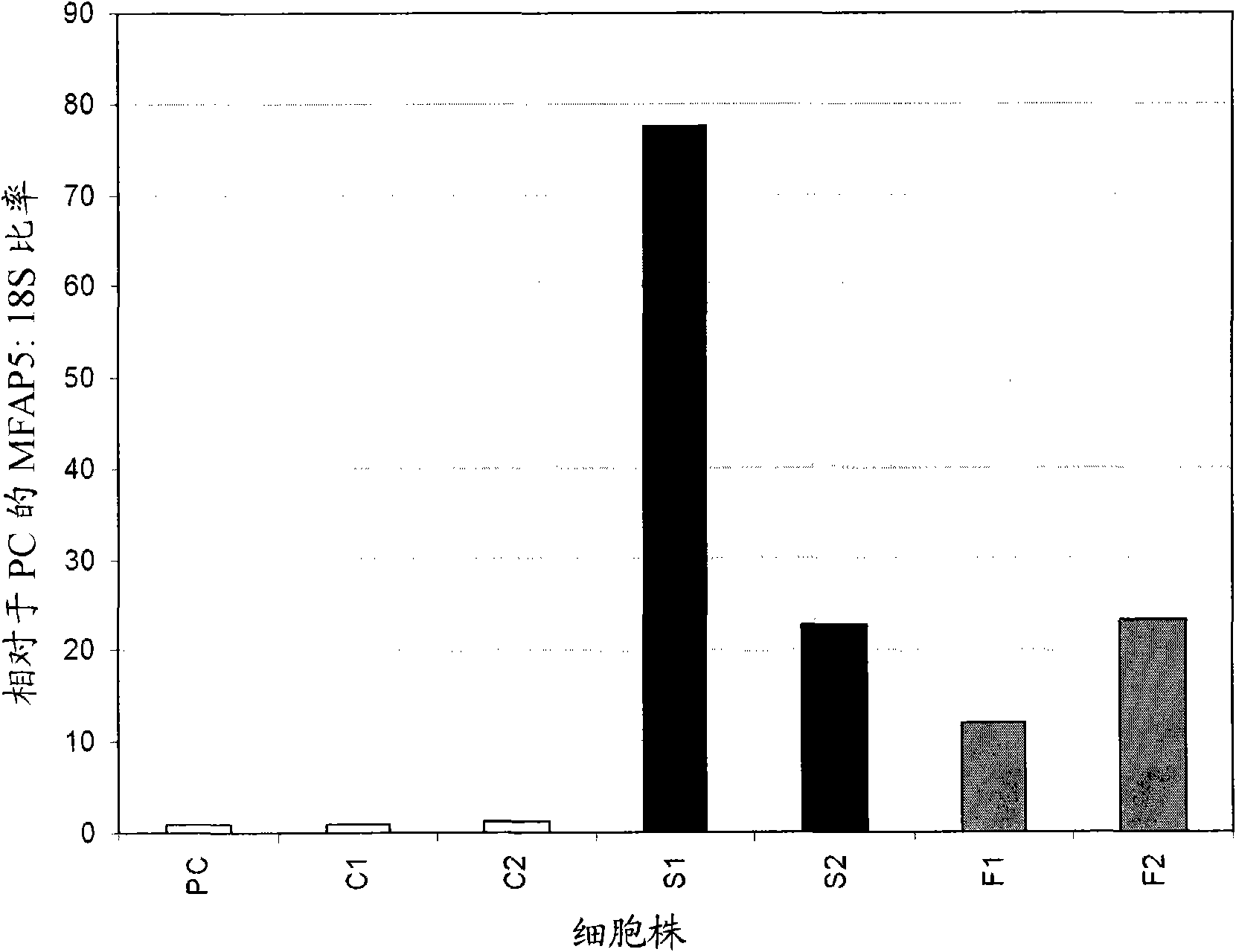
[0101] Example 1: Expression of HAPLN1 and MFAP5 in Chondrocytes, Synoviocytes and Fibroblasts

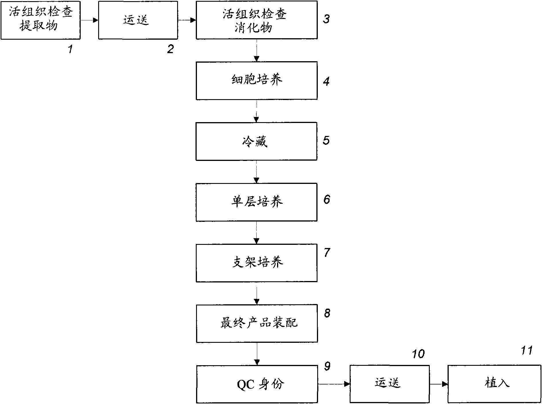
[0102] Cell isolation and culture - Human chondrocyte cultures are used to produce The method of autologous chondrocytes or the proteolytic method used to generate cultured chondrocytes are isolated from cartilage. used to generate For the autologous chondrocyte approach, the cartilage tissue was trimmed away from the bone and synovium, and subjected to a first digestion in which the tissue was enzymatically treated in a collagenase solution at 37°C for 18 hours. Cells released from the first digestion were distributed in tissue culture flasks containing medium (EGHXX) containing fetal bovine serum (FBS) and gentamicin. The cells were then subjected to a second digestion in which the remaining tissue from the first digestion was treated with a collagenase / trypsin solution for 2.5 hours at 37°C. Cells released from the second digestion were distributed among tissue culture flas...

Embodiment 2

[0140] Example 2: Expression of HAPLN1 and MFAP5 in other chondrocytes, synoviocytes and fibroblast lines

[0141] Expression levels of HAPLN1 and MFAP5 were determined in additional cell cultures to demonstrate the fidelity of the method used to differentiate chondrocytes from synovial cell cultures. The cultures used in this example are listed in Table 3.

[0142] Table 3: Cell cultures used in RT-PCR analysis (Example 2)

[0143] cell culture

cell type

cell culture type

PC

Chondrocytes

primary culture

C3

Chondrocytes

second passage

C4

Chondrocytes

second passage

C5

Chondrocytes

second passage

C6

Chondrocytes

second passage

C7

Chondrocytes

second passage

S3

Synoviocytes

second passage

[0144] S4

Synoviocytes

second passage

S5

Synoviocytes

second passage

S6

Synoviocytes

second ...

Embodiment 3

[0153] Example 3: Testing the expression of HAPLN1 and MFAP5 in chondrocytes, synoviocytes and fibroblasts using custom designed primers and probes

[0154] A variety of chondrocyte, synoviocyte, and dermal fibroblast culture assays were performed using primers and probes of known oligonucleotide sequence.

[0155] Cell Isolation and Culture - The cell lines used in this example are listed in Tables 4 and 5 below. As described in Example 1, using the Autologous chondrocyte method to isolate and culture human chondrocyte cell cultures C1, C2, C3, C4, C5, C6, C7, C8, C26, C28, C30, and C34. Human chondrocyte cell cultures C21, C22, C23, C24, C25, C27, C29, C31, C32, and C33 were isolated (using the protease method) and cultured as described in Example 1. Cell isolation and culture methods for human synoviocyte cultures S1, S2, S3, S4, S5, S6, and S7 are described in Example 1 and Example 2. Synovial cell culture S9 was isolated by digesting minced synovial tissue in a soluti...


PUM


Abstract

Methods of evaluating the composition of a cell culture (e.g., to distinguish chondrocytes from fibroblasts) and methods for evaluating the phenotype of an individual cell (e.g., as a chondrocyte) are disclosed. The methods may be used, for example, for assessing chondrocyte cultures used for treatment of cartilage defects. In some embodiments, the invention involves identifying cell culture composition or the identity of a cell based on expression level of a fibroblast marker. In other embodiments, the invention involves comparing expression levels of at least one chondrocyte marker and at least one fibroblast marker in a cell culture sample or in an individual cell. In illustrative embodiments, the chondrocyte marker is hyaluronan and proteoglycan link protein 1 (HAPLN1), and the fibroblast marker is microfibrillar associated protein 5 (MFAP5).

Description

[0001] related application [0002] This application claims priority to provisional application 60 / 910,574, filed April 6, 2007, the entire contents of which are incorporated by reference. field of invention [0003] The present invention relates to methods of determining the composition of cell cultures, and more particularly, to methods of differentiating chondrocytes from fibroblasts. Background of the invention [0004] Articular cartilage lesions have a poor repair rate due in part to a lack of blood supply in the cartilage tissue (Basad et al., in: Hendrich et al., Cartilage Surgery and Future Perspectives, Thieme Verlag, 49-56 (2003)). Trauma to the knee joint can result in, for example, cartilage and osteochondral damage, and such damage can develop into osteoarthritis (Brittberg et al., New England Journal of Medicine, 331(14):889-895 (1994)). In severe cases of osteoarthritis, a total knee replacement may be required. However, artificial prostheses used in knee r...

Claims


Application Information

Patent Timeline
IPC IPC(8): G01N33/50G01N33/68
CPCG01N2333/4722G01N2400/40G01N2800/10G01N33/5044G01N33/6887C12Q1/6844C12Q1/6851
Inventor 斯蒂芬·M·拉普科斯蒂芬·J·杜瓜伊
Owner GENZYME CORP
Why Eureka
Try Eureka