CT imaging contrast agent and preparation method thereof

A technology of CT imaging and contrast agent, which is applied in the preparation of X-ray contrast agent, etc., which can solve the problems of weak X-ray absorption of iodine contrast agent, difficulty in obtaining important information of lesions, and inability to observe lesions for a long time, achieving a good absorption coefficient , good biocompatibility, and uniform shape

Inactive Publication Date: 2010-06-16
XIAMEN UNIV
View PDF0 Cites 14 Cited by
  • Summary
  • Abstract
  • Description
  • Claims
  • Application Information

AI Technical Summary

Problems solved by technology

But existing iodine contrast agent has some defects: (1) iodine contrast agent is not strong to X-ray absorption, and enhancement effect is relatively poor, if increase contrast agent concentration, will cause burden to health; (2) iodine contrast agent is in the

Method used


Image

  • CT imaging contrast agent and preparation method thereof
  • CT imaging contrast agent and preparation method thereof
  • CT imaging contrast agent and preparation method thereof

Examples


Embodiment 1

[0040] 1) Get 0.19, 0.38, 1.9, 3.8ml iohexol solution (300mg I / ml), be dissolved in 22ml ultrapure water respectively, stir evenly;

[0041] 2) Add 3 mL of chloroauric acid solution (0.01M), and stir at room temperature for 10 minutes to obtain a reaction precursor solution;

[0042] 3) Pour the reaction precursor solution into a polytetrafluoroethylene-lined stainless steel reaction kettle, and react at 120°C for 1.5h;

[0043] 4) The reaction system was naturally cooled to room temperature. Centrifuge twice at a speed of 4000 rpm to separate the synthesized gold nanoparticles from excess iohexol in the solution, and then wash twice with ultrapure water.

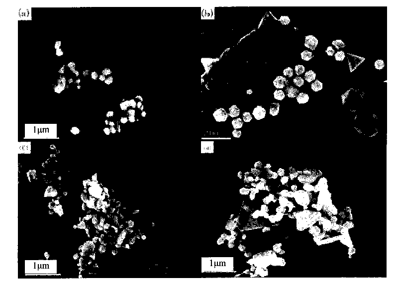
[0044] The scanning electron micrograph of the product obtained under the conditions of iohexol 0.19, 0.38, 1.9, 3.8ml is as follows figure 1 shown.

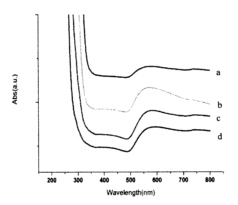
[0045] The ultraviolet diagram of the product obtained under the conditions of iohexol 0.19, 0.38, 1.9, 3.8ml is as follows figure 2 shown.

Embodiment 2

[0047] 1) Get 0.38ml iohexol solution (300mg I / ml), be dissolved in 22ml ultrapure water respectively, stir;

[0048] 2) Add 3 mL of chloroauric acid solution (0.01M), and stir at room temperature for 10 minutes to obtain a reaction precursor solution;

[0049] 3) Pour the reaction precursor solution into a polytetrafluoroethylene-lined stainless steel reaction kettle, and react at 120°C for 1.5h;

[0050] 4) The reaction system was naturally cooled to room temperature. Centrifuge twice at a speed of 4000 rpm to separate the synthesized gold nanoparticles from excess iohexol in the solution, and then wash twice with ultrapure water.

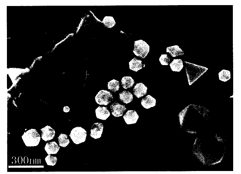
[0051] The scanning electron microscope image of the prepared iohexol-wrapped icosahedral gold nanoparticles is shown in image 3 shown.

[0052] The transmission electron microscope image of the prepared iohexol-wrapped icosahedral gold nanoparticles is shown in Figure 4 shown.

Embodiment 3

[0054] 1) Get 0.38ml iohexol solution (300mg I / ml), be dissolved in 22ml ultrapure water respectively, stir;

[0055] 2) Add 3 mL of chloroauric acid solution (0.01M), and stir at room temperature for 10 minutes to obtain a reaction precursor solution;

[0056] 3) adjusting the reaction precursor solution to pH=7 with Na(OH);

[0057] 4) Pour the reaction precursor solution with pH=7 into a polytetrafluoroethylene-lined stainless steel reaction kettle, and react at 120°C for 1 hour;

[0058] 5) The reaction system was naturally cooled to room temperature. Centrifuge twice at a speed of 4000 rpm to separate the synthesized gold nanoparticles from excess iohexol in the solution, and then wash twice with ultrapure water.

[0059] The scanning electron microscope image of the prepared iohexol-wrapped spherical gold nanoparticles is as follows: Figure 5 shown.


PUM

PropertyMeasurementUnit
Particle sizeaaaaaaaaaa

Abstract

The invention discloses a CT imaging contrast agent and a preparation method thereof, relating to a contrast agent and providing a CT imaging contrast agent with higher chemical stability, better CT contrast effect and biocompatibility and controllable grains and a preparation method thereof. The CT imaging contrast agent contains nano gold and iohexol and is structurally characterized in that nano gold grains are cores, and the iohexol are packed on the peripheries of the nano gold grains. The preparation method comprises the following steps of: dissolving an iohexol solution in hyperpure water to prepare an iohexol solution the iohexol concentration of which is 1.2-6mg I/ml; adding a chloroauric acid solution in the iohexol solution to prepare a reaction precursor solution the chloroauric acid concentration of which is 0.2-2mM; pouring the reaction precursor solution into an agitated reactor for reaction, and cooling reaction system to the room temperature; and centrifuging the cooled system to enable the synthetized nano gold grains to be separated from the surplus iohexol in the solution and then cleaning to obtain the CT imaging contrast agent.

Description

technical field [0001] The present invention relates to a kind of contrast agent, especially relate to a kind of with iohexol and chloroauric acid as raw material, water is solvent, adopt hydrothermal method one-step control synthesis iohexol wrapped nano-gold, mainly used in CT (X-ray Computed tomography) contrast agent can make the display of the image clearer and more accurate, and improve the early correct diagnosis rate of the disease. Background technique [0002] X-ray computed tomography (X-ray computed tomography, CT) refers to a non-invasive imaging in which the X-ray beam scans around a certain layer of the human body, measures the absorption coefficient of each unit volume in this layer, and then reconstructs it with a computer. technology. According to the different absorption and transmittance of X-rays by different tissues of the human body, it uses highly sensitive instruments to measure the human body, and then inputs the data obtained from the measurement ...

Claims


Application Information

Patent Timeline
IPC IPC(8): A61K49/04
Inventor 翁建钟鹭斌李林梅
Owner XIAMEN UNIV
Why Eureka
Try Eureka