Ultrasonic wave tissue-estimating device

An evaluation device, ultrasonic technology, applied in the direction of ultrasonic/sonic/infrasonic Permian technology, ultrasonic/sonic/infrasonic image/data processing, organ movement/change detection, etc. , operability is not strong and other problems, to achieve the effect of eliminating the impact

Inactive Publication Date: 2010-06-23
李干
View PDF0 Cites 11 Cited by
  • Summary
  • Abstract
  • Description
  • Claims
  • Application Information

AI Technical Summary

Problems solved by technology

However, there have been previous studies using ultrasound power Doppler semi-quantitative methods, which were to observe the blood flow distribution in the kidney ultrasound energy map with the naked eye, grade and score the perfusion of renal blood flow, and study the relationship between the score and the glomerular filtration

Method used


Image

  • Ultrasonic wave tissue-estimating device
  • Ultrasonic wave tissue-estimating device
  • Ultrasonic wave tissue-estimating device

Examples


Embodiment 1

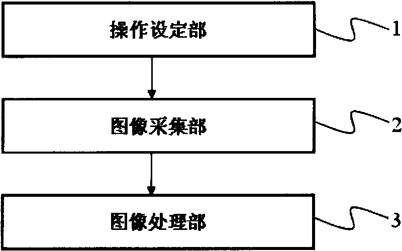
[0027] Such as figure 1 As shown, the ultrasonic tissue evaluation device of the present invention has: an operation setting unit 1 , an image acquisition unit 2 , and an image processing unit 3 .

[0028] The operation setting unit 1 uniformly sets the examination conditions and ultrasonic instruments of all examinees so that the results obtained are comparable. The setting of the examinee's examination conditions includes the examinee's pre-examination preparation, body position and movement during the ultrasonic examination, and the like. Ultrasound instrument settings include depth, probe gray scale frequency, probe color frequency, filtering, dynamic range, etc.

[0029] This is because power Doppler is extremely sensitive to motion. In order to minimize the interference of other factors, it is necessary to unify all controllable inspection conditions during the inspection as much as possible, otherwise different settings will give different results.

[0030] The image ...

Embodiment 2

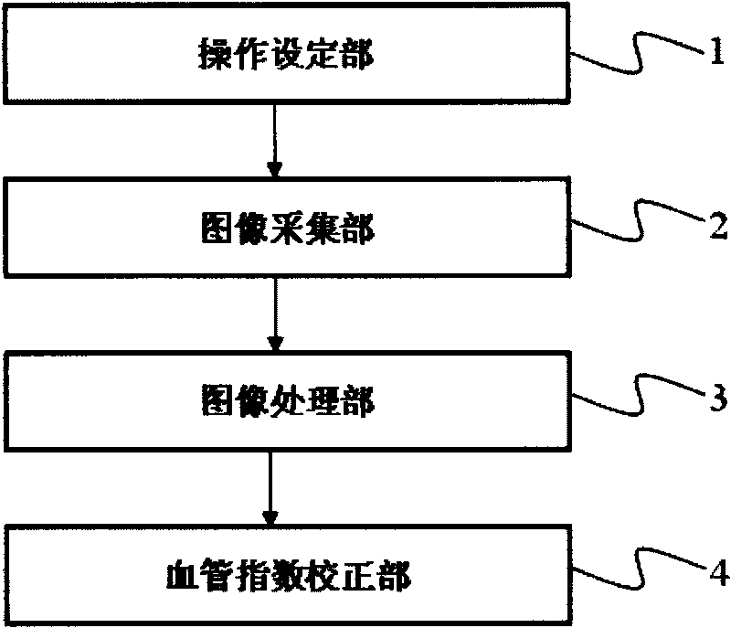
[0041] Such as figure 2 As shown, the ultrasonic tissue evaluation device of the present invention has: an operation setting unit 1 , an image acquisition unit 2 , an image processing unit 3 , and a vascular index correction unit 4 .

[0042] The operation setting unit 1, the image acquisition unit 2, and the image processing unit 3 of the ultrasonic tissue evaluation device of the present invention are the same as those in the first embodiment, and their descriptions are omitted here, and only the differences will be described.

[0043]The vascular index correction unit 4 corrects the vascular index according to the tissue depth d measured based on the collected ultrasound data, because the ultrasonic energy Doppler signal will increase with the increase of the distance between the skin surface and the examined tissue. attenuation. As shown in FIG. 7 , the tissue depth d refers to the average value of the shortest distance d1 and the furthest distance d2 between the inspect...

Embodiment 3

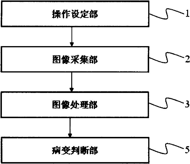
[0045] Such as image 3 As shown, the ultrasonic tissue evaluation device of the present invention has: an operation setting unit 1 , an image acquisition unit 2 , an image processing unit 3 , and a lesion determination unit 5 .

[0046] The operation setting unit 1, the image acquisition unit 2, and the image processing unit 3 of the ultrasonic tissue evaluation device of the present invention are the same as those in the first embodiment, and their descriptions are omitted here, and only the differences will be described.

[0047] The lesion judging part 5 compares the renal vascular index from the image processing part 3 with the standard value stored in the memory (not shown), and obtains the changes of renal function (such as GFR) and pathology.

[0048] GFR (Glomerular Filtration Rate) refers to the plasma flow rate filtered by the kidney per unit time, such as Figure 8 As shown, GFR is positively correlated with renal vascular index VI, and renal VI value can reflect ...


PUM


Abstract

The invention provides an ultrasonic wave tissue-estimating device which can provide an index with a certain help to diagnose the tested tissue according to an ultrasonic gray-scale image and a co-frame color energy Doppler image. The ultrasonic wave tissue-estimating device estimates the vessel distribution of the tissue by ultrasonic energy Doppler and is characterized by being provided with an operation-setting part, an image-collecting part and an image-processing part, wherein the operation-setting part sets a uniform image-collecting condition; the image-collecting part adjusts the color gain, collects the ultrasonic gray-scale image and the co-frame color energy Doppler image of the tissue cross section and outputs cross section image data; and the image-processing part receives the cross section image data from the image-collecting part, defines the interesting area of the tested tissue and sorts the color Doppler signal strength into a plurality of scales according to a color-scale standard map to weight so as to acquire the vessel index of the tissue.

Description

technical field [0001] The invention relates to an ultrasonic tissue evaluation device for quantitatively evaluating tissue by using ultrasonic Doppler energy data. Background technique [0002] In recent years, imaging methods, such as CT, MRI, and radionuclide, have been more and more widely used in the evaluation of visceral function. Ultrasound is mainly used to examine the gross morphology of tissues, and is rarely used to evaluate tissue function and pathological changes. [0003] The emergence of power Doppler provides the possibility to quantitatively study blood flow in tissues. According to the principle of power Doppler, the ultrasonic energy map shows the Doppler frequency shift signal generated by the movement of red blood cells, and its intensity directly reflects the number of red blood cells in the tissue, that is, the blood flow in the tissue. However, there have been previous studies using ultrasound power Doppler semi-quantitative methods, which were to ...

Claims


Application Information

Patent Timeline
IPC IPC(8): A61B8/08
Inventor 李乾
Owner 李干
Why Eureka
Try Eureka